What is this
The smooth contact between the bone tissue of the knee and the adjacent cartilage depends on the condition of the synovial fluid . This substance provides normal nutrition to the knee joint. Any deviation in the volume of the contents of the synovial bursa from the norm indicates the development of pathology. Instability of the knee joint can be caused by both a lack of synovial filling and its excess amount - effusion.
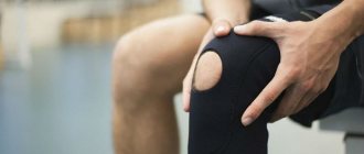
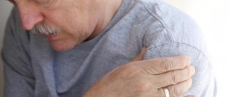
The disease is accompanied by severe pain and movement disorder of the affected joint. At the same time, the person’s general well-being worsens. In the absence of qualified treatment, the disease can cause serious complications, including disability.
Fluid in the knee joint. Treatment without arthroscopy
The accumulation of fluid in the joint cavity causes compression of soft tissues, blood vessels, disrupts the nutrition of cartilage and causes pain. To get rid of this problem, medicine suggests pumping out the fluid using instruments, a mini-surgery, but this injures the joint and does not get rid of the reason why the fluid has accumulated, so over time the operation only leads to increased pain and inflammation.
Fluid accumulation, effusions. Reason for formation
First of all, these are degenerative-dystrophic changes in the joint, which are called osteoarthritis, osteoarthritis, and trauma can also cause effusions. Against this background, inflammatory processes develop in the soft tissues of the knee, bursitis, synovitis, and the synovial membrane lining the inner surface of the articular cavity becomes inflamed. This leads to the active release of synovial fluid, which compresses the surrounding tissues, supporting the development of the inflammatory process, pain and movement disorders.
To relieve pain, doctors suggest surgical intervention - pumping out fluid (pumping out exudate) from the knee joint.
How is the operation performed?
The operation is performed under local anesthesia, usually using arthroscopy. Using a micro-incision (puncture), the instrument penetrates into the joint cavity. Next, the surgeon first pumps out the exudate, then can rinse the cavity with an antiseptic solution, administer medications, antibiotics, and remove elements of the joint (depending on the indications).
After surgery, NSAIDs (non-steroidal anti-inflammatory drugs) are usually prescribed, and immobilization is recommended.
Consequences of joint surgery
Pumping out the fluid does not eliminate the disease that caused it, namely synovitis of the knee, osteoarthritis and arthritis
. Pathological factors that destroy the joint remain. Moreover, surgical intervention further injures and weakens the joint, leading to atrophy of the deep skeletal muscles that nourish and moisturize the knee through a network of blood vessels. Therefore, after the operation, degenerative-dystrophic processes only intensify; over time, the knee begins to hurt and become inflamed again.
Therefore, in order to really cure a joint by eliminating pain, fluid accumulation, and restriction of movement, it is necessary to find and eliminate the root of the problem.
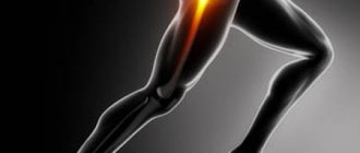
Why does the knee joint begin to deteriorate and arthrosis develop?
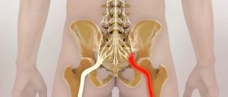
The knee is one of the most complex, vulnerable joints that experiences severe stress when walking. And its health depends solely on the health and full functionality of the muscles that are adjacent to the knee joint: quadriceps, hamstrings, gastrocnemius, semitendinosus, biceps femoris, plantaris, sartorius and subtlest muscles, as well as spinal muscles in the lumbar region and buttocks, which are partially take on the shock load when walking.
If one muscle weakens and stops working when walking, the knee begins to experience increased stress, the adjacent soft tissues spasm, the ligaments of the knee joint become pathologically shortened, and the nutrition and blood supply to the joint is disrupted. As a result, the development of osteoarthritis (destruction of cartilage tissue with subsequent deformation), inflammation of the joint capsule (synovial membrane) that surrounds the joint, stiffness and pain.
How to get rid of fluid and joint pain without surgery
To cure a knee, it is not enough to simply pump fluid out of it or take painkillers, anti-inflammatory drugs, or inject chondroprotectors. The most important thing is to take measures to eliminate the factors that destroy the joint. In the case of the knee, this is unbalanced muscle function, hypotomy (weakness) of individual muscles of the legs, lower back, imbalance of the muscles of the pelvic bones, hernia and protrusion in the lumbar spine
. Each person may have his own reason, and first it must be identified.
This cannot be done using MRI. Therefore, a special muscle kinesiological test (kinesiological testing) was developed, in which a specialist, using various manipulations, evaluates the tone of the muscles of the whole body and checks the ability of the muscle to contract in different positions. And thus discovers which muscles of the body do not work and overstrain the knee joint.
This way, it is possible to identify many disorders even before the occurrence of degenerative changes in the joint (which are diagnosed using MRI and X-rays) and, by taking timely measures, avoid serious problems in the future: injuries, the development of hernias, protrusions, osteoarthritis.
Case Study
.
Complaints: pain in the knee; based on MRI results, a diagnosis of gonarthrosis was made; excess fluid accumulation was found in the articular cavity. Based on the results of a kinesiological test, the doctor discovered that the cause of constant pathological overload of the knee joint was hypotomy of the quadriceps femoris muscle and shortening of the lumbar iliac muscle, the hip extensors
. As a result, the stability of the pelvis and knee joint is disrupted, and the menisci are overloaded, depending on the location of the compensatory overloaded muscle.
After the kinesiologist determines the root of the problem, the next stage begins - the treatment itself. It consists, first of all, in normalizing the tone of the muscles surrounding the knee joint, relieving tension from spasms and returning
full
functionality of weakened, idle muscles.
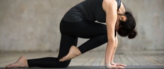
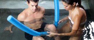
Regular strength exercises are not suitable for this; on the contrary, they are dangerous for new, more serious injuries. This requires special therapeutic movement, with the help of simulators used in rehabilitation. Kinesio simulators allow you to engage deep skeletal muscles without dangerous axial load on the joints and spine. The program of such exercises is drawn up by a doctor based on the results of a kinesiological test.
The goal of treatment is to relieve muscle spasms that cause pain, restore strength and elasticity of muscles, ligaments, tendons of the knee and hip joint, lumbosacral spine, strengthen weakened muscles, and launch natural mechanisms of regeneration of osteochondral tissue. At the same time, posture and pelvic muscles are corrected, correct movement patterns are formed, and a strong muscle corset is developed that nourishes and supports the spine.
Specialist consultation is required.
Doctor’s consultation + diagnostics using applied kinesiology – 1000 rubles.
Call, write! Tel.: (843) 570-55-25 , WhatsApp: 79655968085 or VKontakte group .
Read us on Yandex Zen:
Follow us on Instagram:
Don't miss out on the fun! Subscribe to our news:
Subscribe to news from the Kazan Kinesitherapy Center
Center promotions, therapeutic exercises and useful tips from our specialists on how to independently maintain the health of your joints and spine without medications
Similar articles:
Joints hurt. What is the reason? How to treat?
Osteoarthritis affects 98 percent of the adult population. How to cure without drugs?
Myofascial release and massage in healing the spine and joints
Summary
Article Name
Fluid in the knee joint. Treatment without arthroscopy
Description
Causes of fluid accumulation and effusions in the joint. What to do? Effective methods for healing the knee joint
Author
Spine Health Center
Publisher Name
Kazan Kinesitherapy Center
Causes
Effusion can form for a variety of reasons. Most often, inflammatory fluid accumulates in the presence of the following factors:
- Knee injury.
- Excess weight.
- Gout.
- Rheumatism, arthritis.
- Autoimmune diseases.
- Bursitis.
- Dermatomyositis.
- Ankylosing spondylitis.
According to statistics, the most common cause of knee effusion is injury . This could be a meniscus injury, fracture, severe bruise, etc.
Synovitis of the knee joint
Acute aseptic synovitis
In acute aseptic synovitis, the joint increases in volume within several hours or several days.
Often there is a feeling of fullness, mild pain is possible, aggravated by movement. Upon examination, there is a noticeable disturbance in the shape of the joint, smoothing of the contours and bulging on the sides of the patella. Minor soft tissue swelling may occur. The presence, localization and severity of pain on palpation are determined by the underlying pathology. Fluctuation and voting of the patella are detected: when pressure is applied to the patella, it sinks into the joint, and when the pressure stops, it “floats up”. In some cases, weakness, malaise and a slight increase in temperature are noted. The severity of symptoms depends on the amount of fluid in the joint. When a large amount of effusion accumulates, the pain and feeling of fullness intensify, the swelling increases, the skin of the joint becomes shiny, and sometimes hyperemia appears. The nature of the effusion is usually determined by the cause of the disease. With traumatic injuries and hemophilia, the effusion is hemorrhagic (the fluid is colored with blood), with other aseptic synovitis it is serous (at first the fluid is clear, with a straw-colored tint, later some darkening is observed, the effusion becomes yellow and less transparent).
Chronic aseptic synovitis
Chronic aseptic synovitis occurs in waves, with exacerbations alternating with remissions. During the period of exacerbation, the picture resembles acute aseptic synovitis, but the symptoms are often smoothed out and less bright. In some cases, a small amount of effusion is observed. Pain and limitation of movement in the joint are caused by thickening of the synovial membrane. The severity of symptoms during remission depends on the underlying disease, duration and frequency of exacerbations of synovitis. There may be limited movement, dull aching pain and fatigue when walking.
Acute purulent synovitis
Acute purulent synovitis is characterized by pronounced general and local symptoms. The joint is increased in volume, sharp pain, local hyperemia and hyperthermia are noted. Movements are severely limited or practically impossible due to intense pain. The general condition is disturbed, weakness, chills, weakness, nausea and fatigue appear. Body temperature is elevated to febrile levels. In severe cases, hallucinations, delusions, and confusion are possible. Palpation of the joint is sharply painful. If left untreated, the infection spreads to other structures of the joint, causing acute purulent arthritis.
Symptoms
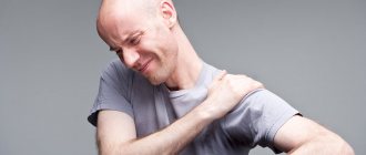
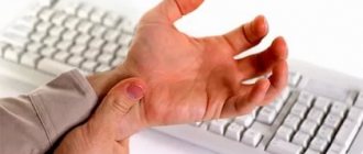
The symptoms of this disease, despite the different causes, are always approximately the same. It all starts with severe pain. At the same time, the pain does not go away for a minute, and its severity does not depend on whether the person is in motion or at rest.
If the effusion contains purulent fluid, then there is throbbing pain .
Swelling is also observed . At the initial stage of the disease, it may be insignificant and disappear from time to time, but then it increases. Due to swelling, the knees become asymmetrical.
The next sign is redness and peeling of the skin at the site of the affected joint. In addition, a person may experience itching in the indicated area.
A characteristic feature of effusion in the knee joint is the inability to bend and straighten the joint normally. When moving, there is a feeling as if something foreign is inside the knee. Sometimes both local and general body temperature may rise.
Treatment of fluid accumulation due to a torn meniscus
If we take a closer look at the case when fluid accumulation is a symptom of a meniscal injury, then the treatment at the Elena Malysheva Medical Center will be as follows.
The meniscus is sutured using arthroscopic minimally invasive intervention. In this way, it will be possible to restore the integrity of the damaged area of tissue.
One of the stages of arthroscopic surgery is securing the fixing element to the cartilage. This will help protect your knee during exercise.
Based on statistics, 90% of meniscus suturing operations are successful, and thanks to minimally invasive intervention, the patient returns to his previous life in the shortest possible time.
Don't endure the pain! Diseases of the knee joints bring great discomfort and unbearable pain. Therefore, do not self-medicate, make an appointment at the Elena Malysheva Medical Center!
Diagnostics
Timely diagnosis of the disease is almost half the success in treatment. So, at the first signs of illness, you need to consult a doctor. Diagnosis always begins with an examination and interview of the patient. Next, the doctor may prescribe the following types of examinations:
- Arthroscopy.
- CT.
- MRI.
- Ultrasound.
- Biopsy and puncture (except for purulent effusions).
Laboratory tests of the synovial fluid itself are also required to determine its composition, as well as urine and blood tests.
Only after receiving the results of all diagnostic measures, the doctor can make an accurate diagnosis and prescribe treatment.
Diagnosis of fluid accumulation in the knee joint
If such symptoms occur, it is recommended to immediately consult a specialist. Fluid accumulation can be a symptom of serious medical conditions that should be diagnosed as early as possible.
At the first consultation, the specialist will palpate the knee joint and prescribe a series of instrumental examinations. Among them:
- Puncture of synovial fluid;
- Radiography;
- Ultrasound examination;
- Magnetic resonance imaging;
- Arthroscopy.
Also during diagnosis, it is important to puncture the accumulated fluid. This will help determine if there is pus or other inclusions in the fluid.
Clinical examinations include a general blood and urine test.
Treatment
The disease described can only be combated with complex treatment. There are four main directions of influence on a sore knee:
- Puncture.
- Drug therapy.
- Immobilization of the knee.
- Operation.
The puncture is performed almost immediately after diagnosis. During this procedure, the doctor pumps out fluid from the knee with a special instrument. At the same time, part of this liquid is sent for laboratory testing.
Drug treatment is carried out with the participation of the following drugs:
- Nonsteroidal anti-inflammatory drugs : Indomethacin, Diclofenac, Ketoprofen, etc.
- Steroid injections : Hydrocortisone, Betamethasone, Prednisone, Diprospen.
- Ointments and gels : Deep Relief, Diclofenac-gel, Voltaren-gel, etc.
Sometimes compresses with Ichthyol or Dimexide . Perform them twice a day for 40-60 minutes.
Treatment of fluid accumulation in the knee joint
Treatment for fluid accumulation in the knee will depend on the underlying cause of the symptom. Treatment will be either conservative or surgical.
Conservative treatment of fluid accumulation
The doctor will recommend taking the following medications.
- Painkillers to relieve the patient's condition.
- Nonsteroidal anti-inflammatory drugs will relieve inflammation. If the inflammation is severe, the doctor will prescribe steroids.
- Immunomodulators or immunosuppressants, in order to influence the functioning of the immune system in one way or another.
- Chondroprotectors are prescribed to normalize the functioning of the joint.
- To get rid of swelling, a specialist will prescribe a course of antihistamines.
- Antibacterial agents are used for infections. The patient can take them orally or inject them into the joint.
In addition to medications, the specialist will prescribe other procedures that will have a beneficial effect on the healing of the joint and accelerate regeneration:
- Reflexology or physiotherapy;
- Physiotherapy;
- Compresses;
- Exposure to dry heat.
Surgery
Modern medicine makes it possible to perform joint operations in a minimally invasive way. That is, using an arthroscope. This instrument is inserted into the body through small incisions.