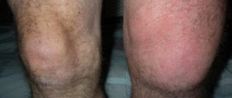
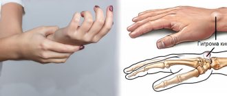
A cyst in the shoulder joint is a round benign tumor. The size of the formation can reach a couple of centimeters. The cystic formation appears as an almost immovable shell around the joint exudate. It should also be noted that the neoplasm is not fused with the subcutaneous tissue on the shoulder, has clear contours and a compacted structure.
A round benign tumor.
Symptoms
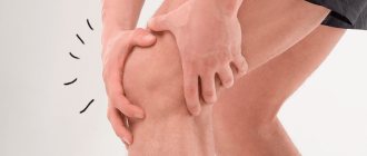
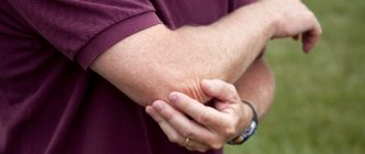
The manifestation of symptoms against the background of a cystic formation in the shoulder is due to the size of the tumor. At the early stage of its formation, the signs are weakly expressed or the disease is completely asymptomatic. As the cyst enlarges, a bulge forms at its location. As a result, the patient complains of:
- discomfort;
- pain (when a person moves a joint);
- tingling;
- numbness.
Under the skin you can determine the formation of a round shape.
When you feel the affected area under the skin, you can determine the formation of a round shape. The joints are functioning normally. Sometimes there is a slight stiffness in shoulder movements.
Bone cyst
Solitary bone cyst
Boys aged 10-15 years are most often affected.
At the same time, earlier development is also possible - the literature describes a case of a solitary cyst in a 2-month-old child. In adults, bone cysts are extremely rare and usually represent a residual cavity after an undiagnosed disease suffered in childhood. As a rule, cavities arise in long tubular bones; the first place in prevalence is occupied by bone cysts of the proximal metaphysis of the femur and humerus. The course of the disease in the initial stages is in most cases asymptomatic, sometimes patients note slight swelling and minor unstable pain. In children under 10 years of age, swelling is sometimes observed and contractures of the adjacent joint may develop. With large cysts in the area of the proximal diaphysis of the femur, lameness is possible, with damage to the humerus - discomfort and unpleasant sensations with sudden movements and raising the arm.
The reason for visiting a doctor and the first symptom of a solitary bone cyst is often a pathological fracture that occurs after a minor traumatic impact. Sometimes the injury cannot be detected at all. When examining a patient with the initial stages of the disease, local changes are not pronounced. There is no edema (with the exception of edema after a pathological fracture), there is no hyperemia, the venous pattern on the skin is not pronounced, there is no local or general hyperthermia. Minor muscle atrophy may be detected.
When palpating the affected area, in some cases it is possible to detect a painless club-shaped thickening with bone density. If the cyst reaches a significant size, the cyst wall may bend when pressure is applied. In the absence of a fracture, active and passive movements are complete, support is preserved. When the integrity of the bone is broken, the clinical picture corresponds to a fracture, but the symptoms are less pronounced than in the case of a normal traumatic injury.
Subsequently, a staged course is observed. Initially, the cyst is localized in the metaphysis and connects to the growth zone (osteolysis phase). With large cavities, the bone at the site of the lesion “swells” and repeated pathological fractures may develop. It is possible to form a contracture of a nearby joint. After 8-12 months, the cyst turns from active to passive, loses connection with the germinal zone, gradually decreases in size and begins to shift towards the metadiaphysis (demarcation phase). After 1.5-2 years from the onset of the disease, the cyst appears in the diaphysis and does not manifest itself clinically (recovery phase). At the same time, due to the presence of a cavity, the strength of the bone at the site of the lesion is reduced, so pathological fractures are also possible at this stage. The outcome is either a small residual cavity or a limited area of osteosclerosis. Clinically, complete recovery is observed.
To clarify the diagnosis, an x-ray examination of the affected segment is performed: x-ray of the femur, x-ray of the humerus, etc. Based on the x-ray picture, the phase of the pathological process is determined. In the osteolysis phase, the image reveals a structureless rarefaction of the metaphysis in contact with the growth zone. In the demarcation phase, radiographs show a cavity with a cellular pattern, surrounded by a dense wall and separated from the growth zone by a section of normal bone. During the recovery phase, images reveal an area of bone compaction or a small residual cavity.
CT stop. Calcaneal cyst on the right (on the left – for comparison – normal).
Aneurysmal bone cyst
It is less common than solitary. Usually occurs in girls 10-15 years old. It can affect the pelvic bones and vertebrae; the metaphyses of long tubular bones are less commonly affected. In contrast, a solitary bone cyst usually occurs after injury. The formation of a cavity is accompanied by intense pain and progressive swelling of the affected area. Upon examination, local hyperthermia and dilatation of the saphenous veins are revealed. When localized in the bones of the lower extremities, a violation of support is noted. The disease is often accompanied by the development of contracture of a nearby joint. With bone cysts in the vertebrae, neurological disorders appear due to compression of the spinal roots.
There are two forms of aneurysmal bone cysts: central and eccentric. During the course of the disease, the same phases are distinguished as with solitary cysts. Clinical manifestations reach a maximum in the osteolysis phase, gradually decrease in the demarcation phase and disappear in the recovery phase. Radiographs in the osteolysis phase reveal a structureless focus with an extraosseous and intraosseous component; with eccentric cysts, the extraosseous part is larger in size than the intraosseous part. The periosteum is always preserved. In the demarcation phase, an area of sclerosis forms between the intraosseous zone and healthy bone, and the extraosseous zone becomes denser and decreases in size. During the recovery phase, radiographs reveal an area of hyperostosis or a residual cavity.
MRI of the pelvis. Bone cyst in the left ilium with a hyperintense signal.
Diagnostics
To identify such a disease in the shoulder joint, diagnostic measures are prescribed. First you need to undergo an ultrasound examination of the shoulder. This procedure is the simplest and cheapest diagnostic method and does not have a negative effect on the body. An ultrasound scan of this area makes it possible to assess the condition of all shoulder elements, muscle and ligament fibers, cartilage tissue and the neoplasm itself, and it is quite informative.
Pathology on X-ray.
The information obtained will allow the doctor to more accurately determine how affected the shoulder is. In addition, if necessary, the doctor will also prescribe an X-ray examination of this area. Thanks to this diagnosis, the specialist will be able to accurately determine the density of the tumor tissue and its location. Then the patient needs to take a blood test and undergo an MRI to understand the nature and etiology of the tumor.
Etiology: causes of shoulder cysts
The disease refers to benign formations. It has a dense shell, clear outlines and compacted texture. A tumor in the area of the head of the bone is a characteristic pathology related to dystrophic fibrous changes. Athletes, loaders and people involved in physical labor are susceptible to it. Doctors identify a number of reasons why the disease occurs:
- arthrosis and osteoarthritis;
- inflammation;
- rheumatoid arthritis;
- domestic and sports injuries;
- tendovaginitis, bursitis, synovitis;
- degenerative changes;
- sudden loads.
Education is more often diagnosed in elderly patients.
The subchondral cyst has the appearance of multiple cavities filled with myxoid substance. Most often it forms in elderly patients with degenerative changes in the joint. Its dimensions are small, 1.5-2 cm. Sometimes the reason for the formation of a neoplasm is the presence of a silicone prosthesis. Subchondral formations arise as a result of increased pressure in the cavity and are secondary in nature.
Treatment
Prescription of appropriate treatment for a cyst of the shoulder joint should be carried out by a doctor in accordance with the research results obtained, as well as taking into account the symptoms of the pathology.
Conservative
For small cystic formations, conservative treatment is usually prescribed. The goal of such therapy is to eliminate synovial exudate from the tumor. A similar manipulation is carried out by puncturing the tumor with a special needle. Once the liquid has been removed, the resulting void is carefully sanitized using an antiseptic. In addition, upon completion of the manipulation, the patient is prescribed a course of treatment with non-steroidal anti-inflammatory drugs in injection form.
Use healing agents in the form of ointments.
Most often, the growth of the tumor stops at the end of the course of injections. After completing all the procedures, a tight bandage and plaster are applied to the shoulder. When the plaster and bandage are removed, the patient is prescribed treatment:
- compresses;
- physiotherapeutic procedures;
- healing agents in the form of ointments.
After some time, due to the inevitable tension of the shoulder, the cavity is refilled with synovial exudate. As a result, a second puncture may be required.
Surgery
During traditional surgery, the tumor tissue is completely removed. The postoperative wound is sutured with a special suture that strengthens the joint tissue of the shoulder.
Rehabilitation and care of the joint after surgery
During the rehabilitation period, the main task is to restore full blood circulation in the joint area and stabilize the motor mode. The increase in load on the joint should be gradual. All exercise therapy activities must be carried out under the supervision of medical personnel. Rehabilitation measures are prescribed by a physiotherapist.
A set of exercises is selected individually for each patient and is aimed at restoring muscle strength and motor activity. As a rule, therapeutic exercises are prescribed from the 2nd day after surgery in a ward. And from 3-5 days they continue classes in the exercise therapy room.
The task of the late rehabilitation period during the treatment process is to fully restore the functionality of the shoulder, which usually lasts about 3-4 months. Most often prescribed: exercise therapy, massage, occupational therapy, swimming pool, physiotherapy, mainly muscle electrical stimulation.
Folk remedies
In case of cystic formation at an early stage, to eliminate the inflammatory process and pain syndrome, you can use alternative medicine. Currently, there are many different recipes:
- The celandine herb needs to be crushed, poured with hot water so that it covers the raw material, and leave for 15 minutes. The infusion is used to apply compresses to the affected area twice a day.
- Aloe juice should be mixed with lemon juice in equal quantities (1 tablespoon each), streptocide should be added (1 gram). The fabric is soaked in the resulting solution and applied to the injured shoulder overnight.
Compresses are made from aloe juice and lemon juice. - Mix cloves with dandelion and add water. The mixture should be boiled over low heat for 20 minutes, then cool slightly. Then add 1 tbsp to the product. a spoonful of alcohol. Soak a cloth in the resulting mixture, apply to the affected area and leave for 3 hours.
- A cabbage leaf should be dipped in boiling water for a couple of seconds, then greased with honey and applied to the location of the tumor. The dressing is left in place for 2 hours. The procedures must be completed within 10 days.
A cabbage leaf is smeared with honey and applied to the tumor. - Soak a piece of gauze in unrefined vegetable oil, apply it to the affected shoulder, and place cling film on top. This bandage should be done 2 times a day.
- To a glass of water you need to add 1 tbsp. a spoonful of apple cider vinegar, 1 teaspoon of honey. Mix everything thoroughly. Take the product half an hour before meals at least three times a day.
But it should be taken into account that treatment only with unconventional methods will not completely eliminate the cystic formation in the shoulder. In any case, you need to consult a doctor and undergo a course of drug therapy.
Healed patients highlighted the most important advantages of Top Ichilov
- The clinic’s specialists are high-level professionals who have undergone extensive training and specialization in the largest medical centers in the world. Working closely with each other, they provide effective diagnosis and treatment of a wide range of bone tumors.
- The clinic is equipped with modern equipment, with the help of which our doctors carry out accurate diagnostics. Cysts are treated using techniques such as puncture, injection of corticosteroids, composite alloplasty, etc.
- The clinic hospitably opens its doors to all foreign patients in need of high-quality and inexpensive medical services. Welcoming and friendly staff, cozy rooms, polite doctors and a personal curator-translator - these are just some of the points due to which Top Ichilov is widely known far beyond the borders of Israel.
- 5
- 4
- 3
- 2
- 1
(3 votes, average: 3.7 out of 5)