The inner thigh often hurts when:
- pathologies of the human genitourinary system, including cancer;
- progressive vascular pathologies;
- inguinal hernia;
- problems with the gastrointestinal tract (constant constipation, obstruction, etc.);
- during pregnancy or before childbirth.
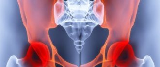
Painful sensations of varying intensity cause defects of the femoral joint:
- injuries (dislocation, fracture, bruise);
- arthritis;
- bursitis;
- synovitis;
- coxarthrosis;
- pelvic abscess and others.
Pain in the inner thigh can cause injury to the adductor muscles, which are responsible for bringing the leg toward the midline of the body. Rupture or stretching can cause:
- intensive sports (football, tennis, hockey, cycling, etc.);
- overload during physical activities, including dancing;
- unnatural jerking movements of the lower extremities.
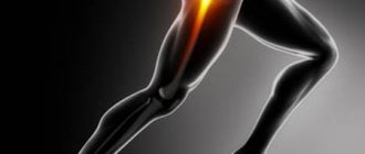
Causes of hip pain
Traumatic injuries
A hip bruise occurs when a blow or fall occurs and is manifested by moderate local pain.
At first the pain is sharp, then dull, aching, intensifying with palpation and movement. Due to the large array of soft tissues, severe swelling often develops. Bruising is possible. The function of the limb is usually slightly limited, support is preserved. A hematoma also forms after blows and falls; its manifestations resemble a bruise. It differs from a bruise in the bursting or pressing nature of the pain, the formation of dense local edema, in the area of which, after some time, an area of fluctuation appears. With deep-lying hematomas, the swelling is nonspecific, fluctuation may be absent. Hematoma can be suspected by a significant increase in swelling in the first days or its persistence for a long time.
Damage to the quadriceps tendon is observed in athletes, but less often develops in everyday life. It is characterized by acute pain along the front surface of the thigh, just above the knee joint, sometimes by a cracking sound at the time of injury. Subsequently, the pain subsides, but remains quite intense. In the area of damage, pain, swelling of soft tissues, and a defect at the site of the rupture are detected. With a complete rupture, support and independent straightening of the lower leg are impossible; with an incomplete rupture, it is difficult.
The cause of a diaphyseal femoral fracture is high-energy impacts: falls from a height, road and industrial injuries. The damage is accompanied by unbearable explosive pain. Subsequently, the pain is very intense and intensifies when attempting to move, palpate, or passively shift the limb. The thigh is swollen and deformed. The leg is shortened. Crepitation and pathological mobility are detected. The general condition is disturbed, traumatic shock is possible.
A pathological fracture of the femoral diaphysis can be diagnosed with bone cysts, tumors, and some hereditary and infectious diseases. It has mild symptoms. The pain is minor or moderate. Displacement, pathological mobility and bone crunch are absent, which is why the injury is often mistaken for a bruise. Distinctive features of a pathological fracture are the long-term persistence of symptoms and the presence of previous pain during exercise or at rest.
Inflammatory diseases
Myositis of the thigh muscles occurs after unusual physical activity: prolonged walking or running, the first workouts after a long break. It is characterized by aching pain in the projection of the affected muscle or muscle group. There is increased pain on palpation and muscle tension. When palpated, a diffuse thickening of the muscle is determined; slight swelling and slight local hyperthermia are possible.
Quadriceps tendonitis is usually found in active people over 40 years of age and manifests itself as vague, low-intensity nagging pain in the lower parts of the front of the thigh. The pain intensifies when the leg is extended. With biceps tendonitis, which is typical for runners, the pain is localized deep in the buttock, spreads down the back of the thigh, and intensifies during running, especially when accelerating. Subsequently, the pain becomes long-lasting, constant, and occurs after minor exertion, at rest, and at night.
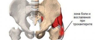
Pain on the outer surface of the thigh is sometimes accompanied by aseptic arthritis of the hip joint of various etiologies (reactive, allergic, post-traumatic). The pain is aching, dull, non-localized, complemented by soreness in the groin and buttock, limitation of movements, and difficulty walking.
Hip pain
Bone infections
Periostitis of the femur develops infrequently, as a rule, it is the result of injuries (bruises, fractures) or purulent lesions (deep infected wounds, abscesses, phlegmon, arthritis). In the first case, the process is aseptic in nature, accompanied by moderate pain, aggravated by palpation. Minor swelling is detected. The general condition is not disturbed.
Sometimes serous post-traumatic periostitis is observed, characterized by significant accumulation of fluid under the periosteum. The pathology is manifested by moderate arching pain, swelling, and deformation. With purulent inflammation of the periosteum, an acute onset with intense twitching, throbbing pain is observed. Local hyperemia, significant swelling of the segment, increased body temperature, chills, and fever are detected.
The femur is one of the most common locations of hematogenous osteomyelitis. Bone inflammation develops in childhood, often due to minor trauma or after an acute infection. It occurs with a sharp increase in temperature, fever, chills, and delirium. A day after the onset of hyperthermia, intense localized deep pain occurs in the thigh. The pain increases, becomes unbearable, forcing the patient to freeze in bed.
There is a local form of hematogenous osteomyelitis, in which the pain syndrome is less pronounced and the general condition is slightly disturbed. In some cases, a severe hurricane course of the disease is observed with a predominance of general symptoms. Post-traumatic and postoperative osteomyelitis are also manifested by pain, swelling, hyperemia, and disturbances in general condition, but the pain appears against the background of suppuration of an open fracture or postoperative wound, progresses more slowly, and does not reach the level of unbearable.
Infections of blood vessels and soft tissues
Pain in the thigh is observed with purulent lesions of soft tissue structures. At first it has a bursting or pressing character. The intensity of the pain syndrome quickly increases, the pain becomes throbbing, tearing, jerking, sharply limits movement, and disrupts night sleep. Swelling and increased local temperature are detected. The severity of general manifestations (hyperthermia, fever, symptoms of intoxication) depends on the extent of the purulent process. The cause of pain is:
- Furuncle.
A limited focus of inflammation, which is a convex, sharply painful formation, covered with bluish-purple skin. In the center of the formation there is a black core surrounded by pus. The general condition is usually not affected. - Carbuncle.
It consists of several closely spaced small foci merging into one infiltrate with a diameter of up to several centimeters. Severe hyperthermia and fever are observed. - Abscess.
A limited purulent focus, the diameter of which can vary from 2-3 to 10 or more centimeters. Accompanied by weakness, weakness, fever to febrile levels. - Phlegmon.
Widespread purulent inflammation without clear boundaries. It is characterized by intense pain throughout the thigh and severe intoxication.
From foci in soft tissues, the infection can spread through the lymphatic vessels. The manifestation of reticular lymphangitis is indicated by increased pain, deterioration of the general condition, and the appearance of a marble pattern around the primary inflammatory focus. With stem lymphangitis, a longitudinal strip appears on the thigh, along which pain on palpation, swelling, hyperemia, and cord-like tissue compaction are determined. When deep vessels are affected, stripes on the limb are not detected; rapidly increasing arching pain and lymphedema of the underlying sections are noted.
With phlebitis and thrombophlebitis of the superficial veins of the thigh, developing against the background of infectious lesions or varicose veins, rapidly progressing pain is detected along the vein. The pain intensifies when walking and is usually localized in the lower third of the segment. Phlebitis and thrombophlebitis of the deep veins are manifested by constant increasing pain, swelling, general hyperthermia, milky white coloration of the skin of the limb.
A special form of venous damage is postpartum thrombophlebitis, which occurs in the first weeks after childbirth. Intense pain on the inner side of the thigh is observed with inflammation of the great saphenous vein, combined with swelling and deterioration of the general condition. When the deep femoral veins are involved, unbearable sharp pain along the anterior inner surface of the thigh, significant swelling, pallor of the skin of the limb, and fever are noted.
Oncological diseases
Benign tumors of the femur are characterized by slow growth and a long, often multi-year course. Osteomas and chondromas manifest themselves as minor, transient, unclear pain; with chondromas in children, deformities and shortening of the limb are possible. With osteoid osteomas, the pain is intense and sharp. The growth of neoplasia is accompanied by increased pain and the appearance of palpable bone density formations.
The most common malignant tumors of the hip include osteosarcoma, chondrosarcoma, and Ewing's sarcoma. Chondrosarcomas are usually localized in the upper part of the bone, osteogenic sarcomas in the upper or lower part, and Ewing sarcomas in the lower part. The pain with such neoplasms is initially dull, unclear, quickly intensifies, becomes unbearable, and is combined with the appearance of a venous pattern, deformation, weight loss, and general weakness.
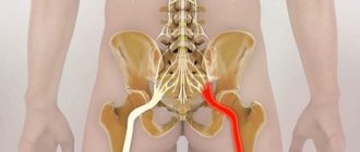
Neurological causes
Hip pain is often caused by neurological diseases. Distinctive features of such pain are shooting, burning, shooting or piercing nature (some patients compare the sensations to the “strike of a dagger”), combination with paresthesia, weakening of muscle strength, and sensitivity disorders. Pain in the hip area is caused by the following pathologies:
- Femoral nerve neuropathy.
There is pain along the anterior inner surface of the thigh and in the groin, which intensifies with extension of the knee joint. - Sciatic nerve neuropathy.
The patient complains of pain along the back of the thigh, which spreads from the buttock to the lower leg and foot, and intensifies when trying to raise a straight leg while lying on his back. - Piriformis syndrome.
Occurs when the nerve trunk is compressed in the infrapiriform foramen. Pain - as with neuropathy of the sciatic nerve, combined with constant pulling, aching, tingling pain in the buttock. - Neuropathy of the external cutaneous nerve.
It is characterized by pain that spreads along the outer side of the thigh, makes walking difficult, and decreases when lying down with bent legs. Due to the pain syndrome, changes in gait are formed that resemble intermittent claudication. - Radicular syndrome.
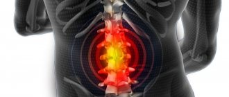
Develops with osteochondrosis, spondylosis, intervertebral hernia, and other diseases of the lumbar spine. It manifests itself as pain spreading from the center to the periphery, intensifying with exertion, sudden movements, coughing, sneezing. The localization of pain is determined by the level of exit of the affected root. - Lumbosacral plexitis.
Manifests with sharp pain in the thigh, buttock, sacrum, lower leg, and groin area. When the lumbar plexus is affected, the pain occurs mainly in the anterior part; when the sacral plexus is affected, the pain occurs mainly in the back part of the leg.
Other reasons
Sometimes hip pain is provoked by diseases of neighboring structures or distant organs, or mental disorders. Possible causes of pain may be:
- Coxarthrosis.
The pain is aching, nagging, occurs at the beginning of movements and after exercise, combined with pain in the hip joint. - Renal cell carcinoma.
The pain syndrome mimics sciatic nerve neuropathy and is caused by compression of the nerve trunk by an enlarging tumor. - Mental disorders.
Pain can appear with depression, hysterical neurosis, and some other conditions. Typical signs are vagueness, unusualness and variability of complaints that do not fit into the picture of a specific somatic disease.
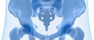
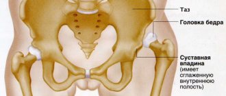
A little about the hip joint
The hip joint is the largest joint in the musculoskeletal system. It connects the lower limbs to the torso. The head of the femur is inserted into the acetabulum of the pelvis. Ligaments and muscles are located around this structure, and synovial fluid is produced inside, which acts as a lubricant.
It is the quantity and quality of synovial fluid that determines how well the joint will function and whether degenerative changes will begin in it. After all, if changes affect the cartilage, they will become inflamed and eventually spread to the bones, which will lead to necrosis of the joints.
It is impossible to live comfortably with a damaged hip joint.
Diagnostics
The examination is usually carried out by orthopedic traumatologists. In case of purulent processes, patients are examined by a surgeon, in case of neurological pathology - by a neurologist. At the initial stage, the doctor finds out the history of life and illness, and conducts an external examination. The following additional studies are prescribed:
- X-ray of the hip.
A basic diagnostic method that allows you to identify all major bone pathologies: fractures, cancer, osteomyelitis, periostitis. In case of lesions of soft tissue structures, it is performed to exclude bone lesions and conduct differential diagnosis. - CT scan of bone.
It is used at the final stage of the examination to clarify the results of radiography, planning conservative therapy or surgical intervention. Makes it possible to accurately determine the size, configuration and structural features of the pathological focus in bone tissue. - Ultrasound.
Sonography of soft tissues in myositis and tendonitis reveals areas of inflammation or degeneration. Ultrasound scanning of the veins of the lower extremities is effective in assessing the condition of the veins and lymphatic vessels in patients with phlebitis and lymphangitis, and in identifying blood clots in thrombophlebitis. - MRI of soft tissues.
Recommended for ambiguous results of other diagnostic procedures. It is carried out to determine the localization of deep hematomas, confirm degenerative or inflammatory changes in tendinitis and myositis. - Electrophysiological studies.
For patients with pain of neurological origin, ENMG, ENG or EMG are indicated to clarify the level of nerve damage and study the condition of nerve and muscle tissue. - Lab tests
. They are performed to determine the severity of the inflammatory process in purulent diseases, assess the condition of organs and systems in malignant neoplasms.
Which specialist should I contact?
Only a doctor can find out the cause of burning and pain in the hip joint after first examining the patient’s symptoms. To make an accurate diagnosis, the following studies are prescribed:
- Ultrasound of the joint;
- X-ray of the hip;
- Magnetic resonance imaging;
- biochemical blood test to detect rheumatoid factor.
After receiving the results of medical studies and identifying the pathology that causes a burning sensation in the hip joint, treatment is prescribed.
Treatment
Help before diagnosis
In case of traumatic injuries, ensure limb rest. In case of fractures, the leg is fixed using two splints: the inner one from the foot to the groin, the outer one to the armpit. Cold is applied to the leg, in case of severe injuries, painkillers are administered, and anti-shock measures are carried out according to indications. The severity of symptoms of myositis and tendonitis is reduced with the help of anti-inflammatory, analgesic gels and ointments. Sharp pain, significant swelling, and problems with general condition are reasons to immediately consult a doctor.
Conservative therapy
Patients with fractures are given blockades, the hip is fixed using skeletal traction, which is replaced with a plaster cast after clinical and radiological signs of fusion appear. Treatment tactics for other lesions are determined based on the nature of the disease. Conservative treatment of most hip pathologies includes:
- Protective mode.
The mode of physical activity is chosen taking into account the type and severity of the disease. Fixation with a plaster splint, the use of splints, crutches or a cane are possible. - Therapeutic exercise.
To maintain muscle strength and joint mobility, prevent complications and improve general condition, individually selected exercise therapy complexes are prescribed. - Physiotherapeutic procedures.
UHF, electrophoresis, laser therapy and other techniques are used to reduce pain, accelerate resorption, improve blood circulation and tissue nutrition, and stimulate healing.
The listed activities are supplemented with massage, sometimes with manual therapy and taping. For purulent diseases, antibiotics are prescribed, for inflammatory pathologies - NSAIDs, for neurological lesions, blockades are performed.
Thigh taping