Interspinous neoarthrosis is a pathology in which another joint, called a false joint, is formed in various parts of the spine.
This additional joint develops due to excessive approximation of the interspinous processes, which often leads to the appearance of a new articular surface.
Typically, neoarthrosis affects men and women over 60 years of age, who also have other degenerative-dystrophic changes in the spine, for example, osteochondrosis. In young and middle age, this disease occurs quite rarely.
Medical description
Any deformation in the vertebral area causes bones to grow, which reduces the distance between them and leads to the creation of fibrous structures. As a result, the patient suffers from severe pain, stiffness and limited motor functions of the spine.
The disease is characterized by the formation of new joints, which is explained by the convergence of the vertebral processes. In official medicine, the pathology is also called interspinous neoarthrosis.
Patients who have suffered serious injuries begin to experience severe pain and discomfort as they age. This is due to the breaking off of the vertebral process and the formation of false articular tissue. If there are problems with the thyroid gland, the formation of an uncovertebral pseudojoint begins. As for additional factors that contribute to the progression of the pathology, these include:
- All kinds of inflammation in the area of the vertebrae and cartilaginous joints.
- Age factor.
- Mechanical damage to joints.
- Degenerative-dystrophic processes.
- Congenital anomalies.
- Other curvatures.
With intense age-related changes, trauma to cartilage tissue cannot be ruled out, leading to breaking off or changing the structure of the vertebrae. As a result of the pathology, neoarthrosis will begin to form in the affected area.
The consequences of such changes manifest themselves in the form of decreased vertebral lability, compression of the spinal cord, as well as disruption of the functionality of individual organs and systems.
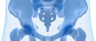
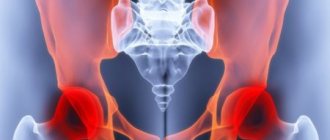
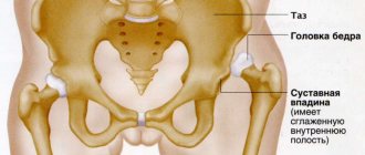
With the progression of the disease and accompanying age-related changes, patients may begin degenerative processes in the hip joints. This is often noticed in women. Due to the destruction of bone tissue in the damaged area, neoarthrosis develops.
It should be noted that neoarthrosis of the hip joint practically does not manifest itself in children and adolescents. Cases of the disease at a young age are extremely rare.
In modern medicine, there is one interesting practice that involves artificially increasing the false joint during the development of certain pathological processes in the hip area. This approach can affect the formation of a new articular head and cavity, allowing you to get rid of purulent-inflammatory complications.
Main symptoms
The first and key sign of the development of neoarthrosis is intense pain in the affected area. Pain often appears after severe physical activity, running, or standing on your feet for a long time. It disappears during sleep and rest. In the early stages, pain is noticed for a short period of time, but then it intensifies and becomes constant. Depending on the location, a number of other symptoms are identified:
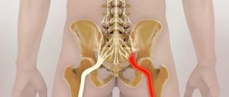
- Deformation processes in the cervical region, leading to migraines, dizziness, unstable blood pressure and other troubles.
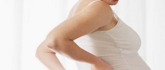
- Throbbing pain in the lumbar region when performing body movements. The syndrome worsens after prolonged sitting.
If the pathology continues to progress and the necessary measures are not taken, the patient may suffer from contracture of the supporting joint, pelvis, hips and knees.
Any complications negatively affect the patient’s ability to work, as a result of which the lower limbs become almost immobile. Constant pain causes a feeling of stiffness, and also increases the risk of developing osteochondrosis, polyarthritis and other more dangerous diseases.
In most cases, neoarthrosis of the lumbar region is associated with natural age-related changes, but there are other causes. Very often, pathology appears after:
- Inflammation in the cartilage tissues of the vertebrae and discs.
- Degenerative changes due to trauma.
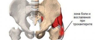
Uncovertebral neoarthrosis of the cervical spine
Uncovertebral neoarthrosis often affects the cervical spine. This disease is characterized by the formation of pathological protrusions on the posterior surfaces of the vertebral bodies. This is the deposition of calcium salts. They come into contact with neighboring vertebral bodies and provoke their destruction due to friction of bone tissue. As a result, false bone processes also form on the opposite side. As a result, they are connected to each other using dense fibrous tissue. The spine loses its usual mobility. Any movement of the head begins to cause pain.
Uncovertebral neoarthrosis of the cervical spine is provoked by the following clinical symptoms:
- frequent headaches localized in the back of the head;
- dizziness, weakness, drowsiness, decreased mental performance, feeling of chronic fatigue;
- crunching in the neck when trying to make circular movements with the head;
- increased blood pressure for no apparent reason.
Neoarthrosis of the cervical spine forms at the age of 30–40 years. At first it doesn't cause any problems. But as the fibrous capsule of the pseudarthrosis thickens, the symptoms increase. There is a constant aching pain in the back of the neck. There is a feeling of tightness, excessive muscle tension in the neck and collar area.
Neoarthrosis of the cervical spine affects the vertebrae C3 - C6, so a disruption of the innervation of the thyroid gland, throat tissue and brachial nerve plexus quickly occurs. Patients experience difficulty swallowing solid food. Paresthesia appears in the upper extremities, a feeling of numbness in the little fingers and thumbs.
If uncovertebral neoarthrosis of the cervical spine is not treated in a timely manner, then gradually osteophytes will completely limit mobility and secondary degenerative destruction of cartilage and bone tissue will begin. For treatment, it is best to contact a vertebrologist. A local therapist in a city clinic is unlikely to be able to prescribe effective and safe treatment. As a rule, patients with such deformities are given an inaccurate diagnosis – osteochondrosis. Accordingly, treatment is prescribed that cannot give a positive result.
Effective diagnostics
As for diagnosing neoarthrosis, it involves drawing up an overall picture after a neurologist has collected an anamnesis. It is important for a specialist to evaluate the time of onset of the pathology, the specifics of the pain syndrome, as well as the list of symptoms that arose after the development of the problem.
Also, during diagnosis, it is necessary to conduct a full medical examination, paying attention to the appearance of pain in the head when changing body position, surges in blood pressure and other accompanying symptoms. To increase diagnostic accuracy, you should apply:
- X-ray examination and computed tomography.
- CT myelography, which involves the use of a contrast agent to determine congenital anomalies or the likelihood of vertebral damage.
- Magnetic resonance therapy. This technique is considered the most effective, since it indicates not only the condition of the bone tissue, but also changes in the area of soft tissue joints.
The type and form of diagnosis are determined by a specialist after obtaining an anamnesis, taking into account the patient’s individual complaints, as well as other data.
A differential diagnosis should also be carried out to exclude sciatica, lumbodynia, cancer and any other forms of arthrosis.
Causes of interspinous neoarthrosis
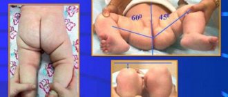
Interspinous neoarthrosis is the most common type of pathology. It is localized in the cervical and lumbar spine. Here, normally, there is a natural anterior deflection of the spine. Due to lordosis, there is a maximum convergence of the spinous processes, which in some cases can come into contact with each other.
The reasons for the development of neoarthrosis in these and other parts of the spinal column are as follows:
- poor posture and curvature of the spinal column in the form of increased lordosis against the background of increased thoracic kyphosis (round back, stoop);
- decrease in the height of intervertebral discs (protrusion) against the background of a long course of degenerative dystrophic disease (dorsopathy);
- spondyloarthrosis and spondylolisthesis – provoke the appearance of cracks in the spinous and transverse processes of the vertebrae, which can cause the formation of a false joint;
- Ankylosing spondylitis or ankylosing spondylitis - provokes the formation of scar tissue between the vertebral bodies, forms false joints and strong adhesions of the spinous processes, and impedes the mobility of the spinal column;
- rheumatoid processes, systemic lupus erythematosus;
- diseases of the endocrine system, for example, neoarthrosis of the cervical spine in most cases provokes thyroid disease;
- excess body weight;
- maintaining a sedentary lifestyle without sufficient physical activity on the back;
- incorrect foot placement with uneven distribution of shock-absorbing load;
- injuries of the back and collar area (impacts, sprains of ligament and tendon tissue, fractures of the vertebral bodies and their spinous processes, bone cracks);
- infections of the spinal cord and tissues of the spinal column (osteomyelitis, tuberculosis, poliomyelitis, etc.).
It is very difficult to exclude all possible causes of the development of this pathology. You need to monitor your weight and posture, properly organize your sleeping and working space, and promptly contact a vertebrologist if you experience any pain in the spinal column.
Treatment methods and preventive measures
As for the methods of treating neoarthrosis, they can be both conservative and surgical. In the first case, therapeutic actions are aimed at combating the causes that caused degenerative processes in the bone tissue. Conservative methods also improve blood circulation and nutrition of joints with valuable substances, and reduce the intensity of pain. If severe pain develops, the patient is prescribed bed rest and complete abstinence from physical activity.
There are other methods that are classified as auxiliary preventive measures. We are talking about balneotherapy, hirudotherapy and visiting the pool. This helps reduce stress on the spine. Regular physical therapy exercises help strengthen the muscle corset, restore natural metabolism in joint tissues, and also stabilize the mobile functions of the vertebrae.
If the disease progresses to an acute stage, conservative treatment methods may be useless. In this case, it is necessary to involve surgical intervention, performing operations to remove osteophytes (overgrown processes of the vertebrae).
During surgery, the patient may additionally need to restore the spinal disc using prosthetic surgery. As a result of surgery, normal joint movement is restored, and pain completely disappears.
Treatment of spinal neoarthrosis
It is necessary to begin treatment of neoarthrosis with a thorough diagnosis. An x-ray in different projections, CT myelography and MRI examination are prescribed. After clarifying the diagnosis, a decision is made on conservative treatment of spinal neoarthrosis or the need for surgical intervention.
For the first and second degrees of development of pseudarthrosis, conservative therapy is recommended. At our manual therapy clinic, a course of rehabilitation treatment may include:
- traction traction of the spinal column, which allows you to restore the physiological position of the vertebral bodies, straighten the intervertebral discs and eliminate compressive pressure on the radicular nerves;
- osteopathy and massage to restore tissue elasticity, enhance microcirculation of blood and lymphatic fluid;
- therapeutic exercises and kinesiotherapy - to stop the formation of the fibrous capsule and increase the range of mobility of the spine;
- reflexology, physiotherapy, laser therapy and much more.
Treatment of uncovertebral neoarthrosis of the cervical spine also includes massage of the collar area and a special course of therapeutic exercises, which is aimed at restoring mobility and increasing the amplitude of head movements.
If you require treatment for spinal neoarthrosis, we recommend that you make a free appointment with a vertebrologist at our manual therapy clinic. Here you will receive comprehensive information on your case of illness from your attending physician.
Pathologies of the temporomandibular joint
Another common form of pathology is TMJ neoarthrosis. It involves the formation of false joints in the lower jaw. In this case, the articular head begins to change its natural position, promoting the development of a new process. In most cases, the problem is caused by all kinds of inflammation and previous injuries.
Experts distinguish three stages of TMJ neoarthrosis, which differ in symptoms and form of progression. At the first stage, the pathology is called osteoarthritis and can last up to six months. Severe inflammation is noticed in the affected area, which leads to the death of bone tissue. The most vulnerable point is the articular cartilage, which is seriously deformed.
Unfortunately, conducting an effective diagnosis and identifying such a disease can be problematic and sometimes impossible, because there are practically no significant degenerative changes in bone tissue. Only with a thorough examination using high-tech X-ray equipment can tissue deformation be noticed.
The problem of TMJ neoarthrosis occurs in children and adolescents. In the absence of effective treatment, the pathology passes from the first stage to the second. In this case, complete destruction of the head of the lower jaw begins, and X-rays already determine a number of changes in the cartilage tissue. The second stage can take from one to three years, while in some young patients the pathology stops in this form and does not progress. As for the mobility of the lower jaw, it becomes very weak.
There is also a third stage of TMJ neoarthrosis. It is called expressed reparation. The development of such a pathology leads to significant deformation of the temporal bone, which can be seen during X-ray examination. Also, the articular tubercle begins to smooth out, becoming like a straight line. The duration of the third stage reaches seven years, and the mobile functions of the joint are practically lost.
The last stage, the fourth, leads to final stiffness of the jaw. It is often noticed in patients during adolescence.
Development of pseudarthrosis
When suffering serious injuries or degenerative changes, the patient may begin to develop pseudarthrosis of the femur. As is known, complete tissue restoration in fractures can take about 4-6 months (the exact period is determined by the complexity of the pathology). During this period of time, the joints begin to fuse, which can be determined using x-rays.
However, sometimes this time period is not enough due to slow consolidation. In this case, after the joints heal, the fracture line becomes almost smooth and invisible. However, after a certain time, it expands, and a bone irritation callus without pronounced contours appears in the formed gap. It is called pseudarthrosis.
Experts distinguish several types of reparative bone tissue regeneration. In the early stages of development, the pathology is called primary. In this case, fusion of bone joints through osteogenesis is observed. This phenomenon causes the formation of an intermediary callus.
When bones fuse without gaps, the primary delayed stage begins, which indicates partial regeneration of bone tissue. Secondary fusion leads to the formation of different types of calluses, as well as an increase in healing time.
Predisposing factors
Predisposing factors
The reasons for the development of pseudarthrosis are very different. It was mentioned above that neoarthrosis can be congenital or acquired. In the case of this pathology, the situation is similar. However, congenital diseases are noticed in rare cases and can occur due to intrauterine changes or failures in bone formation. Genetic disorders also contribute to this.
As for the second factor, it manifests itself with the following provocateurs:
- Experiencing numerous changes and degenerative processes.
- Displacement of bone fragments.
- Violation of osteosynthesis.
- Attachment of a secondary infection.
- Impaired blood circulation.
Also, the problem is often caused by various pathologies that contribute to the deterioration of metabolic processes. This factor manifests itself in people who are overweight.
There are also infectious agents, circulatory disorders and multiple injuries. With combined lesions, the likelihood of developing pseudarthrosis becomes as high as possible.
Clinical picture
To diagnose pseudarthrosis in the sacral or transverse region, it is necessary to have an idea of the clinical picture. The first signs of pathology are as follows:
- The appearance of mobility in those areas where it was absent. The patient may also notice a change in the amplitude or direction of movements. As is known, in healthy people these phenomena are completely excluded, and in patients with pseudarthrosis, the ability to turn an arm or leg 360 degrees was noticed.
- Shortening of the limb. It is the most serious sign of degenerative changes. In this case, the difference between both limbs can reach 10 centimeters.
- Changes in the muscles, decreased strength.
- Deprivation of natural functionality of the limbs, which occurs when performing basic exercises or minimal loads. As the disorder develops, the patient cannot lean on his leg, jump, or even walk.
- Disturbances in the functioning of the true joints due to insufficient load.
Knowing how to diagnose pathology, it is necessary to proceed with appropriate treatment methods. The faster the correct measures are taken, the more effective the treatment measures will be and the time it will take for the damaged bone tissue to heal.
The increase in the operational activity of orthopedic traumatologists in the last 15-20 years. The passion of young doctors for mastering new methods of surgical treatment of patients with fractures of the femoral neck and pelvic bones using submersible metal fixators, to the detriment of the use of previously well-proven conservative methods and methods of hardware treatment of fractures. The expansion of indications for hip replacement and the introduction of this high-tech method, which provides excellent results when used correctly, into the daily practice of surgeons in ordinary trauma departments throughout the country has led to an increase in the number of patients suffering from postoperative osteomyelitis of the bones that make up the hip joint. The number of patients suffering from post-traumatic, gunshot and hematogenous osteomyelitis of the proximal femur and acetabulum is also not decreasing.
Treatment of patients with this severe pathology is extremely labor-intensive, time-consuming and expensive, often associated with the patient’s loss of ability to work, and in some cases ends in death.
According to Hanssen et al. (1996), the cost of treating a patient, excluding the salaries of doctors and nursing staff, with a purulent-inflammatory process that developed in the early stages after hip replacement surgery is $80,000, and a patient with a late infectious complication is up to $140,000 (7 ).
Thus, the problem of surgical relief of the purulent-inflammatory process that has developed in the hip joint with restoration of the supporting function of the limb remains extremely complex and relevant to this day (8)
For the first time, the surgical method of treating patients with purulent-inflammatory processes in the hip joint was used in the 19th century. In 1821 Antoni White performed resection arthroplasty of the hip joint in a patient with septic arthritis. As a result of the operation, neoarthrosis formed, which, due to the death of the patient from consumption, was morphologically studied 5 years later. After the introduction into surgical practice of the antiseptic method, a sharp chisel instead of a hacksaw, and the advent of the radiographic method, resection arthroplasty of the hip joint became the operation of choice for gunshot wounds, tuberculous and nonspecific coxitis (15).
The introduction of blood transfusion in the 20s of the twentieth century, sulfonamide drugs in the 30s and especially antibiotics in the late 40s, as well as the improvement of aseptic and antiseptic methods led to the transition of the operation from casuistic to the category of regularly used interventions.
In the 20-30s of the twentieth century, resection of the proximal femur with its introduction into the acetabulum became known as the Lexer-Whitmann operation. The method involved cutting off the greater trochanter in an oblique direction with the muscles attached to it. After opening the joint cavity, resection of the femoral neck was performed with removal of its head. The remainder of the femoral neck was rounded and covered with a flap of fat, after which it was reduced into the acetabulum. The previously broken section of the greater trochanter was attached below its base to the freshened section of the thigh using screws. The limb was fixed in a hip plaster cast (21, 22).
In 1924, W. Ansch?tz reported on 22 reconstructive operations he performed for various diseases of the hip joint (paralytic loose joints, chronic congenital dislocations, Perthes disease, infectious arthritis, deforming arthrosis, non-union of fractures and pseudoarthrosis of the femoral neck) (16) .
In 1928, GR. Girdlestone developed and introduced his own version of resection arthroplasty of the hip joint for the treatment of patients with septic arthritis and coxitis of tuberculous etiology (20).
The essence of the operation was to create parallel platforms with the largest surface of contact between the femur resected in an oblique direction along the intertrochanteric line and the acetabulum partially resected in the upper and posterior sections. Flaps of scar or muscle tissue are placed between these components in order to create conditions for the formation of neoarthrosis. The limb was fixed in a hip plaster cast for 1 month.
In 1939, PCColonna published a report that the operation he proposed had been performed on 121 patients in America. The percentage of excellent and good results ranged from 70–85% (19).
The author considered the main points of the operation to be the preservation of the fibromuscular layer at the top of the greater trochanter and the transplantation of abductors as low as possible on the femoral diaphysis. The author did not insert the stump of the femoral neck into the acetabulum, but the greater trochanter. The operation was first named Operation Ansch?tz-Colonna, and later as Operation Colonna 2.
Both operations, both Lexer-Whitmann and Ansch?tz-Colonna, involved long-term fixation of the resected end of the femur, embedded in the acetabulum in a hip plaster cast in the position of abduction of the limb at an angle of 40-45 degrees. After 3-5 months, a corrective osteotomy of the femur was performed to eliminate this defective position.
For the first time in the history of medicine, the results of treatment of injuries of the hip joint were studied on a large scale during and after the end of the Great Patriotic War. Thus, M.O. Friedland (1944) rounded the neck during hip resection. The same thing was recommended and implemented 4 times in 1947 with excellent functional results by B.P. Kirillov (2, 14).
An original technique for treating the neck during resection of the hip joint for gunshot wounds was proposed by A.A. Polyantsev (1944). He drew a line for cutting the neck at an angle, from top to bottom and outwards (10).
During the war, M.P. Skrynchenko operated on 36 wounded soldiers and officers with gunshot injuries to the hip joint. Before inserting the greater trochanter into the acetabulum, he shaped the greater trochanter into a cone. Thirty-one patients received a mobile weight-bearing joint, and 5 patients died (13).
In the 40s of the twentieth century, the idea of replacing the articular end of the femur with a metal prosthesis appeared. HKBohlman (1939) was the first to use a pedunculated vitalium prosthesis in the form of a 3-blade nail with a spherical head (17). In 1952, Moor introduced a metal endoprosthesis with a perforated stem.
In the 50-60s of the twentieth century, J. Charnley developed and introduced into surgical practice an endoprosthesis of his own design with a cup made of high molecular weight polyethylene for total hip replacement using bone cement (18).
The foundations of total hip replacement in our country were laid by K.M. Sivash, who made a report on this issue in 1956.
At the first stage of development of the method, the Sivash endoprosthesis was intended for the treatment of patients with ankylosing spondylitis (Strumpell-Bekhterev-Marie disease). The treatment method lived up to the hopes placed on it. Positive experience in treating patients led to an expansion of the range of diseases for which the Sivash endoprosthesis began to be used - rheumatoid arthritis, deforming coxarthrosis (11).
At the same time, the emerging new method of treating patients with hip joint pathology also had its opponents. Thus, E.T. Sklyarenko (1974) expressed the opinion that “biological arthroplasty cannot be replaced by artificial joints. The problem of their use is still far from being resolved. Artificial hip joints are permissible only in cases where biological arthroplasty cannot be performed for some reason, mainly in elderly people” (12).
The method was criticized by G. Kaiser (1974), who in his work about. In young people, the author suggested performing arthrodesis of the hip joint according to indications; in elderly patients, resecting the head and entire neck of the femur to create neoarthrosis (3).
With the introduction of hip replacement into widespread practice, the problem of treating purulent complications arose.
In 1974, K.M. Sivash and V.N. Guryev, based on an analysis of the results of treatment of 830 patients operated on in 1959-1973. CITO identified absolute contraindications for hip replacement surgery: suppurative processes in the joint area; presence of fistulas and post-fistula scars; the presence of fresh elements of active tuberculous granuloma; the presence of a focus of infection in the patient’s body; scar tissue changes in the hip joint as a result of previous operations; coxarthrosis that developed as a result of purulent coxitis suffered by the patient; the absence of a special operating room in which only “clean” operations are performed, and a special department in which patients are treated after total hip replacement (11, 1).
A.A.Pokryvalov (1987) based on a statistical analysis of the results of 766 hip replacements performed at the CITO from 1975 to 1983. proved that “the most significant factor predisposing to the occurrence of purulent complications after endoprosthetics is the presence of surgery on the given joint prior to endoprosthetics” (9).
In the course of analyzing the timing of the development of the purulent-inflammatory process in 115 endoprosthetic patients treated at the CITO from 1975 to 2002, we discovered the following pattern (5).
Of the 13 patients who underwent endoprosthesis replacement in the presence of anamnestic evidence of a previous purulent-inflammatory process in the area of the affected joint, or sepsis, a suppurative process developed within 1 year in 11 patients (84.6%), and in later periods - in 2 (15.4%). The average period of development of suppuration was 5 months.
Of the 36 patients who underwent certain operations on the affected joint, a suppurative process developed within 1 year in 24 patients (66.7%), and in later periods - in 12 patients (33.3%). The average period of development of suppuration is 1 year.
Of the 66 patients who underwent arthroplasty on an intact joint, suppuration developed in 34 patients (51.5%) within 1 year, and in the later stages in 32 patients (48.5%). The average period of development of the purulent-inflammatory process was 2 years 8 months.
That is, the greatest risk, in terms of the development of purulent complications, was associated with hip replacement in patients with a history of one or another purulent-inflammatory process or who had postoperative scars in the joint area.
In other words, the analysis confirmed the well-known thesis of the classics of the method about the need for a differentiated approach to the selection of patients who are indicated for hip replacement with a favorable long-term result.
Currently, in addition to specialized centers for endoprosthetics of large joints with trained personnel, highly qualified surgeons, observing strict selection of patients, operating in operating rooms with laminar air flow with minimal blood loss and in the shortest possible time period, the method has become widely introduced into the practice of trauma departments of ordinary city hospitals countrywide.
According to various authors, from 1 to 1.5 million total hip replacements are performed annually in the world. In Russia in the early 90s, 3,200 such operations were performed per year, while the estimated annual minimum need for endoprostheses in the Russian Federation is 20-25 thousand, and the maximum is 100 thousand (4).
Many surgeons, when deciding on the need for hip replacement, increasingly ignore the presence of a chronic purulent-inflammatory process in the hip joint in the patient or a latent source of infection in the body, not to mention the presence of scar tissue after previous reconstructive surgeries on the hip joint. According to various authors, the percentage of purulent complications after endoprosthetics ranges from 0.2 to 58.5%. (5).
The main methods of treating purulent-inflammatory processes in the hip joint area at present are one or two-stage joint replacement or arthrodesis using various metal fixators or a hip plaster cast (8, 6).
Agreeing with the opinion of the founders of the arthroplasty method about the inappropriateness of its use in the treatment of patients with purulent-inflammatory processes and relying on our many years of experience in treating patients with this severe pathology, we believe that performing hip replacement in patients with purulent-inflammatory processes is extremely dangerous due to the high risk development of both early and late postoperative purulent complications.
We also consider it incorrect to perform hip arthrodesis operations in patients with a purulent-inflammatory process using both submersible and external fixators, including a hip plaster cast. From our point of view, joint arthrodesis in this pathology is fraught with the development of a suppurative process in the area of the fixator with the emergence of the need for its early removal and the remaining unresolved problem of stopping the purulent-inflammatory process, but in worse conditions than before arthrodesis, firstly. Secondly, if ankylosis is achieved, there is a very high risk of developing relapses of the osteomyelitic process with the need for subsequent multiple fistula-sequester necrectomies. Thirdly, even in the absence of infection and destabilization of certain fixators, the risk of arthrodesis failure due to an unresolved purulent-inflammatory process with the need for repeated surgical interventions remains high.
An adequate alternative to both endoprosthetics and arthrodesis of the hip joint in purulent-inflammatory processes, in our opinion, is to perform resection fistula-sequesternecrectomy of the proximal femur and acetabulum with the creation of supporting neoarthrosis, which allows both to stop the suppurative process and to achieve a good functional result for the patient.
From 1985 to 2005, we operated on 75 patients with purulent-inflammatory processes in the area of the hip joint (chronic and acute postoperative , post-traumatic, hematogenous, gunshot osteomyelitis) aged 9 to 75 years. In all patients, it was possible to stop the suppurative process and achieve the formation of supporting neoarthrosis with restoration of the supporting function of the limb almost in full.
In order to improve the results of treatment of patients with this pathology, we have developed and implemented a complex of treatment and rehabilitation measures aimed at stopping the suppurative process with the formation of supporting neoarthrosis in the joint and restoring the supporting function of the limb.
A set of treatment and rehabilitation measures aimed at stopping the purulent-inflammatory process.
- Radical sanitation of the local pathological focus by removing all necrotic tissue, excision of the fistula tracts along the entire length, sequester necrectomy of osteomyelitic areas of bone tissue.
- Establishing adequate irrigation-vacuum drainage of the postoperative wound with prolonged rinsing with antiseptic solutions.
- Adequate antibacterial therapy: initiation of antibacterial therapy 2 hours before surgery with broad-spectrum drugs, followed by their adjustment 1-2 days after surgery based on the culture data of the surgical material obtained both in aerobic and anaerobic conditions, and then based on the data crops separated from drainage.
- Correction of homeostasis, immunocorrection.
- A program of functional treatment of patients with the aim of creating supporting neoarthrosis, which consists of 3 stages (given below).
- Prevention of relapse.
The operation is performed with the patient positioned on the healthy side from an external lateral approach.
It should be noted that in the presence of a functioning fistula, it is necessary to tightly fill the fistula tract with a 2% alcohol solution of brilliant green, after which an incision is made with excision of the postoperative scar and the fistula tract along its entire length. If there are infected metal structures, they are removed, and then a resection sequesterectomy of the proximal end of the femur and acetabulum is performed with excision of all tissues stained brilliant green and questionable in terms of viability. In the fistulaless form of osteomyelitis, resection sequesterectomy of the proximal femur and acetabulum is performed within the healthy (bleeding) bone tissue. Using a rasp and an electric drill, the resected area of the femur is cleaned and all sharp bony protrusions are removed, and an oval shallow notch is cut out at the top of the sanitized proximal end of the femur in the sagittal direction. The notch provides a more stable contact between the femur and the acetabulum. Then the wound cavity is washed generously with solutions of hydrogen peroxide and chlorhexedine, after which it is thoroughly evacuated using suction. The patient is transferred to a horizontal position, the operated limb is abducted to an angle of 40-45 degrees and placed on an additional sterile operating table with the foot fixed in a neutral position in a derotational boot. The wound is drained by two drainages. Single-pole drainage is brought to the acetabulum, and 2-pole drainage is carried out in the longitudinal direction along the length of the entire wound under the muscles, after which the wound is tightly sutured in layers. After completion of the operation, the patient is transferred to the ward without changing the specified abduction angle with the foot fixed in a derotational boot. Such fixation, along with active functional treatment in the postoperative period, ensures a controlled process of formation of supporting neoarthrosis. From the first day after surgery, a comprehensive rehabilitation program begins. Due to the traction of the periarticular muscles, the proximal end of the femur gradually approaches the roof of the acetabulum. These bony structures begin to contact and articulate, the notch created during surgery creates conditions for greater stability, and at the same time scars are formed that hold the proximal end of the femur to the roof of the acetabulum. The time period with the need for bed rest is on average 3-4 weeks and is directly related to the formation of scar tissue in the area of developing neoarthrosis. The main indicator of the formation of sufficiently strong scar tissue is the patient’s free retention of the foot released from the derotational boot in a functionally correct neutral position during the day. At this stage, the patient begins to learn how to sit in bed and stand in the ward on crutches with the leg abducted at an angle of 20 degrees, using shoes with heeled soles to compensate for the anatomically functional shortening.
Program of functional treatment of patients with the aim of creating supporting neoarthrosis.
- A.
Early postoperative period (bed rest until 30 days after surgery). 1-A. Early postoperative period - up to 10 days after surgery. 2-A. The early postoperative period is from 10 to 20 days after surgery. 3-A. The early postoperative period is from 20 to 30 days after surgery. - B.
Late postoperative period (from day 30 until the patient is discharged from the hospital). - B.
The period of rehabilitation on an outpatient basis (up to 6-8 months after the patient is discharged from the hospital).
The essence of these rehabilitation measures is that therapeutic exercises for the muscles of the operated limb and torso begin in the early postoperative period in bed with a minimum number of exercises and are carried out at an increasing pace over time.
Early postoperative period 1-A - up to 10 days after surgery:
in a horizontal position, abduction of the limb at 45 degrees, with the foot fixed in a neutral position with a derotational splint for up to 5-10 minutes, 3 times a day, the following are performed: isometric exercises for the muscles of the operated and healthy lower limb; gentle passively active flexion in the knee joint; sitting up in bed using the Balkan frame; exercises to strengthen the muscles of the arms and healthy leg using a rubber bandage or expander. On days 5-7, exercises are added to actively try to lift the straightened leg from the plane of the bed.
Early postoperative period 2-A - from 10 to 20 days after surgery: against the background of ongoing exercises, the patient learns to sit freely in bed with his healthy leg lowered, and the duration of exercises increases to 10-15 minutes 3 times a day. The complex includes exercises to straighten the torso with lifting of the pelvis from the plane of the bed up to 50-100 times a day.
Early postoperative period 3-A - from 20 to 30 days after surgery:
The duration of gymnastics classes increases to 15-20 minutes 3 times a day. Exercises for active flexion of the leg at the knee joint are added with attempts to lift the straightened operated leg from the bed up to 100 times a day. By day 25, the foot is removed from the derotational boot for some time and fixed between rollers with sand.
B. Late postoperative period - from 30 days until the patient is discharged from the hospital:
The duration of classes increases to 20-30 minutes 3 times a day. Active bending of the leg at the knee joint with attempts to lift it off the bed is increased up to 200 times a day. The patient is taught to stand and walk on crutches with the limb abducted at an angle of 20-30 degrees in shoes with a heel to compensate for the shortening for up to 5 minutes 3 times a day. Add exercises for bending and abducting the leg from a standing position and sitting independently on a chair with an adjustable seat height (pillows are placed) up to 10-15 exercises 3 times a day.
B. The period of rehabilitation of the patient on an outpatient basis:
against the background of the ongoing rehabilitation complex from a lying and standing position up to 45 minutes 3 times a day, the number of all exercises is increased to 100 per day. The patient walks on an abducted limb for up to 30 minutes a day with a load on the leg up to 10-15% of the norm, with a monthly increase in the load by another 10-15% and the walking duration is 30 minutes. From the moment (on average 3-4 months after surgery) when the patient can freely lift his leg from the plane of the bed and hold it in this position for some time, the leg can be brought into a functionally correct position with a corresponding decrease in the height of the heel to compensate for the anatomical shortening. The patient gradually begins to walk with one crutch or cane. On the day when the patient gets up and forgets about the need to pick up a crutch or cane (an average of 4-6 months), he will be able to gradually begin walking without additional support.
CONCLUSIONS:
The method we developed and implemented for the formation of supporting neoarthrosis is qualitatively different from the methods of resection arthroplasty Lexer-Whitmann, Anschutz-Colonna and GR.Girdlestone, since, firstly, it allows us to achieve relief of the osteomyelitic process in patients, usually with advanced, severe forms of osteomyelitis. Secondly, it does not require long-term additional fixation of the operated limb in a hip plaster cast, but on the contrary implies an active rehabilitation program aimed at restoring joint function. Thirdly, the formation of supporting neoarthrosis does not require the use of plastic materials (fat, muscle and other types of tissue), since during treatment the resected bone fragments are covered with their own connective tissue. Fourthly, with a successful outcome of treatment according to the method we propose, the patient gets rid of the inflammatory process, pain in the joint, almost completely restores the supporting and motor function of the joint and completely eliminates the need for repeated surgical interventions.