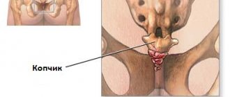
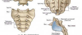
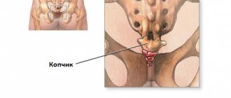
The coccyx is the lowest part of the human spinal column. It consists of five rudimentary vertebrae that are fused together. The coccyx does not have an internal lumen of the spinal canal. The spinal cord does not pass through it. However, next to it is the dura mater and the cauda equina nerve plexus.
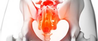
Displacement of the coccyx leads to excessive stretching of the dura mater and compression of the cauda equina. This gives not only a pronounced pain syndrome, but also a number of neurological manifestations. In particular, displacement of the tailbone can provoke disruption of the bladder and large intestine. It is not uncommon that when the coccyx vertebrae are displaced, the patient develops weakness in the legs and loses the ability to walk independently.
In the absence of timely treatment, if the coccyx is displaced, negative consequences may develop. Most often, the internal organs of the abdominal cavity and pelvis are affected. The nerve plexus located in the coccyx area is responsible for their innervation. Any dislocation of this node leads to disruption of intestinal motility. Against this background, various pathologies can develop, ranging from irritable bowel syndrome to the formation of polyps and ulcers in the mucous membrane of this digestive organ.
Displacement of the coccyx vertebrae can occur due to traumatic impact or due to increased pressure from the pelvic organs. For example, in women, a deviation of the rudimentary part of the spine may appear during pregnancy. After childbirth, the natural position of the tailbone is not always restored. This provokes the development of vascular pathologies. Varicose veins of the lower extremities and pelvic cavity, atherosclerosis of arteries and arterioles are just some of the possible diseases that develop when the tailbone is not positioned correctly.
Proper treatment of the pathology can be carried out only after the coccyx has been realigned. This is done by an osteopath or chiropractor. In our clinic, an individual course of treatment is developed for each patient.
If you suspect that your tailbone is in an incorrect position, we recommend that you sign up right now for a free initial consultation with a vertebrologist at our manual therapy clinic in Moscow. Experienced doctors work here. During the initial examination, they will be able to make an accurate diagnosis. You will receive safe and effective medical care.
To make an appointment with a vertebrologist, fill out the form located at the end of this page. Or call the phone number listed on this page.
Causes of coccyx displacement (trauma, fall, pregnancy)
The main reason for the displacement of the coccyx is a traumatic effect on this part of the spinal column. The coccyx connects to the sacrum via the coccygeal-sacral joint. This articulation of bones has a very small range of mobility. When exposed to it from the outside or inside, the integrity of the joint capsule is violated. It stretches, causing subluxation or dislocation of the coccyx.
The bend or displacement of the rudimentary part of the spinal column is fixed due to inflammatory processes in the articular capsule. It will be very difficult to restore the normal position on your own. The help of an osteopath or chiropractor is required.
Many patients who have suffered a fall on the coccyx area, after eliminating the pain syndrome, continue to live with a bend in the lower part of the spinal column. This negatively affects the functioning of the intestines and bladder, leading to improper distribution of the load on the rest of the spinal column. All this contributes to the rapid destruction of intervertebral discs in the lumbosacral region.
Displacement of the tailbone after a bruise requires mandatory consultation with a vertebrologist. Meanwhile, displacement of the coccyx during a fall is not the most common cause of the development of this pathology.
Let's consider other risk factors for bending of the rudimentary part of the spinal column:
- violation of posture and curvature of the spinal column in the lumbar and thoracic regions - compensatory bending and displacement occurs;
- destruction of the hip joints with compensatory excessive muscle tension in the lumbar and sacral areas, the gluteal area;
- incorrect placement of the foot in the form of flat feet or club feet;
- inflammatory processes and tumor growth in the pelvic cavity;
- pathologies of the rectum (hemorrhoids, proctitis, paraproctitis, fissures, etc.);
- pregnancy and childbirth;
- hard physical labor.
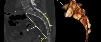
A bruise of the coccyx with displacement of the vertebrae is recorded using an x-ray. Therefore, if there was a fall, impact, accident, emergency braking, unsuccessful movement, then you should immediately visit a traumatologist. This doctor will order an x-ray examination. In this way, it is possible to exclude not only a possible bend with deformation of the coccygeal-sacral joint, but also a crack or fracture.
Displacement of the tailbone after childbirth occurs in women who do not listen to the recommendations of their doctor. It is necessary to follow all recommendations throughout pregnancy, use a prenatal bandage, do special gymnastic exercises and avoid high-heeled shoes. Bending can occur during labor if the muscles of the perineum and lumbosacral region are weakened. Therefore, during the entire period of bearing a child, you should pay maximum attention to your physical condition.
In approximately 7% of patients, the cause of the bend is a congenital disorder of the formation of cartilage tissue. They are born with a predisposition to the incorrect position of the rudimentary part of the spinal column. The pathology begins to develop when the baby is getting on his feet and is finally formed by 7-10 years of age.
Another important negative impact factor is the habit or need to sit on a hard surface for a long time. There is a gradual displacement of the coccyx, which begins to give clinical symptoms after stretching of the dura spinal membrane.
Diagnostics
If a subluxation of the coccyx is suspected, the doctor palpates the injured area. The procedure involves inserting fingers into the rectum, which allows you to determine the degree of injury and the level of joint displacement.
The diagnosis is carried out by a traumatologist. Even if the injury is minor and there is no severe pain, this is not a reason to cancel diagnostic measures.
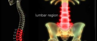
MRI will allow you to make an accurate diagnosis
After palpation, an X-ray examination of the affected area is performed. If necessary, magnetic resonance imaging of the vertebrae and spinal cord is prescribed. Such a diagnostic event will help make an accurate diagnosis and identify complications.
What is a displacement of the coccyx in front and to the left?
In the process of primary diagnosis, it is very important to establish the type of pathology. Depending on the direction of the bend, the dural sheath of the spinal cord, the cauda equina nerve plexus, or individual nerves passing through the soft tissue adjacent to the distal spine may be affected.
The following types of pathology occur in orthopedic practice:
- posterior deviation of the vertebrae relative to the central axis;
- direct right-sided displacement without anterior or posterior deviation;
- left-sided without anterior or posterior deviation;
- displacement of the coccyx anteriorly and to the left;
- displacement of the coccyx to the left and posteriorly;
- distal segmental displacement of the coccyx.
Women who have recently given birth to a child know best about the displacement of the tailbone in front to the left. As the fetus passes through the birth canal, the coccyx deviates. If, due to incorrect obstetric care, an injury occurs to the joint located between the coccygeal and sacral sections of the vertebral column, then the bend may persist for several months after birth. This causes a lot of trouble. The woman is forced to sit and lie in a certain way. Any movement increases the pain. Stool and urination are impaired.
If there is pain in the tailbone area, you should not endure it. See a vertebrologist or chiropractor as soon as possible. These specialists will be able to restore the normal position of the rudimentary part of the spinal column and eliminate pain.
Treatment
– In the case of a dislocation in the sacrococcygeal region, conservative treatment : it is recommended to avoid stress on the tailbone, sit less often, use special pillows that reduce the load in a sitting position, prescribe painkillers and anti-inflammatory drugs, positional physiotherapy and even a course of osteopathy for the tailbone area. – In particularly severe cases, when the pain does not decrease and severely limits the patient’s usual activity, treatment may be surgical . Neurolysis of the nerves in the affected area or removal of the coccyx is performed. According to the Filum System® method: With a forward dislocation or fracture in the area of the sacrum, coccyx or sacrococcygeal region, Neuro-Craniovertebral Syndrome is formed: the ligament, called the filum terminale, which connects the spinal cord to the vertebrae at this level, is over-tightened. An abnormal tensile force appears on the spinal cord, followed by tension in the entire central nervous system. Therefore, after years of clinical observation of similar cases, our Institute offers surgical treatment using the Filum System® method. This treatment involves minimally invasive cutting of the filum terminale using our unique technique.
Symptoms of coccyx displacement
Clinical symptoms of coccyx displacement appear when its physiological position is disrupted. Characteristic features include:
- sharp pain in the lower part of the spinal column, which intensifies when sitting and standing;
- unpleasant sensations may occur when trying to make deep bends back and forth and left and right;
- pain can radiate to the groin area, rectum, trochanter of the hip joint;
- when straining during defecation, the pain intensifies many times;
- Problems with bowel and bladder emptying begin.
If medical care is not provided in a timely manner, then various neurological pathologies associated with the innervation of the tissues of the abdominal organs, pelvis and lower extremities arise.
To carry out diagnostics, you should consult a vertebrologist or traumatologist (if there was a traumatic impact on the spinal column, the Doctor will prescribe an x-ray in different projections. During this examination, you can assess the position of the rudimentary part of the spinal column, its condition. Ultrasound and MRI are also prescribed as necessary and CT examinations.
Why you can’t carry out the procedure yourself
Subluxations and dislocations do not have specific signs. Their clinical manifestations are similar to those of fractures, bruises, and tumor processes. An attempt to put the tailbone back in place on your own can aggravate the condition and cause irreparable harm to the muscles and ligaments that fix the described joint in the correct anatomical position.
Even if you know exactly what the cause of severe pain is, reduction manipulation involves simultaneous use of both hands on the pathological area. It is impossible to do this without outside help. Wrong actions can provoke undesirable phenomena.
Consequences of coccyx displacement
The negative consequences of a displaced coccyx can occur over a long period of time. If the patient receives prompt and complete medical care, the only consequence may be a habitual dislocation of this part of the spinal column. A full course of rehabilitation will help restore the full elasticity of the joint capsule and the performance of the ligamentous apparatus. The doctors of our manual therapy clinic will be able to develop it.
Other negative consequences of coccyx displacement include the following pathologies:
- vascular disorders associated with obstruction of blood vessels, their compression;
- the formation of blood clots in the deep veins, which is a life-threatening condition;
- dysfunction of the bladder in the form of hyperfunction or urinary retention;
- intestinal atony or spastic colitis when the process of innervation of the muscular wall of this organ of the digestive system is disrupted;
- weakness in the muscles of the lower extremities, the appearance of paresthesia effects.
All consequences of coccyx displacement can be eliminated using manual therapy methods. We will discuss methods of effective treatment later in the article.
Dissection of the filum terminale using the Filum System® method
Advantages
1. Eliminates the cause of Neuro-Cranio-Vertebral syndrome associated with a dislocation or fracture in the sacrococcygeal region.
2. Thanks to the minimally invasive technique of ICSS, surgical time is about 40 minutes. Short stay in the clinic. Local anesthesia. Short recovery period without restrictions. No blood transfusion.
3. Its use has a 0% mortality risk, without consequences or serious complications.
4. Improves symptoms, stops the development of symptoms and damage to the nervous system and spine.
How is a displaced coccyx treated?
It is necessary to begin treatment for a displaced coccyx by restoring its normal position. The easiest way to achieve this effect is by visiting an osteopath. It will gently and painlessly restore the position of the vertebrae. Then it is necessary to undergo a course of restorative therapy to eliminate the risk of re-bending.
There is a certain difference in how coccyx displacement is treated in a city clinic and in a manual therapy clinic. Official medicine does not strive to restore the normal state of tissues. All efforts of doctors are aimed at restoring the patient’s performance. It is important to relieve pain and restore body mobility. For this purpose, anti-inflammatory and painkillers are prescribed.
What to do if the coccyx is displaced if you want to restore its normal position? Contact a manual therapy clinic. Symptomatic medications will not be prescribed here. An osteopath will restore the normal position of the tailbone. Then the vertebrologist will develop an individual rehabilitation course. It may include therapeutic exercises, kinesiotherapy, osteopathy, massage, physiotherapy, etc.
Make an appointment for a free appointment with a vertebrologist right now using the special form located further on the page.
Complications
The main risk with a dislocation in the sacrococcygeal region is a long healing period, accompanied by pain that limits quality of life and usual activity . – According to Filum System®: At our Institute, we observe that the majority of patients with anterior dislocation in the sacrococcygeal region, and in more rare cases of fracture in this area, develop Neuro-Craniovertebral Syndrome (NCVS) . In this case, the injury results in non-congenital tension on the filum terminale . This occurs due to the lever mechanism due to the flexion of the coccyx, to the first vertebrae of which the filum terminale is attached, which leads to increased tension on this ligament. The development of the clinical picture of neuro-craniovertebral syndrome can aggravate the consequences of dislocation, worsening the quality of life of patients. In this case, the symptoms consist not only of pain and inflammation in the coccyx area, but also symptoms may appear in other parts of the spine and in the central nervous system, as happens with filum terminale disease.
But maybe it’s still inflammation?
Here it is necessary to clearly establish that the reason is precisely the physical impact and curvature of the tailbone, and not inflammation and muscle damage. In the second option, the pain is usually constant, aching, dull, and may intensify with movement.
In case of inflammation, the most effective method is to take medications. We go to the doctor, get a prescription, buy medicine. Usually the appointment lasts from 2 days to two weeks, during which time we try to give maximum rest.
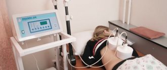
Complications may include muscle spasms caused by inflammation. Spasmed muscles can distort the spine, touch nerves and serve as a source of pain. A severe spasm will not go away on its own. To relieve spasms, use the Sacrus device. This will be discussed below.
First aid
If there is severe pain in the coccygeal region, it is recommended to call an ambulance or take the victim to the hospital on your own.
Before the doctor arrives, you need to numb the injured area. To do this, apply a cold compress (you can take any product from the refrigerator), which helps reduce the intensity of the syndrome in a few minutes. If the pain is severe, you can take a painkiller tablet.
It is prohibited to take a sitting or semi-sitting position. In this case, bone displacement becomes even more difficult. It is allowed to lie on your side and remain in this position until the ambulance arrives.
Important! Self-medication, namely bone realignment, is prohibited. Before such a procedure, it is necessary to accurately determine the location of the displacement, its angle, and associated damage, which is only possible based on the results of instrumental diagnostics.