Author
Posysoeva Margarita Alekseevna
Leading doctor
Ultrasound diagnostic doctor
until October 31
Appointment with a doctor based on the results of instrumental diagnostics with a 50% discount More details All promotions
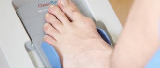
Ultrasound of joints
and periarticular (periarticular) tissues is a modern informative diagnostic method. Different body tissues reflect ultrasound differently, so ultrasound examinations provide images of soft tissue (muscles, ligaments, tendons), not just bones, and this compares favorably with radiography. In addition, ultrasound is completely safe and can be performed as often as necessary to monitor the situation.
Which joints can be examined using ultrasound?
Ultrasound can be used to examine:
Ultrasound of joints
- shoulder joints;
- elbow joints;
- knee joints;
- ankle joints with foot joint;
- hip joints;
- wrist joints with hand joints;
- mandibular joints.
Both joints are examined at once (for example, two elbows or two knees), even if only one hurts. In each person, the structure and development of tissues in the area of the right and left joints are approximately the same, and at the same time unique. Comparing a diseased joint with a healthy one allows us to evaluate the pathological changes that have occurred.
What does the diagnosis show?
An ultrasound of the joints shows what is happening to them at the moment. With the help of an examination, injuries, pathologies, cysts, degenerative and inflammatory processes can be identified. Let's look at each point in more detail.
Pathologies in the joints
Ultrasound of joints is also done to identify pathologies. These can be transformations after operations, inflammatory processes, dystrophic changes.
Based on the results of the examination, heterogeneity of the structure, hyperechoic formations, edema, displacement of ligaments and tissues can be revealed. These changes may indicate degenerative processes.
Detection of a cyst
A cyst is a hollow tumor, usually filled with fluid. Using ultrasound, you can identify a cyst in the joints and evaluate its development. Most often, cyst removal is only possible through surgery. If left untreated, it can grow and/or become malignant.
Injuries
Joint injuries can occur for various reasons: a fall, high load, awkward movement. During ultrasound diagnostics, it is possible to identify the consequences of these injuries: rupture of ligaments, tendons, hematomas.
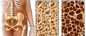
Degenerative processes
Factors in the development of degenerative diseases are osteoporosis (decreased bone density) and wear and tear of the joint with subsequent destruction of cartilage. Mostly elderly people are susceptible to these processes. Subsequently, various types of arthrosis arise. Ultrasound clearly shows these processes and the condition of the joints (for example, narrowing of the gap between the joints, irregularities).
Joint inflammation
Inflammation is a normal reaction of the body to the ingress of pathogenic bacteria. For any manifestations of inflammatory processes in the joints (pain, swelling, redness, limited mobility, crunching, fever), you must immediately consult a doctor to undergo an examination and receive the necessary treatment. Neglected inflammatory processes lead to chronic diseases (gout, various arthritis) and even disability. During an ultrasound of joints and limbs, inflammation is indicated by free fluid in them.
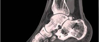
Ultrasound of the hip joints in newborns
Newborn babies are recommended to have an ultrasound of the hip joints
. Such an examination allows us to identify cases of congenital inferiority of joints, some orthopedic pathologies, dislocations and pre-dislocations received during childbirth.
Indications for ultrasound of the hip joints in newborns are:
- breech presentation;
- prematurity;
- asymmetry of the legs or skin folds;
- the presence of corresponding pathologies in the family.
Content
- What can you see on an ultrasound?
- What do they look for on an ultrasound?
- Indications for ultrasound diagnostics
- Cost of ultrasound examination
- Frequently asked questions to the doctor
Ultrasound diagnostics helps to see the condition of the joints of the upper and lower extremities and the spine. Procedures that require special precision in drug administration are carried out under ultrasound control. For example, intra-articular injections of drugs.
Ultrasound of joints is used for:
indications for ultrasound of joints
- identifying the presence of fluid in the joint cavity;
- diagnosis of injury (ligament ruptures, knee meniscal tears, etc.);
- diagnosis of arthrosis (destruction of joint tissue as a result of metabolic disorders);
- diagnosis of inflammatory diseases such as:
- arthritis or synovitis (inflammation of the synovium of the joint);
- bursitis (inflammation of the joint capsule);
- tendinitis (inflammation of the tendon);
- ligamentitis (inflammation of the ligament), etc.
Indications for the procedure
The main types of injuries, as well as pathological processes for which this examination is prescribed:
- mechanical injuries,
- diagnosis after suffering from Lyme disease,
- arthrosis,
- arthritis,
- overweight,
- incessant pain
- endocrine disorders,
- inflammatory processes,
- immobility.
In addition, ultrasound examination can also be prescribed for preventive purposes after 45 years.
Using this method you can identify:
- Synovitis, ruptures of the synovial membrane, hemarthrosis;
- Various inflammations and damage to ligaments (chronic tendonitis, rupture of tendons or ligaments);
- Damage to the meniscus (cysts, ruptures of its horns);
- The presence of loose bodies in the joints;
- Various bone pathologies;
- Tumors.
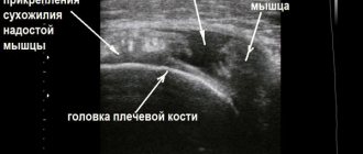
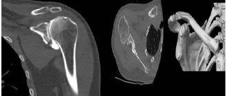
Soft tissue neoplasms on ultrasound
Task. Vascular malformation of the knee
Task. 6 months of anterior knee pain with background history of ACL reconstruction. pain in the tibia area. They did recall an occasional smaller area of swelling appearing on the front of the lower leg in recent months. There was full range of motion of the knee. There was some localized tenderness medial to the tibial tuberosity, directly where the ACL scar was. the patellar tendon and tibial tuberosity insert were intact. There was no evidence of deep infrapatellar bursitis. There was an anechoic, noncompressible lesion medial to the tibial tuberosity that appeared to communicate with the calf cortex in the area of the ACL scar. No vascularity was visible. It was not compressible and there seemed to be a clear relationship between cortical irregularities. The lesion was painful under pressure from the probe, reproducing the patient's symptoms. When viewing the images, a collection of fluid was visible on the front tibia. opinion about the pretibial ganglion. Conclusion: Pretibial hygroma.
Tags: lectures joints ultrasound
Rear access
With this approach, the neurovascular bundle of the popliteal fossa, the medial and lateral heads of the gastrocnemius muscle, the distal part of the fibers of the semimembranosus tendon, the posterior horn of the internal meniscus and the posterior horn of the external meniscus, and the posterior cruciate ligament are visualized.
The patient is in a prone position. The sensor is located transversely to the long axis of the limb in the popliteal fossa. The neurovascular bundle is displaced laterally in the popliteal fossa. The popliteal artery is located behind the vein; the muscle bundles of the popliteal muscle are visualized below. With a panoramic scan using energy mapping, the course of the popliteal artery can be traced.
The tendons of the medial and lateral heads of the gastrocnemius muscle arise from the corresponding condylar surfaces of the femur. The semimembranosus tendon inserts on the posteromedial surface of the proximal tibia.
Between the semimembranosus tendon and the medial head of the gastrocnemius muscle is a small bursa that usually contains the neck of a Baker's cyst. Landmarks for visualizing this bursa during transverse scanning are: the posterior surface of the medial condyle of the femur, covered with hyaline cartilage, the semimembranosus tendon, and the fibers of the gastrocnemius muscle.
When scanning the popliteal fossa longitudinally, the sensor is shifted laterally and rotated according to the plane of the joint cavity. In this case, the posterior horn of the external meniscus is visualized. The posterior cruciate ligament is also visualized from this position, with the probe rotated 30 degrees counterclockwise when examining the right limb and 30 degrees clockwise when examining the left limb. The posterior cruciate ligament, like the anterior ligament, is partially visualized. Due to the anisotropy effect, its fibers are hypoechoic.
To assess the posterior horn of the medial meniscus, it is necessary to move the transducer medially in the popliteal fossa and image the tendon fibers of the medial head of the biceps femoris muscle, which attach to the medial epicondyle of the tibia. From this position the body of the medial meniscus is visualized.
From the posterior approach, one can also evaluate the peroneal nerve, which, leaving the lateral part of the sciatic nerve in the distal thigh, follows laterally and downwards along the posterior surface of the distal biceps femoris tendon before passing to the popliteal region, then around the head of the fibula to the anterior surface of the leg. In this area, nerve damage often occurs between the fibers of the fibrous tunnel.
Popliteal bursitis (Baker's cyst) is located on the back of the knee in the popliteal fossa. On the inside of the knee there is a popliteal bursa known more commonly as a Baker's cyst. Visible only when the knee is bent; as soon as the leg is bent, the cyst disappears. Rarely occurs as an independent cavity, it usually communicates with the joint cavity through a valve connection, the fluid can flow only in one direction, from the joint to the bag, where it is usually absorbed. With this bursitis, there is pain in the knee, which intensifies when squatting, and there will be limited mobility in the knee joint.
Baker's cyst on ultrasound
Task. Patient with discomfort in the popliteal fossa. Ultrasound shows a Baker's cyst with septations, increased vascularization around the perimeter. Conclusion: Baker's cyst.
Preparation
The described technique is one of those diagnostic options that does not require special training. The person does not need to fast, interrupt medication therapy, or take medications.
However, questions still arise:
- Sometimes patients do not understand why it is necessary to perform a procedure for two symmetrical joints if there is pain in only one. For example, when an ultrasound scan of the joints of the lower extremities is prescribed. Two ultrasounds are not performed randomly: this is necessary to see the difference and make a comparative assessment between healthy and affected joints. In addition, diseases of the rheumatic type are characterized by certain pathological changes that do not manifest themselves in any way (there is no pain or other unpleasant sensations).
- Why is it necessary to perform ultrasound of the same joints during treatment? The answer is obvious: to establish dynamics. Rheumatic pathologies have the specificity of modifying the level of severity of inflammation, sometimes even the localization of the lesion changes. For example, osteochondrosis is complicated by synovitis. In other cases, Baker's cysts form when fluid accumulates in the popliteal bursa. The need for re-checking is determined by the attending physician.
Where in Khabarovsk can you get an ultrasound of the knee or shoulder joint?
Medical offers its patients all types of ultrasound diagnostics at reasonable prices. We can perform an ultrasound of the ankle, shoulder, knee or spine directly on the day of your visit. You don't even need a referral from your doctor to do this!
To make an appointment, call 8 (4212) 50-30-038;. You can also order a call back absolutely free by leaving a request on our website. The clinic administrator will contact you as soon as possible and arrange an appointment at a convenient time for you.
Medical is located at: Khabarovsk, st. Panfilovtsev, 38. Detailed directions are presented on our website.
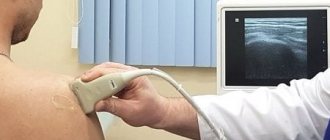
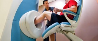
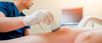
How is ultrasound performed?
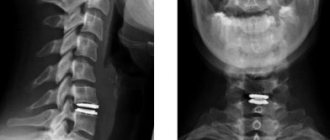
Ultrasound of the knee and other joints in the Art Medic clinic is carried out according to standard international protocols. Typically, the study is carried out in a resting position, then functional diagnostics are shown in dynamics (flexion - extension). For example, during a spinal ultrasound, the patient is asked to tilt their head forward and back to examine the cervical vertebrae.
A special conductive gel is applied to the skin of the body, which provides an airless environment. This is necessary so that the ultrasonic waves do not attenuate. The doctor moves a special sensor over the patient’s body in the place where the joint is projected onto the surface. The image is displayed on the screen of the ultrasound machine.
Ultrasound is often performed not for diagnosis itself, but as a treatment control. For example, when administering drugs intra-articularly, the doctor focuses on the image on the monitor of the ultrasound machine, so as not to damage the surrounding tissue with the syringe needle.
Advantages of this diagnostic method
Diagnostics using ultrasound has a great advantage compared to other types of studies, therefore it is prescribed most often. Among the advantages it is worth highlighting:
- The procedure is completely painless.
- Non-invasive - for diagnosis there is no need to make punctures, incisions or injections.
- No exposure. If we compare X-rays and ultrasound, the latter technique has zero radiation exposure. This advantage also allows for repeated testing during treatment.
- The ability to examine not only bones, but also soft tissues. If it is possible to prescribe an ultrasound of the knee, then in terms of information content the doctor will prefer it to an x-ray.
- High diagnostic accuracy, which is improving as modern ultrasound diagnostic stations are created.
- Low price. This indicator compares ultrasound of joints favorably with MRI or tomography.
The combination of positive qualities allows the use of sonography, including for diagnostics in children, and the procedure can be repeated as many times as desired.