Causes of development of autoimmune diseases
The causes of the development of autoimmune diseases are:
- Infection by a microorganism, the antigenic determinants (epitopes) of the most important proteins of which resemble the antigenic determinants of normal tissues of the host body. By this mechanism, autoimmune glomerulonephritis develops after streptococcal infection or autoimmune reactive arthritis after gonorrhea.
- Destruction or necrosis of tissues or a change in their antigenic structure, so that the altered tissue becomes immunogenic for the host. According to this mechanism, autoimmune chronic active hepatitis develops after hepatitis B.
- Violation of the integrity of tissue (histohematic) barriers that normally separate some organs and tissues from the blood and, accordingly, from the immune aggression of the host lymphocytes. Moreover, since normally the antigens of these tissues do not enter the blood at all, the thymus does not destroy autoaggressive lymphocytes against these tissues. Autoimmune thyroiditis develops through this mechanism.
- Hyperimmune state (pathologically enhanced immunity) or immunological imbalance with a violation of the “selective” function of the thymus that suppresses autoimmunity or a decrease in the activity of the T-suppressor subpopulation of cells.
The mechanism of development of many autoimmune diseases (systemic scleroderma, periarteritis nodosa, acquired hemolytic anemia, etc.) is not clear. Most of them develop as delayed-type allergic reactions with the participation of immune lymphocytes. In case of autoimmune blood lesions, antibodies circulating in the blood are of paramount importance.
Proteins, nucleic acids, phospholipids, sugars, and immunoglobulins themselves can act as autoantigens (rheumatoid factor is autoantibodies to IgG). Natural autoantibodies, usually found in the blood in small quantities, usually of the IgM class, do not cause pathological processes, but stimulate tissue regeneration.
Autoimmune diseases can be divided into:
- Organ-specific (with damage to the thyroid gland, adrenal glands, stomach, pancreas - Hashimoto's thyroiditis (Hashimoto's), primary myxedema, thyrotoxicosis, pernicious anemia, Addison's disease, type I diabetes mellitus, etc.).
- Organ-nonspecific (with damage to the skin, kidneys, joints, muscles - dermatomyositis, systemic lupus erythematosus, scleroderma, rheumatoid arthritis, etc.).
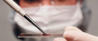
The detection of various autoantibodies in the blood serum is sometimes of decisive diagnostic importance for confirming a particular disease, is closely related to the activity of the disease, or can determine the prognosis. The laboratory tests used are an important tool in choosing a treatment method and monitoring the effectiveness of the therapy.
Changes in the level of autoantibodies, both in the direction of increasing titers and decreasing them, have prognostic significance. Most autoantibodies are not specific for any disease and are found in various combinations.
“There are two directions in which the pathology of the immune system can develop:
- Immunodeficiency is the inability of one or more components of the immune system to respond to a pathogen through a defense mechanism
- Autoimmune disease is an erroneous response to the structures of the host body, or, one might say, a loss of immune tolerance to them
Traditionally, autoimmune diseases have been considered rare and, according to epidemiological studies, occur in 3-5% of people, with the most common being chronic autoimmune thyroiditis (targeting the thyroid gland) and type 1 diabetes mellitus.
There are approximately 100 different autoimmune diseases, both locally affecting a target organ (for example, autoimmune hepatitis) and systemic in nature with multiple lesions (a typical variant is systemic lupus erythematosus). A clear increase in the risk of developing autoimmune diseases has been described in the presence of such in first-degree relatives, as well as in monozygotic twins. In general, almost all such diseases are more common in women (in a ratio of 1:1 to 10:1), with the exception of one of the inflammatory bowel diseases - Crohn's disease, which is approximately 1.2 times more common in men. Data also varies by geographic region. Thus, the autoimmune disease of the central nervous system - multiple sclerosis - is registered tens of times more often in Asia than in Europe, and type 1 diabetes mellitus is most common in the USA and is practically not observed in China. Until now, the causative factor in the occurrence of autoimmune diseases is known only in isolated cases. The most actively discussed roles are the intestinal microbiota, infectious agents, combustion products of tobacco, drugs, hormones, exposure to ultraviolet radiation, heavy metals, and collagen/silicone implants. A classic example is the long-known phenomenon of molecular mimicry (imitation of the antigenic structure of the cell’s own component) of the M-protein of streptococcus pyogenes (a common causative agent of acute tonsillitis) and “human” lysoganglioside and myosin, resulting in the appearance of cardiotoxic T-lymphocytes.
Today, despite the fact that for most autoimmune diseases the key trigger causative factor has not been established, we know through which parts of the immune system the mechanism of progression of the immune attack against one’s own body (pathogenesis) is realized. Basically, this process does not involve any single component of the immune system, but a group of such components, and for each specific disease (or at least a “subclass” of diseases), the mechanism may differ. This aspect determines the exceptional importance of correct diagnosis verification at the earliest possible stage. At the very least, it is necessary to understand the general nature of the pathogenesis that underlies the totality of the patient’s symptoms, and to begin a universal treatment that acts on the key “points” of the disease mechanism.
The process of confirming the diagnosis of an autoimmune disease in modern realities is an assessment of the patient’s combination of clinical symptoms, laboratory changes (including the determination of autoimmune antibodies of various types), comparing them with data from a morphological study of biopsy material (the most important diagnostic step in many autoimmune clinical situations) and with the results of the trial treatment, if any. Next, this complex information is compared with sets of diagnostic criteria that have been developed by expert communities for almost every disease, and if the number of necessary criteria is reached, this makes it possible to confirm (verify) the exact diagnosis. Verification of the diagnosis, in turn, already makes it possible to use therapy that targets the incorrectly functioning parts of the immune system.
In recent decades, thanks to the successes of pharmacology in the treatment of autoimmune diseases, without unnecessary exaggeration one can claim a breakthrough associated with the emergence of so-called genetically engineered biological drugs. Unlike traditional synthetic drugs, such drugs are biological substances (mainly antibodies), the purpose of which is to “point” block exactly those components of the immune system that are most involved in the mechanism of development of a particular disease. Thus, firstly, several lines of immune defense do not “switch off” at once and high efficiency of treatment is ensured with a significantly lower frequency of specific and nonspecific adverse events, although, of course, they are present in the form of nuances for such medicinal agents. It is also worth noting that currently genetic engineering biological therapy is successfully used in oncology, oncohematology, allergology, cardiology, and even in the treatment of infectious diseases (here, in fact, we should mention effective approaches to blocking the cytokine storm in severe cases of coronavirus infection using antibodies to interleukin-6, one of the main mediators of immunity that triggers systemic inflammation).
To summarize, it should be said that autoimmune diseases remain one of the most complex areas of internal medicine, and working with them requires the doctor to pay exceptional attention to the individuality of each patient, carefully analyze and synthesize the diagnostic concept, apply the principles of evidence-based medicine and deep factual knowledge, constantly updated therapeutic approaches to the treatment of such diseases."
Major systemic and “localized” autoimmune diseases
- Vessels and blood system: local leukocytoclastic vasculitis, idiopathic thrombocytopenic purpura, antiphospholipid syndrome
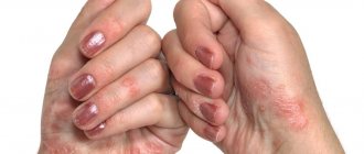
- Skin: psoriasis, vitiligo, pemphigus
- Pancreas: type 1 diabetes mellitus, autoimmune pancreatitis
- Nervous system: acute disseminated encephalomyelitis, acute and chronic inflammatory demyelinating polyneuropathy, multiple sclerosis, myasthenia gravis
- Thyroid and parathyroid glands: Graves' disease, autoimmune hypoparathyroidism, autoimmune Hashimoto's thyroiditis
- Hearing organ: autoimmune inner ear disease
- Heart: rheumatic fever
- Liver: autoimmune hepatitis, primary sclerosing cholangitis, primary biliary cholangitis (primary biliary cirrhosis)
- Gastrointestinal tract: ulcerative colitis, Crohn's disease, celiac disease, autoimmune atrophic gastritis
- Adrenal glands: Addison's disease
- Reproductive system: autoimmune orchitis and oophoritis
- Connective tissue and joints: rheumatoid arthritis, systemic lupus erythematosus, Sjogren's disease, systemic scleroderma, mixed connective tissue diseases, ankylosing spondylitis, psoriatic arthritis, dermato- and polymyositis
Rheumatic diseases
Rheumatic connective tissue diseases include:
- systemic lupus erythematosus (SLE)
- systemic scleroderma
- Sjögren's syndrome
- mixed connective tissue disease
- polymyositis (myositis) or dermatomyositis
- rheumatoid arthritis (RA)
- myasthenia gravis
- multiple sclerosis (MS)
- sometimes antiphospholipid syndrome and periarteritis nodosa, although in most recent publications this disease “opens” the group of systemic vasculitis
Laboratory diagnosis of rheumatic diseases
Laboratory diagnosis of rheumatic diseases includes determination of:
- autoantibodies
- immunoglobulins
- circulating immune complexes
- components of the complement system
- acute phase proteins of inflammation
- indicators of endothelial dysfunction/damage
- genetic markers
- bone metabolism markers
Rheumatoid factor (RF)
Rheumatoid factor (RF) is an antibody against the Fc portion of IgG. Most often these antibodies belong to IgM, but IgG, IgA, and IgE antibodies are also found. Rheumatoid factor (RF) is present in 75-80% of patients with rheumatoid arthritis; it is also found in Sjogren's syndrome, scleroderma, dermatomyositis, hyperglobulinemia, and B-cell lymphoproliferative diseases. The presence of rheumatoid factor is considered an important prognostic sign of rapidly progressive destructive joint damage and systemic manifestations in rheumatoid arthritis.
Anti-cyclic citrullinated peptide (anti-CCP) antibodies
Anti-cyclic citrullinated peptide (anti-CCP) antibodies belong to a heterogeneous group of autoantibodies that recognize antigenic determinants of filaggrin and other proteins containing the atypical amino acid citrulline. These antibodies are present in 79% of sera from patients with rheumatoid arthritis. Anti-CCPs are detected at a very early stage of RA. In addition, the test allows you to differentiate between erosive (anti-CCP-positive patients) and non-erosive (anti-CCP-negative patients) forms of RA.
Antibodies to modified citrullinated vimentin (anti-MCV)
Vimentin is a cytoskeletal protein of various cell types, such as mesenchymal and endothelial cells, fibroblasts, chondrocytes and osteocytes. It is used as a marker for soft tissue tumors. Vimentin is synthesized and modified by macrophages under the regulation of proinflammatory and anti-inflammatory cytokines and is found in the synovial fluid of patients with rheumatoid arthritis. About half of RF-negative patients are detected using anti-MCV.
Circulating immune complexes (CIC)
The formation of circulating immune complexes (CICs) is a physiological defense mechanism leading to the rapid elimination through the reticuloendothelial system of either endogenous or exogenous antigens (microorganisms, viruses, parasites, plant antigens, fungal antigens, pollen or food).
High levels of circulating immune complexes (CICs) in serum and/or other biological fluids are observed in many inflammatory and malignant diseases, which can cause the development of pathology.
Determination of circulating immune complexes (CICs) in serum is an important marker for assessing disease activity, especially in autoimmune diseases.
Frequency of antibodies (%) in autoimmune diseases
Disease | anti-dsDNA | anti-osDNA | anti-histone | anti-SS-A (Ro) | anti-SS-B (LA) |
| Systemic lupus erythematosus (SLE) | >90 | >90 | 30-50 | 10-30 | 30-50 |
| Drug-induced lupus erythematosus (DLE) | — | 30-50 | 50-90 | — | — |
| Sharpe's syndrome/mixed connective tissue diseases | 10-30 | 10-30 | — | — | — |
| Rheumatoid arthritis | — | 30-50 | 30-50 | 10-30 | — |
| Sjögren's syndrome | 10-30 | 10-30 | — | >90 | >90 |
| Scleroderma | 10-30 | 10-30 | — | 10-30 | — |
| Photosensitive dermatitis, dermatomyositis | 10-30 | 10-30 | — | — | — |
Disease | anti-Sm | anti-RNP/Sm | anti-Scl-70 | anti-Jo-1 |
| Systemic lupus erythematosus (SLE) | 10-30 | 10-30 | — | — |
| Drug-induced lupus erythematosus (DLE) | — | — | — | — |
| Sharpe's syndrome/mixed connective tissue diseases | — | >90 | — | — |
| Rheumatoid arthritis | — | — | — | — |
| Sjögren's syndrome | — | — | — | — |
| Scleroderma | — | — | >90 | — |
| Photosensitive dermatitis, dermatomyositis | — | — | — | 50-90 |
New algorithms for diagnosing autoimmune lesions of the spine and joints
Seronegative spondyloarthritis or spondyloarthropathy (SSA) is a group of interrelated inflammatory diseases that have similar clinical features, as well as common genetic predisposition factors. This group includes ankylosing spondylitis (AS) (Bechterew's disease, Marie-Strumpell disease), spondyloarthritis associated with ulcerative colitis or Crohn's disease, reactive arthritis associated with intestinal or urogenital infection, psoriatic arthritis, juvenile spondyloarthritis, "undifferentiated spondyloarthritis ", as well as probably Behçet's disease and Whipple's disease. AS was described at the end of the 17th century, although there is reason to believe that its post-mortem signs are present in some Egyptian mummies.
Symptoms last for years
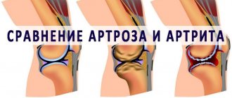
Differential diagnosis of SSA in the early stages is not always possible. This is due to the slow progression of the process and the gradual, sometimes over several years, manifestation of symptoms. Historically, SSA has been differentiated primarily from rheumatoid arthritis (RA). In contrast to the latter, they are all characterized by sacroiliitis with the development of back pain, asymmetric peripheral arthritis, the absence of rheumatoid factor, subcutaneous nodules, enthesitis (inflammation at the sites of attachment of ligaments or tendons to the bones), as well as extra-articular manifestations in the form of inflammatory lesions of the eyes, heart, lungs and skin.
Enthesitis in SSA, especially at the insertion of the Achilles tendon on the calcaneus, is considered to be as pathognomonic an early symptom as synovitis in RA. Facts of family predisposition to SSA, as well as the association of SSA with the HLA-B27 antigen, have long been described. The similarity of the clinical picture and genetic predisposition suggest a common pathogenesis of this entire group of diseases.
The incidence of AS in the general population is 0.3-0.8%, while the overall incidence of SSA is comparable to the incidence of rheumatoid arthritis. Just like RA, seronegative arthritis is characterized by articular and extra-articular manifestations. Recognition of musculoskeletal lesions is most important in clinical practice.
The most common symptom is back pain
Back pain is the most common symptom of SSA. Unlike degenerative-dystrophic lesions of the spine, pain in SSA is not acute, occurs in the morning, progresses gradually, is accompanied by morning stiffness for at least 30 minutes, and radiates to both buttocks. These symptoms improve after moderate exercise. When the disease manifests itself, it may be accompanied by fever and weight loss.
Modern criteria for pain in SSA include: 1) concomitant morning stiffness for more than 30 minutes; 2) feeling better after exercise, but not after rest; 3) night back pain; 3) intermittent pain in the buttocks.
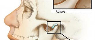
The disease begins in the lumbosacral spine, spreading to its other parts. Damage to ligaments and joints is pathognomonic for SSA and occurs in 30-50% of patients. Enthesitis is inflammation at the sites of attachment of tendons, ligaments, and joint capsule to bones, often beginning with erosions and ending with ossification of the ligaments. The most typical location of enthesitis in SSA is the attachment of the Achilles tendon to the calcaneus, the attachment of the plantar fascia to the calcaneus or metatarsal heads, and the base of the fifth metatarsal. “Tuberositas tibiae” - the tuberosity of the tibia, the attachment points of the ligaments to the upper and lower poles of the patella, the area of the ischial tuberosity, as well as the costal cartilages, the epicondyles of the ulna.
Enthesitis can be accompanied by quite intense pain at the site of inflammation, especially in the morning, after getting out of bed. SSA is also characterized by arthritis of large joints - the hip, knee, acromioclavicular joint and, in the first place, the sacroiliac joint.
There is a need for new algorithms
In cases where the manifestation of symptoms is typical, recognizing SSA is not difficult. In 1991, the European Study Group (ESSG) developed criteria for their differential diagnosis.
However, in the early stages of the disease with a history of 1 year or less, the sensitivity of these criteria leaves much to be desired.
Due to the fact that in recent years immunotropic biological products, primarily tumor necrosis factor inhibitors, whose effectiveness and safety are expressed precisely in the early stages of autoimmune inflammation, have been introduced into the practice of treating SSA (as well as other autoimmune diseases), there is a need for new diagnostic algorithms for SSA. , which increase the reliability of diagnosis in the early stages.
In the diagnosis of spondyloarthritis (SSA), more and more attention is paid to the search for subclinical signs of inflammation in the sacroiliac joint, which can be detected by magnetic resonance imaging, which was not previously included in the diagnostic criteria for SSA. This set of signs makes it possible to optimize the diagnosis of SSA, which is important for the timely and early prescription of therapy using inhibitors of pro-inflammatory cytokines.
In this regard, a group of experts of the International Society for the Assessment of Spondyloarthritis (ASAS) in 2010 developed new criteria suitable for use at the onset of the disease, as well as in the case of so-called undifferentiated spondyloarthritis, in which extra-articular manifestations and positivity for the HLA-B27 antigen are significantly detected less often.
Considering that differentiation of individual forms of SSA in the early stages has virtually no effect on treatment tactics, these algorithms are aimed at establishing the reliability of SSA diagnoses as such, dividing them into central (axial, associated with spinal lesions) and peripheral forms. Algorithms for diagnosing central and peripheral forms of SSA, which were discussed at the congresses of the European (Rome, June 2010) and Asia-Pacific (August 2010) Leagues of Antirheumatic Societies in 2010, are presented in Fig. 1 and 2.
As can be seen from them, in the diagnosis of SSA, more and more attention is paid to the search for subclinical signs of inflammation in the sacroiliac joint, which can be detected by magnetic resonance imaging, which was not previously included in the criteria for diagnosing SSA. This set of signs makes it possible to optimize the diagnosis of SSA, which is important for the timely and early prescription of therapy using inhibitors of pro-inflammatory cytokines.