Spinal dysplasia
Dysplasias are endogenous disorders of bone formation.
Spinal dysplasia is a conditional generalizing concept used to designate various variants of the malformation of the vertebrae. Unlike vertebral anomalies related to embryo- and fetopathy, spinal dysplasia can develop over a long period of time, including in the postnatal period and after the end of growth. Clinical manifestations of dysplasia largely depend on the conditions of formation and existence of the organism.
The localization of the zone of impaired bone formation in dysplasia is determined according to the X-ray anatomical scheme Ph. Rubin (1964), distinguishing the following sections of tubular bone:
- epiphysis
- physis or germinal zone proper
- metaphysis
- diaphysis
According to these zones M.V. Volkov identified epiphyseal, physeal, metaphyseal, diaphyseal and mixed lesions. In addition, taking into account the systemic nature of the pathology, dysplasias that occur with the obligatory presence of vertebral syndrome (systemic spondylodysplasia) and dysplasias in which damage to the spine is possible, but not necessarily, are distinguished. Table 24 shows the characteristic features of vertebral syndrome in various types of systemic dysplasia.
Table 24. Types and clinical and radiological features of skeletal dysplasias
| Type of dysplasia | Clinical and radiation features of vertebral syndrome |
| I. Epiphyseal dysplasia | |
| Spondyloapitheal dysplasia - Morquio-Brailsford disease | Gentle kyphosis with a short torso. A typical radiological sign is total platyspondyly - flattening of all vertebrae, more pronounced in the thoracic region. |
| Multiple epiphyseal dysplasia (Fairbank disease) | Scoliosis is possible; radiographically, the spine is normal or resembles Scheuermann's disease. |
| Multiple deforming articular dysplasia | The presence of vertebral syndrome is not typical. |
| Hemimelic form of epiphyseal dysplasia | The presence of vertebral syndrome is not typical. |
| Diastrophic dysplasia - Lamy-Maroteaux disease | Scoliosis or kyphoscoliosis (in 1/3 of cases), severe lumbar hyperlordosis (50% of cases). X-ray: the vertebral bodies are unchanged or secondarily deformed, in the lumbar region there is a decrease in the interpedicular distance in the caudal direction (a picture reminiscent of achondroplasia). |
| Pseudoachondroplasia | Clinically - a normally developed spine with sharply shortened limbs. The X-ray picture depends on age: at 4-6 years old the vertebrae are oval in shape, with pronounced defects of the apophyseal angles. The central part of the vertebral body has a “beak” appearance due to the expanded vascular canal. The changes are more pronounced in the lumbar region. With age, the shape of the vertebral bodies normalizes, but the decrease in the interpedicular distance in the lumbar region remains. |
| II. Physeal dysplasia | |
| Achondroplasia | Thoracolumbar kyphosis with sharp lumbar lordosis. X-ray: the vertebral bodies are cuboid-shaped, in the area of the thoracolumbar junction there are 1-2 posterior wedge-shaped vertebrae, a gradual narrowing of the interpedicular distance of the lumbar vertebrae in the caudal direction (spinal canal stenosis). |
| Hypochondroplasia | The presence of vertebral syndrome is not typical; a flat back may be present; rarely, thoracic lordosis or scoliosis. |
| Exostotic dysplasia | The presence of vertebral syndrome is not typical. |
| III. Spondyloepimetaphyseal dysplasia with predominant damage to the spine and hip joints | |
| Congenital spondyloepimetaphyseal dysplasia | Scoliosis may be present; thoracic hypokyphosis and accentuated lumbar lordosis are more typical. X-ray: up to 5-6 years, the vertebral bodies are round in shape, reduced in height in the thoracic region, uneven contours, depressions, irregularities, defects of the anterior angles. The discs are narrowed, the endplates are loose. Gradually (over 5-6 years) the contours of the bodies are smoothed out, the bodies are flattened, the anterior sections of the thoracic vertebrae acquire a tongue-like shape, the lumbar vertebrae are cuboid-shaped or slightly changed. Thinning of the arches of the lumbar vertebrae, more pronounced in children than in adults. |
| Late spondyloepimetaphyseal dysplasia, Kozlovski type | Slow body growth; manifesting itself after two years and slowly progressing kyphoscoliosis. X-ray: widespread platyspondyly, kyphosis, scoliosis. |
| Metatropic dysplasia | Clinically: at birth - a normal trunk with shortened limbs, subsequently - rapidly progressing kyphoscoliosis. The vertebral bodies are flattened throughout, in the thoracic region - in the form of narrow “tongues”. Typical “constriction” in the supraacetabular part of the body of the ilium. |
| Kniest's disease | Widespread platyspondyly with lengthening of the anteroposterior size of the bodies, up to the appearance of tongue-shaped vertebrae in the thoracic region |
| Parastremmatic dysplasia | Possible kyphoscoliosis |
| IV. Metaphyseal dysplasia | |
| Chondrodysplasia of Jansen, Schmid, McKusick, etc., dyschondroplasia (“enchondromatosis”) of Ollier, Pyle’s disease, etc. | The presence of vertebral syndrome is not typical; increased lumbar lordosis is possible. |
| V. Diaphyseal dysplasia | |
| Hereditary hyperostoses | The presence of vertebral syndrome is not typical. |
| Osteogenesis imperfecta - Lobstein-Vrolik disease - early and late forms. | Shortened and deformed torso. The vertebral bodies are biconcave in shape - “fish” vertebrae. |
Mayer's disease
Now about Mayer's disease (dysplasia). The Austrian orthopedist Mayer in 1964 first described the atypical (non-characteristic) clinical and radiological picture of Perthes disease. Its cause is a disruption in the transition of precursor cells (mesenchymal tissue and osteoblasts) of the femoral head in children into a mature bone cell - an osteocyte.
The risk group for its development are children under one year of age who are registered with an orthopedic doctor with a diagnosis of epiphyseal dysplasia (maturation delay) of the hip joints. The disease is most often registered in boys under 5 years of age.
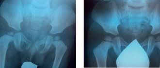
X-ray of patient K., 9 months old, diagnosis: Epiphyseal dysplasia of the right hip joint.
The clinical picture in children with Perthes and Mayer's disease is almost the same. However, in patients with Mayer's disease, the leading symptoms are less pronounced. Painful sensations in the groin area may persist for about 2 to 3 months and will stop spontaneously. Gait disturbance is not a permanent lameness; it is also short-term in nature, without restriction of movements in the affected hip joint.
To clarify the diagnosis and treatment tactics, the orthopedic doctor is obliged to recommend an X-ray examination and CT scan. The X-ray picture of these diseases in the initial stages is very similar. Only a CT scan can clarify the volume, location and depth of osteonecrosis. Its focus is most often located in the central or anterior part of the femoral head, with a lesion depth of 15–25%, combined with a decrease in the height of the femoral head.
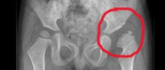
X-ray and CT examination of patient K., 4 years old, diagnosis: Mayer's disease on the right before treatment (25% osteonecrosis of the affected head).
Treatment in the initial stages of Perthes and Mayer's diseases should be the same: protective regime, unloading of the affected lower limb, drug treatment - destruction of the focus of osteonecrosis using Alflutop and physiotherapeutic procedures before repeated x-ray examination.
Parents should know that according to Russian and foreign scientific literature, about 40–50% of Mayer’s disease without treatment develops into Perthes disease within 4–6 months.
Further treatment tactics can be assessed based on the results of repeated X-ray monitoring after 4–6 months. A decrease in the focus of destruction will indicate Mayer's disease. It does not require the use of orthopedic products and plaster casts - spacers that sharply limit movements in the hip joints. Dosed loads on the sore leg, swimming, cycling, and physical therapy are allowed. With this treatment, replacement of osteonecrosis with normal bone tissue occurs after 6–9 months.
X-ray and CT examination of patient K., 5 years old, diagnosis: Mayer's disease on the right (complete replacement of the focus of osteonecrosis).
Then the child’s load can not be limited. True, X-ray monitoring is required 2 times a year for another 2 years, to assess the shape of the femoral head, as well as CT examination once a year until complete skeletal maturation.
Microperforated hymen.
The microperforated hymen is a thin membrane that almost completely covers the opening in a young woman's vagina. Menstrual blood can usually leak from the vagina, but the opening is very small. A teenager with a microperforated hymen may not realize that the hole is very small. While she can place a tampon in her vagina, she won't be able to remove it once it becomes filled with blood. Treatment for this condition is minor surgery to remove excess tissue from the vaginal opening to create a normal-sized opening for menstrual blood to flow out.
Rokitansky-Küster-Mayer syndrome
Rokitansky-Küster-Mayer syndrome is a complete absence of the vagina (except for the introitus - the entrance to the vagina) and the uterus. It is often genetic in nature. A congenital condition requiring the creation of a vagina from a section of intestine, skin or peritoneum (colpopoiesis). Pregnancy with this anomaly is impossible, but the ovaries function normally and therefore procreation is possible (surrogate mother). Genetic consultation is required. This condition is often associated with renal abnormalities or accompanies a cloacal anomaly or rectal and anal atresia.
Divided hymen by septum
A hymen with a septum occurs when the thin membrane of the hymen (the thin membrane that surrounds the opening of the vagina) has a strip of extra tissue in the middle that divides the opening of the vagina into 2 small ones instead of one. Teens with a septate hymen may have difficulty inserting a tampon. Divided hymen treatment is a minor surgery to remove the excess tissue and create a normal-sized vaginal opening.
Vaginal atresia
Vaginal atresia is a transverse vaginal septum - a more or less extended section of tissue that forms during the development of the fetus in the womb and blocks the vagina. Menstrual blood collects in the upper part of the vagina (hematocolpos) and the uterus (hematometra). Sometimes there is a small hole in the transverse vaginal septum that causes a woman to have regular menstrual periods, but these periods may last longer than the usual four to seven to ten days. Transverse vaginal septum requires surgery to remove fibrous tissue blocking the vagina and restore the integrity of the organ. This can be a rather complicated operation if the vaginal septum is located high in the upper and middle third of the vagina.
Overcoming infertility caused by uterine aplasia
Women suffering from Rokitansky-Küstner syndrome are unable to carry a pregnancy to term on their own. However, they have normally functioning ovaries in which eggs mature. In this regard, to give birth to a genetically native child, the surrogacy method is used: embryos obtained after fertilization of the patient’s eggs with her husband’s sperm are transferred into the uterine cavity of the surrogate mother. Puncture of the ovaries is carried out in this case either laparoscopically, through a puncture of the abdominal wall, or through the neovaginal fornix, if colpopoiesis was previously performed.
Nova Clinic specialists have extensive successful experience in conducting surrogacy programs for patients with Rokitansky-Küstner-Mayer syndrome, including full medical and legal support for the procedure up to the receipt of a child’s birth certificate from the registry office.
Imperforate (overgrown) hymen
A condition occurring before birth in which the hymen (the thin membrane that surrounds the vaginal opening) does not open and therefore completely closes the opening of the vagina, blocking the flow of blood during menstruation. Typically, teenage girls with an imperforate hymen do not get their period but experience pain in the lower abdomen and pelvic area, and some may also have pain with bowel movements and difficulty urinating. Treatment for imperforate hymen is surgical - the hymen is cut to create a normal-sized vaginal opening so that blood can flow out. A closed hymen is most often diagnosed after puberty in girls with otherwise normal development.
Vaginal agenesis or aplasia
A birth defect in which the vagina stops developing. Some patients may have a shorter vagina, part of the vagina, or no vagina at all. Occurs in one in 5,000 women. Patients with vaginal agenesis sometimes have other abnormally formed parts of their reproductive tract, such as an absent uterus or a small (vestigial) uterus, in addition to kidney abnormalities or problems affecting the spine, ribs, or limbs. Surgical treatment - restoration (reconstruction) of the vagina or the creation of part or all of the organ from the skin, part of the intestine or peritoneum.