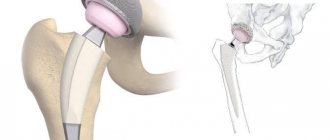
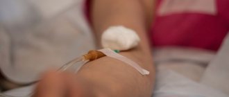
Injections into the hip joint (HJ) are made directly into the joint cavity. They not only help to quickly relieve pain and inflammation, but also improve the condition of cartilage tissue. Today this is one of the most progressive and effective methods of treating inflammatory and degenerative joint pathologies.
Injections in the hip joint are a kind of first aid for the joint. Injection of drugs quickly relieves pain and returns normal mobility. To achieve maximum results, it is important to choose the right medicine.
Advantages:
Anesthetic injections for pain in the hip joint are a way to relieve severe pain in the shortest possible time. The effect of chondroprotectors and other therapeutic agents lasts for a long time. A course of intra-articular injections allows you to delay and even avoid surgery, reduce the dosage and amount of medications taken, which is very important for problems with the digestive organs. Injection therapy can be repeated many times, since adverse reactions and complications are rare. The procedures have a beneficial effect on cartilage, accelerating healing and slowing down destructive processes.
Features of the course of the disease and distinctive features.
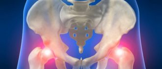
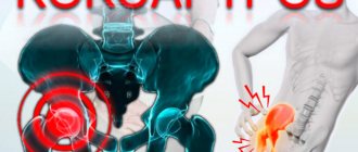
In terms of frequency of occurrence, coxarthrosis (arthrosis of the hip joint) ranks second among all degenerative joint diseases. It is difficult, with the patient losing the ability to work and becoming disabled in the absence of timely treatment. Like any other, it has a chronic course, with gradual progression. At the same time, specialists are most often turned to when conservative therapy alone may be ineffective. That is why knowledge of this problem and the use of prevention opportunities is important to prevent an increase in the number of patients with this pathology.
Injections into the hip joint: treatment with drugs
The choice of drug depends on the diagnosis given to the patient. During the course of treatment, the doctor may change medications, extend or stop the course ahead of schedule.
For injections into the hip joint, drugs of several groups are prescribed:
- anesthetics;
- glucocorticosteroids (hormones);
- vitamins;
- chondroprotectors;
- hyaluronic acid.
Attention! Anesthetics and vitamins are administered in combination with glucocorticosteroids.
Injections with hormones are indicated for intense inflammation, when a person suffers greatly from pain. In tablets, such drugs also have an anti-inflammatory effect, but directly at the source of pain they work much better. The most popular in this category are Celeston, Kenalog, Diprospan, Flosterone and Hydrocortisone. Anesthetics with which they are often combined are Lidocaine, Novocaine, Trimecaine. Glucocorticosteroids effectively relieve inflammation, relieve swelling and relieve pain. Hormonal injections have no effect on the condition of cartilage tissue.
Causes.
Coxarthrosis is not hereditary, but can occur at a young age. The causes can be congenital or acquired.
Congenital ones include:
- Congenital hip dislocation.
- Hip dysplasia.
- Metabolic disorders.
- Hereditary connective tissue diseases.
Acquired reasons include:
- Traumatic injuries.
- Endocrine system disorders.
- Infectious joint lesions (including rheumatoid arthritis).
- Aseptic necrosis of the femoral head.
Predisposing factors also include:
- Loads on joints in professional athletes.
- Overweight.
- Circulatory disorders.
- Age-related changes.
- Physical inactivity.
- Scoliosis, different lengths of the lower limbs.
Chondroprotectors
Dystrophic changes in joints due to arthrosis and arthritis lead to the destruction of cartilage. The synovial fluid loses its lubricating properties, and instead of sliding between the bones, friction occurs. The cartilage tissue is subjected to additional trauma, which causes pain to intensify.
Chondroprotectors can correct the situation. They speed up metabolism and activate the synthesis of their own collagen, which makes up cartilage. A noticeable effect from their use does not occur immediately, but after several weeks and even months. But it lasts a long time. After a course of procedures, walking becomes easier, movements cause much less discomfort. Chondroprotectors can relieve unpleasant symptoms only in the first three stages of arthrosis, when the cartilage has not yet been completely destroyed. For stage 4 deforming arthrosis, such drugs are useless.
Symptoms
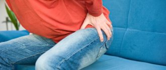
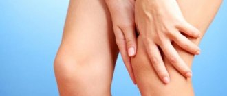
The clinical picture of the disease is determined by the stage of its development. The main symptom of arthrosis is pain in the joint area. At first it appears only during movement or walking. As the pathological process develops, the pain intensifies, it becomes constant, and difficulties with movement arise.
Stiffness of movement occurs in the morning after waking up. This is the starting pain. In the initial stages of development, such pain goes away on its own after the person begins to move.
The localization of pain is the groin area, the outer part of the thigh, buttocks, the front surface of the thigh, it radiates to the knee and lower leg. The severity of pain affects the range of motion. At first, simply painful spasms appear in the muscles, after which the joint capsule contracts.
The intensity of pain affects gait. A person tries to avoid putting stress on a sore limb and begins to limp. Over time, a persistent contracture forms and the limb shortens.
Hyaluronic acid
Injections with hyaluronic acid replenish the deficiency of synovial fluid characteristic of joint pathologies. It is called “synovial prosthesis”, since the consistency and composition of hyaluronic acid is identical to natural synovium. If there is a lack of it, the articulating surfaces of the bones begin to rub against each other when walking and injure the cartilaginous layer. Hyaluronic acid returns synovium to its normal qualities, at the same time relieving inflammation and stimulating the regeneration of damaged areas.
How many injections need to be given?
The effect of corticosteroids can last from 2 weeks to 3 months. Injections with hyaluronic acid are prescribed taking into account the stage of the musculoskeletal system disease. 1 course consists of 3-5 procedures, which are carried out once a week. In the early stages this is enough. If the joint is very bothersome and arthrosis has already entered the 2-3rd stage, the courses are repeated every 6 months. Names of drugs with hyaluronic acid:
- gialgan;
- hialubrix; (hormones);
- Hyalon.
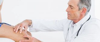
Injections into the joint are made with a syringe with a very long needle, which should fall exactly into the joint space. The procedure is performed under sterile conditions and under ultrasound control.
After intra-articular injections, it is recommended to limit thermal and water procedures for 24 hours; there are no restrictions on physical activity. Intra-articular injections are a very responsible procedure, which should only be performed by qualified specialists. If you have problems with the hip joint - it has become difficult to walk or perform various movements - contact our center. You will receive competent advice: the doctor will assess the severity of the problem and prescribe a course of treatment procedures, selecting the most suitable drug.
Coxarthrosis in systemic diseases
Gouty coxarthrosis
Etiological phenomena
The etiological mechanism of arthritis has not yet been fully elucidated, but certain patterns of the onset of the disease have been studied in detail. In general, there are two areas of causes leading to this pathology:
- infectious lesion;
- endogenous factor of an autoimmune or allergic nature.
Infectious lesions are caused by the action of the following pathogenic microorganisms on the body:
- intestinal infection (salmonellosis, shigellosis, yersiniosis);
- infection of the urogenital tract (chlamydia, mycoplasma, ureaplasma);
- Group A and B streptococci (cause rheumatoid arthritis).
In this case, the infection extremely rarely penetrates the joint itself, but damage to the body is sufficient, and then the autoimmune mechanism is activated. Quite often the cause is diseases such as meningitis, Lyme disease, tuberculosis, brucellosis, rubella, and gonorrhea.
Inflammation in the joint can occur without infection. In this area, allergic reactions, metabolic disorders, hereditary diseases with damage to the immune system, ankylosing spondylitis and Crohn's diseases, and malignant formations are highlighted. The following factors provoke the activation of the etiological mechanism:
- hereditary predisposition;
- frequent and prolonged physical overload of the joint;
- injuries in the hip area;
- hypothermia;
- diseases of the musculoskeletal system;
- hormonal disorders;
- taking certain medications.
Symptomatic manifestations
Symptoms of hip arthritis depend on the type of pathology, but in general they have common features. There are several categories of symptoms:
- manifestation of an inflammatory reaction: pain, swelling, redness, increased temperature in the area of the affected joint;
- signs of structural disorders: limited mobility, up to complete immobilization; stiffness; joint deformity; lameness;
- signs of general intoxication of the body: fever, general weakness, fatigue, headache, loss of appetite.
Coxitis can develop in acute, subacute and chronic forms. Depending on this, the intensity of symptoms also changes. The acute and subacute stages of the disease are characterized by a sharp, unexpected manifestation of pain, swelling, and blocking of movement. The general condition of the sick person worsens significantly. The untreated acute form becomes chronic. This development of pathology is characterized by alternating phases of exacerbation and complete remission. There is a slow destruction of joint tissues, which can lead to loss of joint mobility.
The most characteristic symptom of hip arthritis is pain. As a rule, pain is concentrated in the groin, buttocks, trochanteric area of the thigh, as well as along the anterior femoral surface, spreading along the lower limb to the knee. The intensity of pain depends on the type of lesion and its severity. As the pain syndrome progresses, it becomes the main factor limiting mobility. At the initial stage, restriction of movement is associated with pain due to increased muscle tone, and then with disorders in the joint capsule.
Pain syndrome is directly related to mechanical stress. The most severe pain appears in the morning, immediately after getting up, and also after being in a sitting position for a long time. After a short walk they calm down. In general, the manifestation of pain increases with the progression of the disease. If at the initial stage it has the appearance of the indicated “starting pain” or appears only during physical activity and disappears after rest, then in the advanced stage the pain is fixed and at rest can continue in an intensive mode for up to 2 - 3 days.
It is pain that gradually leads to a change in gait. A sick person instinctively seeks support so that the discomfort is minimal, which gradually becomes a habit. The so-called Trendelenburg gait is quite typical for the pathology, when the pelvis, when moving, falls more towards the leg opposite the lesion. In the last stages of coxitis, pronounced signs of joint deformation appear. Stable contractures are formed, and shortening of the lower limb is observed.
Diagnosis of gouty arthritis
The key point of diagnostic procedures is the detection of sodium urate crystals in the synovial fluid of the joints, both during an attack and during remission. Synovial fluid for analysis can be taken from any large joint, even one that has never been subject to inflammation, for example, from the knee. Also, the contents of the tophi or any other biological material can be taken for research.
Hyperuricemia (increased levels of uric acid in the blood) in combination with periodic inflammation of the joint of the big toe is not considered a confirmation of gout, it is only a marker of impaired purine metabolism. Many people with hyperuricemia do not have gout.
With a long course of the disease, it makes sense to conduct an x-ray examination. At the early stage of the disease there are no characteristic changes. Then, on x-rays, signs typical of gout appear: destruction of cartilage, defects in the end sections of bones, punctures.
When gout develops on the upper extremities, it is quite difficult to differentiate it from other joint diseases: rheumatoid arthritis, osteoarthritis, etc.
Causes of gouty arthritis
The etiology of the disease is not fully understood. The main risk factors for its occurrence include:
- Hereditary predisposition;
- Poor nutrition: excessive abuse of meat products, sausages, chocolate, strong coffee and tea, alcohol. (Gout used to be called “the disease of aristocrats”);
- The presence of concomitant diseases, such as heart failure, hemoblastosis, kidney disease, hormonal abnormalities;
- Use of certain medications: drugs for high blood pressure, diuretics, cytostatics, etc.
There are also primary and secondary gouty arthritis:
- Primary gout develops as a result of a combination of genetic predisposition and high intake of purines from the foods listed above;
- Secondary gout occurs due to the presence of these diseases and medications.
The accumulation of sodium urate microcrystals in the joint cavity can occur asymptomatically for a long time, until some factor provokes an acute attack: physical fatigue (long walking), injury, infection, stress, hypothermia, fasting or consumption of large amounts of “purine” foods in combination with alcohol.
Preventive actions
In order to avoid or reduce the likelihood of developing pathology, especially for people after 35 years of age, one should remember about preventive actions. Prevention of gout involves simple steps, for these purposes you should:
- control body weight;
- avoid colds and infectious pathologies;
- prevent joint injuries;
- wear comfortable shoes that do not restrict movement;
- monitor blood pressure;
- Avoid drinking alcoholic beverages, especially beer;
- adhere to proper nutrition;
- lead an active lifestyle and do physical exercise;
- systematically take a biochemical blood test to monitor urea levels;
- Do not take medications without supervision.