Immaturity of the hip joint in newborns is a pathological condition in which the joint does not form quickly enough. When a child is born, the main part of his hip joints consists of cartilaginous tissue. Ossification nuclei are localized in the area of the heads of the femoral bones of the joints, and their nucleus sizes are 3-6 mm. Sometimes such zones appear later, often before six months of age.
Immaturity of the nuclei of the femoral bones is often combined with various forms of dysplasia. Previously, these concepts were identified, highlighting immaturity as an early form of dysplasia. Now immaturity of the hip joints is considered as a separate pathological condition, despite the similarity of diagnosis and treatment. Only conservative methods are used in therapy - physiotherapeutic and massage procedures, wearing orthopedic devices. And to eliminate severe forms of dysplasia, surgery is required.
Characteristic features of the pathology
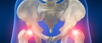
Underdevelopment of the hip joint (HJ) is diagnosed in more than 20% of newborns, and four times more often in girls than in boys. Unlike dysplasia, with immaturity, the hip joints are able to form correctly, but this happens much more slowly than normal. Since children's joints and spine consist of a large amount of cartilaginous tissue, their temporary immaturity is quite physiological. The diagnosis is made on the basis of delayed development of ossification nuclei. Other signs of immaturity are considered normal:
- large size of the acetabulum;
- their flat shape;
- increased elasticity of the elements of the ligamentous-tendon apparatus.
But with the combination of such structural features and the immaturity of the hip joint, the development and progression of dysplasia is possible - a disease dangerous with its serious consequences. Therefore, pediatric orthopedists do not wait for the formation of foci of ossification, but take measures to ensure the full development of the hip joint.
How to forget about joint pain?
- Joint pain limits your movements and full life...
- You are worried about discomfort, crunching and systematic pain...
- You may have tried a bunch of medications, creams and ointments...
- But judging by the fact that you are reading these lines, they did not help you much...
But orthopedist Valentin Dikul claims that a truly effective remedy for joint pain exists! Read more >>>
loading…
Source
Causes and provoking factors
In some cases, immaturity of the hip joints in a newborn can be predicted due to a complicated pregnancy. If, while carrying a child, a woman’s chronic diseases worsen or she is diagnosed with an acute form of infection, medication is required. Drugs of certain clinical and pharmacological groups (antibiotics, immunomodulators, cytostatics) often provoke adverse reactions. One of them may be a slowdown in the formation of ossification nuclei. Other causes of physiological immaturity of the hip joints in newborns:
- breech presentation of the fetus;
- severe toxicosis throughout most of pregnancy;
- lack of sufficient amounts of microelements, fat- and water-soluble vitamins in the expectant mother’s diet;
- sharp fluctuations in hormonal levels;
- complicated childbirth.
In premature babies, hip joint immaturity is almost always diagnosed, and there is a logical explanation for this. The child was born prematurely, so the ossification nuclei are only at the stage of formation. Often physiological immaturity is provoked by insufficient supply to the fetus of nutrients and biologically active substances necessary for the proper development of the musculoskeletal system. But there are also genetic prerequisites, usually identified at the stage of pregnancy planning.
The delayed formation of ossification nuclei is influenced by the hormonal background of the expectant mother. For example, before childbirth, the ovaries and placenta begin to produce an increased amount of relaxin. This hormone helps to relax the ligaments of the pubic joint of the pelvic bones, expand the pelvis, and normalize childbirth. But selectivity is not typical for this hormone. Therefore, at the same time, the bone structures of the fetus soften, causing underdevelopment of the hip joints.
Complications
Treatment started in a timely manner (up to 3 months from the moment of birth), as a rule, does not lead to the development of complications. If joint immaturity is not detected in time and not corrected, quite serious undesirable consequences can develop:
- Lameness.
- Pain in the joint when walking.
- Atrophy of the muscles of the sore leg.
- Compensatory hypertrophy of the muscles of a healthy limb.
- Poor posture (lordosis, scoliosis).
- Vertebral displacement.
In children after 2 years of age, it is usually not possible to correct disorders using conservative methods. The issue of surgical treatment is being decided.
Clinical picture
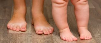
Underdevelopment of the hip joints in newborns is sometimes discovered in the maternity hospital during the first examination by a pediatric orthopedist. But unlike dysplasia, hip joint immaturity does not manifest itself with severe symptoms, especially during the first days of a child’s life. Signs of delayed ossification and improper formation of joints usually appear after 3 months. What parents or a pediatrician may notice during the next examination:
- shortening of the thigh;
- decreased muscle tone;
- asymmetrical arrangement of skin folds;
- the occurrence of an obstacle when attempting to abduct the joint;
- a characteristic click when the hip joint is abducted.
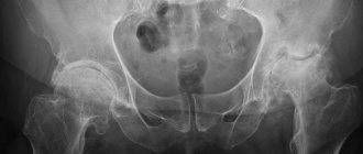
The earlier the pathology is diagnosed, the faster and more successful the therapy. Underdevelopment can be detected by external examination, complaints from parents, and functional testing. The results of ultrasound and x-ray examinations help confirm the diagnosis. Although radiography is considered the most informative method, its use is contraindicated in children under 3 months. The degree of maturity of the hip joint is determined according to the parameters of Graf’s ultrasonic classification. For example, Graf type 2a is an immature dysplastic joint.
Ossification of the femoral heads occurs at approximately 7 months of age in girls, 9 in boys. If treatment for immaturity of the hip joints is carried out before the child reaches six months, then later they form within normal limits.
| Forms of underdevelopment of the hip joints | Short description |
| Acetabular form | This is the name for congenital underdevelopment of the acetabulum in newborns. It is characterized by changes in the centralization of the femoral head and increased elasticity of the ligaments. The deviations are minor and can be easily corrected with therapeutic massage and gymnastics. In the first months of life, a similar condition of varying degrees is detected in most infants. Whether it will become a prerequisite for incorrect formation of the hip joint can only be determined by subsequent examinations, which are usually carried out every month |
| Dysplastic changes of the proximal femur | This congenital form of underdevelopment is revealed when measuring the neck-shaft angles. The parameter is calculated using lines connecting the centers of the necks and heads of bones, and diaphyseal lines. In children older than 3 months, underdevelopment of the femoral heads is determined by radiographic images |
| Rotational underdevelopment | A developmental disorder characterized by changes in the angles between the axes of the hip and knee joints in horizontal planes. If the obtained values deviate from the norm (in newborns - about 35°), a violation of the centering of the hip joint in the acetabulum is diagnosed |
Delayed development of the hip joint
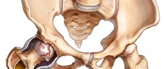
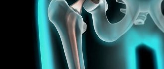
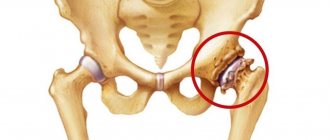
Impaired development of the hip joint , manifested in the form of its delay, is called dysplasia.
The pathology is characterized by a discrepancy between the femoral and pelvic components of the joint. In other words, the full contact and relationship between the head of the femur and the acetabulum of the pelvis is disrupted. At the same time, almost all elements of the joint are underdeveloped. Vertebroneurological specialists at the Clinic of Dr. Ignatiev in Kyiv observe the phenomenon of dysplasia more often in newborn girls. The left joint is predominantly affected. Typically, pathology is detected approximately a month after birth during a medical examination and requires the intervention of a specialized specialist. The clinic's vertebrologists examine patients after prior appointment.
Treatment methods
When diagnosing hypoplasia (underdevelopment) of the hip joints in newborns, its form is determined. This allows pediatric orthopedists to quickly determine the necessary treatment methods to correct further formation of the hip joint.
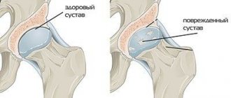
Dysplastic changes often accompany immaturity of the hip joints. The most dangerous is congenital dislocation of the hip joint, which, if left untreated, leads to clubfoot, lameness, and poor posture. Pre-luxations and subluxations are detected more often, but are also much easier to correct using conservative methods.
Conservative therapy
Treatment of mild underdevelopment of the hip joints in newborns begins with massage. The procedure is performed only by a specialist with a medical education who is well acquainted with the anatomy of small children. Subsequently, after the child has recovered, massage can be performed by parents to prolong the clinical effect and for preventive purposes.
Description of exercises:
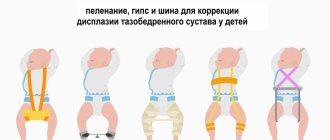
The treatment of hip joint immaturity, complicated by dysplastic changes, requires a slightly different approach. For some time, the hip joints are fixed to ossify in a physiological position. This can be done by properly swaddling a newborn:
- the diaper is folded so that a triangle is formed, pointing downwards;
- put a diaper on the baby and place it on the diaper so that its upper ends are located on both sides of the belt;
- bend your legs at an angle of 80°;
- wrap the sides of the diaper around the legs, and fasten the lower part to the belt.
If swaddling is done correctly, the baby ends up in the “frog” position. In some cases, to achieve this position, wearing a Freik pillow, coxite bandage or other orthopedic devices is indicated. They are used for several months, and when diagnosing serious dysplastic changes - 1-2 years.
Daily therapeutic exercises contribute to the correct formation of the hip joint. A set of exercises is developed by a physical therapy doctor, who conducts the first classes. He shows parents how to bend their baby's legs, rotate their feet, and stretch their heels. The “bicycle” exercise is very useful, during which all joint structures are involved.
If the child has already learned to walk, then pediatricians recommend purchasing an orthopedic mat. Its surface imitates river pebbles, large pebbles or sand. Walking on such a mat stimulates blood circulation in the legs and promotes faster recovery.
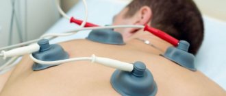
Pharmacological drugs are rarely used in the treatment of underdevelopment of the hip joint in newborns. Children's orthopedists recommend taking vitamins that accelerate regeneration processes. Physiotherapeutic procedures, usually electrophoresis, are also prescribed. Tampons soaked in medicinal solutions are applied to the hip joint, and metal plates are placed on top. An electric current is passed through them, under the influence of which the drug molecules penetrate and are evenly distributed in all tissues of the joint. When carrying out medical procedures, a calcium solution is usually used to accelerate the formation of ossification nuclei.
Surgical intervention
If conservative treatment is ineffective, the doctor may decide to perform surgery. When diagnosing ordinary immaturity, this method of therapy is not used. Surgical intervention is necessary if concomitant dysplasia is detected - congenital dislocation of the hip joint. If the child is over one year old, the doctor prescribes bloodless reduction of the joint using the Adolf Lorenz method. Under anesthesia, the surgeon returns the head of the femur to a physiological position. Then the pelvis and legs are immobilized for about six months. To do this, a coxite plaster cast is applied to fix the child’s legs in an extended position.
Treatment history:
Open operations are performed less frequently. This is the reduction of a congenital dislocation with dissection of the articular capsule, and sometimes deepening of the acetabulum. An osteotomy is performed to give the correct configuration of the femur. When choosing the best method, the surgeon takes into account the degree of deformation of the pelvic cavity and the elasticity of the ligaments. During the rehabilitation period, massage and physiotherapeutic procedures, daily therapeutic exercises are indicated.
Therapy for hip joint underdevelopment is carried out immediately after diagnosis, since the child is growing and ossification nuclei are not formed. There is too much elastic cartilage tissue in the joints that does not turn into bone. Therefore, one of the main tasks of modern pediatric orthopedics is the early detection of a pathological condition.
Diagnostics
A preliminary diagnosis is made by an orthopedic surgeon during the initial examination. It is especially important to visit an orthopedist for premature babies, as they have a high risk of developing dysplasia. The doctor will conduct a series of clinical tests to detect mobility problems in the hip joint. In addition, he will find out how the pregnancy proceeded and whether there were cases of congenital joint pathology in the family. The final diagnosis is made after an ultrasound examination.
Ultrasound diagnostics in newborns
This method is absolutely safe and at the same time very informative. Until the baby is 3 months old, ultrasound is the only method to confirm the diagnosis. An important advantage is that ultrasound diagnostics can determine all types and stages of hip immaturity and dysplasia.
Preventive measures
The following tips will help minimize the likelihood of developing problems with the hip joints:
- monitor the healthy course of pregnancy;
- use special car seats to transport a child;
- hold the baby correctly in your arms;
- use the free swaddling technique;
- regularly massage your child and do gymnastics;
- attend routine examinations with an orthopedist.
It is possible to reduce the risk of symptoms of the disease even if the baby has a hereditary predisposition. Systematic implementation of preventive measures can significantly improve the child’s condition.
The baby’s recovery directly depends on the degree of parental responsibility. The sooner they detect signs of pathology and consult a doctor, the more conscientiously they carry out the procedures prescribed by the orthopedist, the sooner the child’s hip joints will recover.