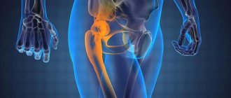
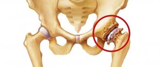
Coxarthrosis - or (as it is also called) arthrosis of the hip joint - is a progressive pathological condition that is characterized by the destruction of the cartilage and bone of the hip joint.
Coxarthrosis of the hip joint usually develops after fifty years. In most cases, women are affected by this disease. According to statistical data, in the vast majority of cases (about 70%), the symptoms of coxarthrosis of the hip joint and the need for its treatment arise due to the aging of the body. Other cases of occurrence are usually associated with excessive physical activity, injuries and excess weight.
You can undergo a course of treatment for coxarthrosis at the CELT multidisciplinary clinic.
At CELT you can get a consultation with a traumatologist-orthopedic specialist.
- Initial consultation – 3,000
- Repeated consultation – 2,000
Make an appointment
Causes
Depending on the cause of the onset and development of the disease, it is customary to distinguish different forms of coxarthrosis:
- Primary coxarthrosis - the cause has not been established; development occurs after 50 years and is characterized by bilateral damage;
- Secondary coxarthrosis - appears against the background of existing pathologies, usually at a young age.
As for pathologies that can lead to the appearance and development of coxarthrosis, they include the following:
- various injuries;
- defects of cartilage tissue, which are characterized by the formation of loose intra-articular bodies;
- excessive physical activity due to professional activities;
- Perthes disease;
- numerous microtraumas of the joint;
- presence of rheumatoid arthritis;
- pathologies characterized by spinal curvature;
- obesity degrees III-IV;
- flat feet;
- hip dysplasia.
How to treat coxitis?
Medicines:
- Antibacterial and sulfonamide drugs (take up to 2 months);
- Non-steroidal anti-inflammatory drugs;
- Glucocorticosteroids in the form of injections;
- Antibiotics, which are prescribed for purulent forms.
Intra-articular debridement and surgical intervention are prescribed if drug treatment does not work. Prosthetics or hip replacement are prescribed. The following types of surgical procedures are distinguished:
- Arthroplasty;
- Necrectomy;
- Intra-articular resection;
- Extra-articular resection - the operation gives a lesser result than with intra-articular resection;
- Corrective osteotomy – returns the physical position.
go to top
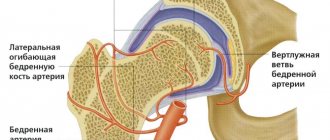
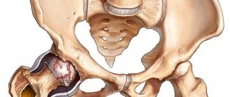
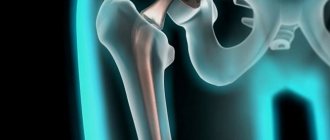
Anatomy of the hip joint
The hip joint is formed by the acetabulum of the pelvic bone and the head of the femur. During movement, the femoral head moves in several directions, but the acetabulum remains fixed. The ball and socket joint provides full mobility of the hip and all actions: rotation, extension, adduction, abduction.
The movement of the surfaces of the articular bones is ensured by sliding, which is provided by hyaline cartilage. It has high strength and elasticity and protects the femoral head and acetabulum from friction. Hyaline cartilage absorbs shock and eliminates friction, and most importantly, it is responsible for evenly distributing the load during any movement.
Hyaline cartilage is nourished by articular fluid, which eliminates friction. The outer part of the joint is protected by a strong capsule, which is surrounded by the gluteal and thigh muscles. Muscle tissue also ensures normal functioning and performs a shock-absorbing function, protecting the hip joint from injury.
Coxitis in children
Coxitis in children often manifests itself in one form - tuberculosis. It affects boys more often than girls. In adults, this type of coxitis is rare - in 5 out of 100 cases. The cause of tuberculous coxitis is an infection that is transmitted from other organs affected by tuberculosis. The infection penetrates the synovial bursa and joint endings, where it begins to multiply and secrete pus. Purulent exudate breaks through and affects soft tissues that are nearby. This leads to abscesses and fistulas. The joint gradually collapses, which provokes the possibility of dislocation.
Symptoms do not appear immediately. It all starts with pain that radiates to the knee, as well as atrophy of the femoral and gluteal muscles, accompanied by thickening of the subcutaneous tissue of the thighs. This set of symptoms is called the “positive Alexandrov symptom.” The child begins to limp. A blood test reveals changes in the number of lymphocytes and ESR. X-rays show foci of tuberculosis, mainly in the area of the acetabulum and femoral head.
If parents neglect to treat their child, then after a couple of years acute coxitis goes away and the pain subsides. However, after this the muscles remain atrophied and the joint is deformed. A pathological type of hip dislocation occurs.
go to top
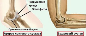
Mechanism of development of coxarthrosis
At the initial stage, the consistency of the joint fluid changes - it becomes viscous and thick, which leads to the gradual drying of the hyaline cartilage. Because of this, it gradually collapses and cracks appear on the surface. Due to the thinning and drying of cartilage tissue, pathological deformations begin in the hip joint. Further development of the pathology leads to rubbing and deformation of the bones, as the load on the bone tissue increases. At a severe stage, diagnostics reveals not only the destruction of cartilage and bone tissue, but also muscle atrophy.
Coxitis in adults
In adults, coxitis also occurs due to, often, infection from already diseased organs of the body. Neglect or improper treatment of underlying diseases leads to the spread of infection throughout the body. The hip joints may be affected.
A man may experience dislocations when working in hazardous and heavy industries. Physical work and heavy physical activity can provoke coxitis as an independent disease. In women, this disease occurs during heavy physical activity - when carrying heavy bags, playing strenuous sports. There may be cases of inflammation when walking in high heels for a long time.
go to top
Clinical manifestations
Symptoms of coxarthrosis of the hip joint depend on what stage of development the disease is at. In accordance with this, it is customary to distinguish three degrees of coxarthrosis.
1st degree
This stage of development of the disease is characterized by pain in the joint, which appears after prolonged physical exertion. Apart from this, no symptoms are observed: the patient retains his normal gait, and the thigh muscles are not atrophied.
2nd degree
The 2nd degree is characterized by more severe symptoms, namely:
- Painful sensations appear more vividly and become constant not only during movement, but also at rest;
- The pain radiates to the thigh and groin area, so the patient begins to limp;
- There is limited movement of the hip.
3rd degree
The following clinical manifestations are characteristic of this stage of disease development:
- constant severe pain;
- difficulty moving independently, the need to use a cane or crutch;
- significant limitation of hip movements;
- shortening of the leg due to atrophy of the gluteal, calf and thigh muscles.
Orthopedic therapy:
- Fixation of the hip joint in order to immobilize it and give it rest;
- As recovery progresses, the plaster cast is removed, allowing the patient to walk first with crutches and then without them;
- Massage;
- Physiotherapy;
- Therapeutic and recreational gymnastics.
The last procedures are carried out only after the swelling has been removed and the symptoms of acute coxitis have passed.
Diet does not play a significant role in the treatment of coxitis. The immune system is strengthened, which is beneficial when consuming vitamin-rich foods.
After all procedures, it is recommended to visit medical and sanatorium institutions, where additional preventive treatment will continue and rest will be provided.
go to top
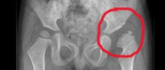
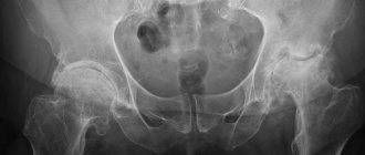
Diagnostics
Before starting treatment for coxarthrosis of the hip joint, CELT specialists conduct a comprehensive diagnosis, which makes it possible to identify the cause of the development of the pathology and determine the degree of its development. Diagnostics includes the following:
- examination by a specialist and medical history;
- radiography, which often allows one to determine not only the degree of pathology, but also the cause of its occurrence;
- MRI and, which make it possible to determine pathological changes in bone and soft tissues.
The symptoms of coxarthrosis are similar to osteochondrosis and gonarthrosis. At the second and third stages of the pathology, acute pain syndrome is observed due to muscle atrophy. The pain is localized not only in the hip, but also in the knee joint. Therefore, a specialist must conduct a visual examination and palpation of the knee joints, and then give a referral for an MRI or CT scan.
Pain from pinched nerve roots due to osteochondrosis can also affect the hip joint, but it only appears during physical activity. Differential diagnosis includes palpation to determine the location of the discomfort. Mobility in the hip joint is not limited, and with coxarthrosis a person cannot move the leg to the side. Since both pathologies can occur simultaneously, a comprehensive diagnosis is required.
It is necessary to differentiate the pathology from trochanteric bursitis, since similar symptoms are observed. But with this disease there are no restrictions on mobility, acute pain is observed during physical activity, and the main difference is that the development of trochanteric bursitis occurs in a maximum of 14 days.
With ankylosing spondylitis and reactive arthritis, similar symptoms appear. The pain goes away while walking, and stiffness in the hip joint is observed. The main difference is that pain appears at night, and joint mobility is restored during prolonged walking or exercise.
Types and forms
There is a detailed classification of types and forms of the disease.
Coxite is divided into the following categories:
- Primary synovial. The inflammatory process begins in the synovial bursa, causing an increase in the volume of fluid produced. Subsequently, coxitis spreads to the bone elements of the articulation. Mostly caused by infection or rheumatoid arthritis.
- Primary bone. The disease originates in bone tissue, and then expands its influence on the synovial membranes and adjacent structures. This group includes tuberculosis and nonspecific forms of the disease.
- Reactive. It develops rapidly and is most often a consequence of an infectious disease. Inflammation spreads to the ligaments, internal organs and skin may be affected.
- Transitional. It is predominantly a consequence of traumatic injury or another inflammatory process in the hip joint area, but the exact cause is quite difficult to establish.
- Spicy. The symptoms manifest themselves quite intensely, and the patient experiences severe pain. The duration of the acute phase is 2 - 4 months.
- Chronic. It is characterized by alternating periods of active manifestation of symptoms and their subsidence. It develops in the absence of quality treatment in the early stages of the disease.
The specificity of the course of the disease and the variety of provoking factors determine the division of coxitis into the following types:
| Title and photo | Short description |
| Infectious
| It develops against the background of an infectious disease, most often childhood infections (measles, rubella, etc.). |
| Pyogenic
| Accompanied by suppuration of the joint cavity, it can lead to blood poisoning and death. |
| Allergic
| It is characterized by rapid development against the background of an allergic reaction, for example, during preventive vaccination. |
| Tuberculous
| It belongs to a specific type of infectious coxitis, is a consequence of infection with tuberculosis, and is characterized by a slow progression with mild symptoms. |
| Gonorrheal
| It is a side disease of gonorrhea; the characteristic symptoms are severe pain and large effusion. |
| Syphilitic
| It appears as a side symptom in secondary and tertiary syphilis. |
| Brucellosis
| It is a consequence of human infection with a zoonotic infection – brucellosis. |
| Dysenteric
| It develops as a complication of dysentery and is classified as an infectious arthritis. |
| Psoriatic
| A disease accompanying psoriasis as a developed complication. |
| Rheumatoid
| It is a consequence of gene mutations and occurs in autoimmune disorders. |
| Aseptic
| It is considered one of the most severe forms of the disease, as it provokes a heart attack of the hip joint, that is, necrosis of the femoral head |
Treatment
The treatment tactics for this disease directly depend on the degree of its development. The initial stages are usually treated with conservative methods. They allow you to stop the development of pathology, but do not completely eliminate it. For the first and second degrees of coxarthrosis, drug treatment is used, which consists of taking the following drugs:
- non-steroidal anti-inflammatory drugs;
- chondroprotectors;
- symptomatic.
In addition, you need:
- minimize stress on the affected joint;
- engage in physical therapy;
- get rid of excess weight;
- have a full rest.
CELT specialists resort to surgical treatment only in extreme cases, when the patient has grade 3 coxarthrosis, leading to immobilization. In this case, endoprosthetics surgery is performed, which involves replacing the joint with a prosthesis.
Depending on the stage of the disease, we choose one or more treatment methods:
Osteopathy
Soft technique of working with the spine, joints, muscles, ligaments, internal organs. Eliminates pain and starts the self-healing process.
Therapeutic massage, osteopathy, manual therapy
Helps bones and joints take the correct physiological position, relieves pain and spasms, relaxes muscles.
Acupuncture
Work on biologically active points. It affects the affected area and the body as a whole. Eliminates the cause of the disease and removes the symptoms.
In addition, according to indications, the following are used: taping, pharmacopuncture, FormTotix insoles, exercise therapy with an instructor and other methods. The choice of procedures depends on the current condition; taken together, they act faster and give a more lasting result.
Moscow osteopath
Orthopedics and traumatology services at CELT
The administration of CELT JSC regularly updates the price list posted on the clinic’s website. However, in order to avoid possible misunderstandings, we ask you to clarify the cost of services by phone: +7
| Service name | Price in rubles |
| Appointment with a surgical doctor (primary, for complex programs) | 3 000 |
| X-ray of the hip joint | 2 400 |
| MRI of hip joints (2 joints) | 8 000 |
| MSCT of the hip joint | 7 500 |
All services
Make an appointment through the application or by calling +7 +7 We work every day:
- Monday—Friday: 8.00—20.00
- Saturday: 8.00–18.00
- Sunday is a day off
The nearest metro and MCC stations to the clinic:
- Highway of Enthusiasts or Perovo
- Partisan
- Enthusiast Highway
Driving directions
Tuberculous drives
The primary focus is localized mainly in the condyle of the femur, less often in the tibia and in the patella. A parietal lesion may be accompanied by synovitis and is clinically more likely to manifest itself than deep and central lesions.
A feeling of fatigue and incomprehensible pain after exercise, which disappears with rest, may be the first signs and require further X-ray and laboratory examination of the patient. On a radiograph, primary osteitis can have different sizes and shapes (usually round), and sometimes a sequestration. The transition of the tuberculosis process from the primary focus to the joint may be barely noticeable, since nonspecific reactive synovitis initially dominates. Only when all elements of the joint are involved in the process does the classic clinic of tuberculosis drive appear. The knee joint is swollen, severe flexural antalgic contracture, muscle atrophy.
Palpation can detect pain and increased local temperature, slight fluctuation in the joint, infiltration of the joint capsule (superior inversion), Alexandrov's symptom. All these symptoms are especially pronounced at the height of the tuberculosis process. With a significant destructive process, cold flowing abscesses appear, which can break through to the surface with the formation of a fistula.
At the attenuation stage, all clinical symptoms of inflammation gradually disappear. The joint becomes painless, cold, dry. Destructive processes stop and reparative processes predominate, but muscle atrophy, contractures and anatomical and functional disorders in the joint remain to varying degrees.
Treatment of tuberculous gonitis
Treatment of patients with tuberculous gonitis does not differ from the general principle of treatment for osteoarticular tuberculosis of other localizations. Antibacterial therapy is an important means of specific treatment, and a plaster circular gonitis bandage grips the pelvis and foot, creates proper rest at the end, and prevents the occurrence of contracture in an unfavorable position of the limb. Restoration of joint function can be counted on only when treatment of the patient in the prearthritic phase is started in a timely manner. For this purpose, in addition to conservative means, extra-articular early necrectomy of foci is used, which gives better results. In the arthritic phase, it is necessary to eliminate contractures and subluxations of the leg; surgical treatment is indicated: synovectomy, necrectomy, joint resection, etc.