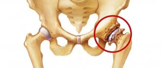
Osteoarthritis (OA) of the hip joints refers to serious degenerative-dystrophic changes that occur in the cartilaginous tissues of the articular surfaces of the hip joint. Osteoarthritis with this localization is considered one of the most common joint diseases. The pathology, which mercilessly destroys the hip joint, ranks second in occurrence after gonarthrosis of the knee joints. In terms of the risk of rapid disability in the general structure of joint diseases, OA ranks first. Another specific name for the disease, which refers specifically to damage in a given area of the musculoskeletal system, is coxarthrosis.
This is what an advanced diagnosis looks like on an x-ray.
Approximately 80% of elderly people (60 years and older) have radiographic evidence of hip OA to one degree or another, and a quarter of them experience intense pain and severe motor limitations. Osteoarthritis of TB joints is diagnosed almost 2.5 times more often in women. According to statistics, a sharp increase in the incidence rate is observed in the population over 45 years of age. According to authoritative medical sources, among the established causes of the development of degenerative disease of the hip joints, congenital dysplasias lead. That is, violations of the anatomical shapes of the ends of the bones that form the joint, already present from birth.
On the left is the healthy surface of the femoral head, on the right is affected by disease.
The pathology to which the entire article will be devoted is a severe variant of a medical problem with a complex orthopedic outcome. A disproportionate number of patients with this diagnosis face permanent disability accompanied by excruciating chronic pain. People very often become dependent on outside help, lose the ability to move normally, and are unable to go to work or perform usual household chores.
The most unpleasant thing is that conservative treatment does not always bring relief; it is often necessary to take extreme measures - to relieve the patient of suffering through surgery. Fortunately, in the modern age of orthopedics, surgeons have unique innovations that provide great prospects for gaining full functionality of the problem leg, which previously could only be dreamed of. We will introduce you to all the intricacies of osteoarthritis, including treatment, in order.
Knee replacement in the Czech Republic: guarantees, prices, rehabilitation, reviews and statistics.
Find out more
Minimally invasive endoprosthetics in the Czech Republic: doctors, rehabilitation, terms and prices.
Find out more
Causes of osteoarthritis of the hip joint
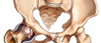
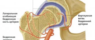
The hip joint is a large, articulated synovial joint, which is called the most powerful because it bears a greater range of loads. It is formed by the spherical head of the femur and the cup-shaped cavity of the pelvic bone (acetabulum), the surfaces of which are covered with smooth hyaline cartilage. Together they represent a hinge, where their musculoskeletal contact interaction occurs.
Every day, this unique mechanism performs the most important functions, which include maintaining the weight and position of the body in an upright state (standing, sitting), ensuring motor activity during movement (walking, running, jumping, etc.), and moving parts of the body relative to each other.
The forces passing through the hip joint apparatus are quite significant. For example, in the usual position of “standing on two lower limbs” the joint experiences a load of 1/3 of a person’s weight, while standing on one limb - 2.5 times higher than the body weight, when walking and running - a load of 2-6 times exceeds the person's original weight. Experts definitely attribute the fact of significant loads to one of the explanations for the high susceptibility of the joint to destabilization and wear, and, consequently, to the development of osteoarthritis. However, to say that the cause of the disease is the natural functional physiology of the joint is fundamentally incorrect; without an aggravating factor, the pathological process is impossible.
And another important point: for the hip joint to function well, it must have a sufficient range of movements with very good stability. And this, in turn, is possible provided there are strong ligamentous muscle groups that control movement and support functions in the joint, as well as a strong articular capsule that protects and holds the bone joint. The depth of insertion of the spherical element of the femur into the acetabular bed of the pelvis also plays a huge role. Failure of the designated structures and/or incorrect anatomical parameters can clearly lead to degeneration of the articular surfaces.
This is what the joint cavity looks like through an arthroscope.
All of the above is general introductory information, it is also important to know and understand. Of course, we will not leave readers without an answer to the burning question: what specific reasons cause such a complex diagnosis? Experts name the following root causes that most often lead to osteoarthritis of the hip joints:
- genetic defects in the development of the hip joint, characterized by congenital disorders of the development and growth of bone, cartilage, ligamentous, and muscle structures of the joint (dysplasia);
- old age, since against the background of age-related changes, cartilage hydration, muscle elasticity, blood circulation in joints, etc. decrease;
- systemic pathologies, where osteoarthritis is one of the consequences (gout, rheumatism, collagenosis, diabetes, complex types of allergies, etc.);
- various chondropathy leading to modifications of bone structures related to the joint;
- chronic arthritis (long-term inflammatory phenomena in the joint due to infection, autoimmune and metabolic disorders);
- avascular osteonecrosis of the femoral head (death of bone tissue) as a consequence of local circulatory disorders of traumatic or non-traumatic origin;
- hormonal imbalance, especially in women during menopause, when the level of estrogen, which protects joints, sharply decreases;
- obesity of any stage (excess weight is the worst enemy, since it significantly increases the load on the TB department and on the limbs in general);
- previous injuries localized to the pelvis and femur (fractures, dislocations, bruises, etc.), as well as a history of surgical manipulations on the hip region;
- orthopedic disorders, in particular valgus and varus deformities of the limbs, flat feet, spinal curvatures;
- a sedentary lifestyle, which causes a deficiency of muscle mass, ligament laxity, poor blood supply and limited supply of nutrients to the hip joint, which contributes to the onset and progression of osteoarthritis;
- constant overload of the hip region and lower extremities due to intense sports training, performing work while standing with heavy lifting, long monotonous poses (especially in static positions “standing” in one place, “sitting”), long walking forced by the nature of the activity.
It is impossible not to ignore the fact that smoking and alcohol abuse also have an adverse effect on the condition of the joint. Toxic products of nicotine and alcohol lead to a critical disruption of blood circulation around the musculoskeletal organ, to depletion and irreversible death of osteochondral tissues.
Symptoms and diagnosis of the disease
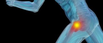
In a patient with hip osteoarthritis, the range of motion in three anatomical and physiological directions of the joint decreases: inward/outward rotation, flexion/extension, adduction/abduction. However, the most important early symptom to suspect the disease is pain in the groin area. The pain can also spread down the leg, radiating to the buttock, front or side of the thigh. Sometimes, which often hinders timely diagnosis and treatment of the TB joint, painful phenomena are localized only in the knee area for a long time. The intensity and duration of pain depends on the stage and physical activity.
Painful areas are marked in red.
For the first manifestations of osteoarthritis of the hip joint, another important distinguishing sign is stiffness in the joint after prolonged rest, for example, with morning activation after a night's sleep. Patients initially notice that after separation, stiffness and pain go away on their own. Later, as the disease progresses, the pain syndrome becomes brighter and more persistent, and noticeable limitations in range of motion, mobility, and stability begin to appear:
- difficulty moving the leg laterally;
- problems with pulling the limb to the chest by bending, especially when you need to put on shoes, socks, stockings, etc.;
- difficulty in adopting the position of “sitting astride a chair”;
- limited range of wide leg extension to the sides;
- problematic going up/down stairs;
- sudden jamming of the joint with sharp pain;
- increased pain during and after physical activity, in severe cases the pain does not leave throughout the day, even at rest;
- unsteadiness and uncertainty of gait;
- in the last stages, muscle atrophy develops, shortening of the affected limb and lameness, pelvic distortion, and with bilateral lesions, abnormal waddling of the body from side to side when moving (“duck gait”).
Along with pain and limited mobility in the area of the hip joint, swelling, redness, and hyperthermia of the skin often appear. One of the symptoms, more characteristic of the late stage, may be the appearance of crepitus (grinding, frequent clicks in the joint) at the time of movement.
Osteoarthritis can affect one of the hip joints or both the right and left joints at the same time. In practice, bilateral lesions of approximately the same severity occur infrequently, mainly against the background of pathology of rheumatoid etiology.
Photo of the femoral head after its removal during surgery.
But there are cases when, in the absence of a rheumatoid clinic, the patient initially develops pain in one joint, and a year or later the second one begins to bother him. As a rule, such injuries are caused by advanced arthrosis in the initially diseased joint, which necessitated the need to spare the diseased limb, transferring colossal loads and body weight to the hip joint of the opposite leg. From increased exploitation, incommensurate with the physiological reserves of the once non-problematic segment, its structures are subject to degenerative-dystrophic metabolism.
Changing the pelvic axis.
Primary diagnosis of OA is carried out using radiography (conventional or CT). With its help, fundamental criteria are determined that indicate the presence of morphological changes in the articular cartilage of the hip joint of a degenerative type:
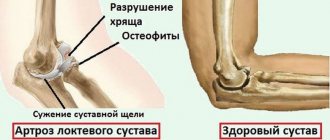
- narrowing of the joint space;
- subchondral osteosclerosis;
- bone growths (osteophytes);
- disturbed mutual position and distorted shapes of the head, femoral neck, and acetabulum.
Deformation of the right hip joint.
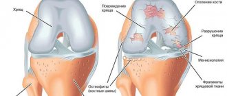
The cartilage itself, lining the surface of the femoral head and acetabulum, is not visualized by x-rays. It is also not possible to see other soft tissues (ligaments, muscles, capsule, neurovascular formations, etc.) on an x-ray. Therefore, to obtain more detailed information about the clinical picture, after receiving unsatisfactory X-ray results, specialists refer the patient for an MRI. Magnetic resonance imaging allows you to obtain the most complete information about the condition of:
- hyaline layer (cartilaginous covering) of articular surfaces (localization, scale, degree of destruction of cartilage tissue, nature of thinning and fiber disintegration);
- the peripheral part of the epiphysis of the articular bones, located under the hyaline layer (osteophytes, foci of sclerosis and cystic formations of the subchondral bone plate);
- bone marrow (edema of the intraosseous medulla);
- fibrous and synovial membranes of the joint capsule (various damage and inflammatory areas, excess/lack of synovial fluid);
- ligaments, muscle bundles, tendons, blood vessels, nerve structures.
During any imaging examination (CT, X-ray, MRI), not one, but two homogeneous joints are always scanned. In addition to the listed diagnostic methods, it is mandatory to conduct special physical tests to check the quality of the motor capabilities of the hip joint. Sometimes ultrasound examination (arthrosonography) is prescribed.
According to statistics, during diagnosis more often (in 60% of patients) damage to the most vulnerable and most loaded area of the hip joint is detected - its upper pole with the definition of superolateral dislocation of the femoral head. In 25% of cases, damage is found in the medial pole with the femoral head displaced medially and its abnormally deep insertion into the acetabulum due to protrusion of the latter. The concentric form of osteoarthritis of the hip joint, in which the entire articular apparatus is affected, is detected less frequently, in only 15% of cases.
Clinical picture
Manifestations of pathology depend on the stage of the lesion. The first sign of damage to the hip joint is pain, it occurs directly in the joint and intensifies when walking. At first it has a pulling character, with the development of degenerative processes its intensity increases. The pain syndrome is especially painful in damp weather. There is discomfort after sleep and prolonged sitting. Damage to the hip joint is accompanied by the following symptoms:
- limited mobility of the joint;
- dysfunction of the pelvic organs;
- pain radiating to the lower back, knee, groin;
- when moving, a crunching sound occurs;
- muscles get tired quickly.
Most often, subchondral sclerosis of the roof of the acetabulum of the hip joints is observed. The localization of the pathology is associated with high loads on this area. The patient, trying to protect the limb from tension, transfers the main load to the healthy leg. This is a direct path to the development of degenerative processes in the second joint. You should not wait for serious manifestations of pathology; it is better to stop the development of sclerosis at the initial stage.
Stages of osteoarthritis of the hip joint
The disease is classified into 3 stages, depending on the X-ray and magnetic resonance imaging. Let's look at what features are characteristic of each stage.
- It is extremely rare to catch stage 1 in practice, since patients usually do not go to a medical facility for examination in the initial period, not counting periodically mild discomfort in the joint as a serious pathology. But it is an achievable task to detect early pathogenesis using X-ray examination. The clinical picture on x-ray is as follows: slight narrowing of the joint space, small calcifications (pointwise) along the outer and inner edges of the acetabulum, sharpening in the area of the fossa of the spherical component of the femur. No serious deformations of the articular surfaces are detected, the condition of the hyaline cartilage is satisfactory.
- At stage 2 of osteoarthritis of the hip joint, a progressive reduction in the level of the joint space is determined (the clearance decreases by 45% of normal). The second stage is also characterized by subluxation of the femoral head and moderate subchondral sclerosis. Additionally, pronounced osteophytosis with large bone growths along the marginal line of the acetabulum and along the periphery of the femoral head, significant tuberosity and roughness of cartilage tissue, protrusion of the pelvic bed, exposure and moderate deformation of the articular surfaces are diagnosed. In some cases, bone cysts and chondromic bodies free in the joint are detected.
- For stage 3, a characteristic sign is a critically small lumen of the joint space or complete closure of the space between the articular surfaces. This stage is extremely severe, disables the person, and therefore surgical treatment is unconditionally required. Based on the results of radiography and other leading diagnostic methods, generalized necrosis of the head, maximum disappearance of cartilage tissue from the surfaces of the joint, giant osteophytes of various shapes and sizes, advanced sclerosis and cystic reconstructions of bone tissue are detected. The next signs are a critical condition of the acetabulum with an abnormally large depression, which is caused by severe osteophytosis; subluxation/dislocation of the hip, reduction in size and deformation of the femoral head with loss of rounded shape.
Dynamics of disease of large joints of the lower extremities.
Many people are interested in how the concept of “deforming osteoarthritis” differs from the concept of “osteoarthrosis”? We answer: nothing, both medical diagnoses refer to the same pathology, the outcome of which is deformation of the joint and periarticular structures. That is, the word “deforming” simply emphasizes the deforming properties typical of pathology.
Classification of pathology
Doctors classify pathology according to several criteria.
Development form:
- primary – sclerosis appears in a healthy joint as a result of injury or increased load;
- secondary - the lesion accompanies arthrosis and other joint diseases.
Based on the extent of the formation of bone growths, four degrees of the disease are distinguished:
- at the first stage there is a slight proliferation of osteophytes along the edge of the acetabulum;
- second stage - pronounced bone growths, the joint space is narrowed;
- third stage - the gap inside the joint is barely noticeable due to the formation of large osteophytes;
- fourth stage - complete deformation of the head and acetabulum occurs.
The degree of damage is determined by X-ray examination.
Treatment of the disease: conservative and surgical methods
Today, not a single conservative method, unfortunately, is capable of either completely stopping or reversing the pathological process of this disease. Conservatively, it is only possible to slow down the rate of progression of degenerative pathogenesis. Medication and physiotherapeutic methods, which are used in a non-invasive approach, are designed for symptomatic treatment and prevention of the accelerated rate of tissue destruction of the hip joints. In the final stages, it is useless to treat a pathologically changed joint conservatively.
Therapy methods
When treating pathology, therapy is aimed at getting rid of the underlying disease that caused subchondral sclerosis of the articular surface. The doctor's prescriptions depend on the situation. Non-steroidal anti-inflammatory drugs help stop the pathological process: Nimesulide, Meloxicam, Nise. Medicines of various spectrums of action are used in therapy:
- antihistamines;
- antibacterial;
- analgesics;
- hormonal drugs;
- chondroprotectors;
- vitamin complex.
A significant component of treatment is exercise therapy – exercises to restore the functionality of the joint. The complex is selected by the doctor, based on the individual needs of the patient. Exercises in water give effective results.
To speed up metabolism, increase muscle tone and blood supply, physiotherapy is prescribed:
- electrophoresis;
- laser therapy;
- exposure to magnetic pulses.
Acupuncture, massage, and mud applications help improve your general condition.
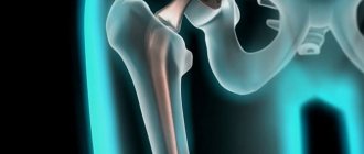
Surgical treatment is performed for complete deformation of the articular surfaces. The destroyed organ is replaced with a prosthesis.
Treatment with medications
To combat pain and inflammation in the hip joint, drugs from the NSAID group are used. There is no best remedy that is equally suitable for everyone, so the drug is selected individually. In some cases, doctors prescribe analgesics. But for unbearable chronic pain, a certain drug from the class of corticosteroids may be prescribed, perhaps intra-articular injection of an anesthetic or the same corticosteroid. However, it is important to say: when it comes to the use of corticosteroids and intra-articular injections, this is the first signal that the patient already urgently needs surgery.
For arthralgia (joint pain), Paracetamol, Diclofenac, Ibuprofen, Ketoprofen and their analogues in the form of tablets and ointments are widely used in medical practice. Neutralization of the pain syndrome will allow an additional increase in human activity.
There are also special medications that have a chondroprotective effect; they can improve nutrition in preserved cartilage and protect them, as far as possible, from rapid destruction. Moreover, they help improve the sliding of articular surfaces relative to each other. However, chondroprotectors are ineffective for degenerative-dystrophic changes that have reached stage 3; in some cases, even at stage 2, the clinical value of such drugs is low. Therefore, their use may be justified either at stage 1, or as a preventive measure for the appearance of arthrosis if the patient is at risk. Well-known medications with chondroprotective properties include Structum, drugs Dona, Teraflex, Elbona, Aflutop.
To the attention of patients! Chonroprotectors do not restore damaged areas of cartilage, but only nourish viable tissues and increase lubrication in the joint. And even such an effect should not be too hopeful.
Diagnostics
Comprehensive diagnostics makes it possible to determine the degree of damage to the articular surfaces and the reasons that caused the pathology. The main method is radiography. From the image, the doctor can assess the size of the joint space and bone outgrowths. Additionally, an ultrasound is prescribed.
Subchondral sclerosis can be detected at an early stage using MRI. The patient also needs to undergo a general and biochemical blood test. An informative diagnostic method is a subchondral bone density test.
Physiotherapy therapy
Physiotherapeutic procedures are aimed at increasing blood circulation in the periarticular tissues, activating metabolism and delivery of valuable substances to joint tissues, improving lymphatic drainage, and preventing muscle atrophy. Along with the effect of physical methods, it is sometimes possible to achieve a permanent elimination of pain and inflammation, and an increase in the performance of the limb. Common physical therapy treatments with the best potential for effectiveness include:
- electromyostimulation;
- laser treatment;
- magnetic therapy;
- ultrasound treatment;
- mud and paraffin treatment;
- therapeutic mineral baths.
In advanced stages, physiotherapeutic sessions, including massage techniques, can have the opposite effect, so they should be used with caution. Orthopedic doctors emphasize that physiotherapy has the greatest benefit at stage 1, and at the beginning/middle of stage 2. If there is a final form of stage 2, any stage of 3 tbsp. physiotherapy will give productive results after surgery.
Complications
If sclerosis of the acetabulum roof is not treated, the bone tissue grows greatly. When it extends beyond the joint, the tissue affects inflammatory processes in the subcutaneous tissue. In advanced cases, a necrotic or purulent process occurs that can be transmitted to internal organs.
For diagnosis, special techniques are used to visualize the structure of the joints:
- MRI;
- X-ray;
- CT scan.
To conduct a differential study, laboratory techniques are used.
Treatment with gymnastics
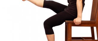
Performing a complex of therapeutic exercises, individually developed by a competent specialist for a specific patient, will help increase the range of movements in the problem area, improve the functions of the TB department, and correct muscle balance. Special physical training in an adequate mode will allow you to competently relieve the sore area of the musculoskeletal system, reduce the severity, duration and frequency of pain, increase vitality and generally improve well-being.
Exercise therapy courses should be conducted in a rehabilitation gym under the supervision of medical personnel regarding the recovery of patients with musculoskeletal disorders. In any case, until the person perfectly masters the techniques of rehabilitation exercises recommended by the doctor. Patients are recommended to have at least a certain range of aerobic training with and without machines, but with the exception of force loads on the affected joints. Loads are selected strictly taking into account the main diagnosis, concomitant diseases, weight and age of the patient, and his physical capabilities.
Activities organized in the pool - therapeutic aqua gymnastics, swimming - provide invaluable benefits. Measured walking and gentle exercise on an exercise bike are encouraged.