Diseases of the musculoskeletal system worry more and more people every year, and their development is increasingly observed at a young age. This is facilitated not only by changes in lifestyle, but also by an increase in the level of injuries, which is largely interconnected. One of the most common pathologies of the musculoskeletal system is arthrosis of the hip joint, which is characterized by progressive pain and limited mobility. Ultimately, the disease can lead to complete joint immobility and disability. To avoid such undesirable consequences, it is important to begin treatment for arthrosis as early as possible. And if in the early stages of development it can be stopped using conservative methods, then in case of severe changes, restoration of the functions of the hip joint and elimination of unbearable pain can only be achieved with the help of high-tech surgery.
What is arthrosis of the hip joint
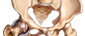
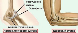
Arthrosis of the hip joint is a chronic degenerative disease in which gradual destruction of the hip joint occurs. At the same time, all its components are gradually involved in the pathological process, but hyaline cartilage is especially affected, which leads to a narrowing of the joint space and deformation of its other components. More often, pathological changes occur in only one hip joint, although it is also possible to affect both at the same time.
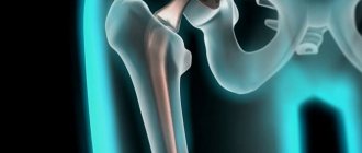
The hip joints are the largest joints in the human body because they bear the most stress during the day. Each is formed by the head of the femur and the acetabulum, which is a cup-shaped depression in the pelvis. Both surfaces are covered with smooth, moderately elastic hyaline cartilage. It is this that ensures the smoothness and unhindered sliding of the head of the femur in the natural recess and thereby makes it possible to perform movements in various planes.
The movement of the hip joint is provided by a group of muscles connected to it by fascia. It is also surrounded by ligaments, the tasks of which are to limit its mobility within physiological limits and ensure the stability of its position.
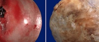
The entire joint is surrounded by a joint capsule covered with synovium. Its main task is the synthesis of synovial fluid, which lubricates the contacting parts of the hip joint and at the same time acts as a carrier of nutrients for it. It is from the synovial fluid that the hyaline cartilage covering the head of the femur and the surface of the acetabulum constantly receives components for the formation of new cells, i.e., regeneration. This is extremely important for this cartilaginous formation, since with every movement of the hip it is abraded, but normally it is immediately restored. But when injured or under the influence of other factors, this does not happen, which leads to the development of arthrosis of the hip joint, i.e. thinning and destruction of its hyaline cartilage.
As a result, deformed areas are formed in ideally smooth cartilage, increasing as the pathology progresses. As it wears away, the surfaces of the bones that form the joint are exposed. When they touch, a characteristic crunch and severe pain occurs. This provokes the formation of osteophytes and at the final stages of development the head of the femur completely fuses with the acetabulum, making any movement in the hip joint impossible.
At the same time, arthrosis of the hip joint can provoke the development of various inflammatory processes inside the joint, including:
- bursitis - inflammation of the synovial bursa;
- tenosynovitis - an inflammatory process in the sheath of the muscle tendon sheath;
- tunnel syndrome - compression of nerves, causing radiating pain along the pinched nerve.
Pharmacological drugs
Most medications used in the treatment of coxarthrosis are prescribed to patients to eliminate symptoms. For pathology of 1st degree of severity, medications are used in the form of tablets and (or) ointments. And to reduce the severity of severe pain in coxarthrosis at stages 2 or 3, solutions for intramuscular injections, periarticular, periarticular, and intraarticular blockades are used.
Elimination of pain
Nonsteroidal anti-inflammatory drugs (NSAIDs) are the first choice drugs for eliminating the pain that occurs when the hip joint is destroyed. These are Piroxicam, Indomethacin, Diclofenac, Ketoprofen, Ibuprofen. NSAIDs are not intended for long-term use, since their active ingredients negatively affect the liver, kidneys, gastrointestinal tract, and also suppress the ability of cartilage tissue to regenerate.
Often, NSAID ointments or gels are prescribed along with tablets:
- Nurofen;
- Fastum;
- Ketonal;
- Finalgel;
- Nise.
This method of treatment allows to reduce the pharmacological load and minimize the likelihood of damage to internal organs. An analgesic effect also occurs when using warming ointments - Finalgon, Capsicam, Apizartron, Nayatox. Their components have a pronounced local irritating and distracting effect, stimulating the acceleration of blood circulation.
And Menovazin solution and Espol cream relax skeletal muscles, eliminating painful muscle spasms.
Improving blood supply to the joint with nutrients
The use of drugs that accelerate blood circulation in it helps to stop further destruction of the hip joint. These are Stugeron, Cinnarizine, Trental, Pentoxifylline, Xanthinol nicotinate. A course of medications helps eliminate the deficiency of nutrients and bioactive substances and prevents spasms of small blood vessels.
If necessary, the therapeutic regimen includes muscle relaxants (Mydocalm, Sirdalud, Tizanidine) - drugs that relax striated muscles and reduce the severity of pain.
In the treatment of pathologies accompanied by destruction of the hip joints, chondroprotectors are necessarily used: Artra, Structum, Teraflex, Alflutop, Dona. Their active ingredients are glucosamine and (or) chondroitin - substances that stimulate the restoration of damaged hyaline cartilage. But taking chondroprotectors is effective only for coxarthrosis of 1st degree of severity. Experienced orthopedists prescribe them to patients either in tablets or in solutions for intramuscular administration. The ability of chondroitin and glucosamine to penetrate into the hip joints from ointments has not yet been clinically confirmed.
Causes
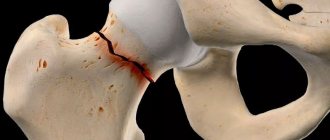
One of the common causes of arthrosis of the hip joint is mechanical damage, not only direct injuries, but also microdamages caused by the destructive effect of excessive loads on it. One of the most common causes of the disease is a fracture of the femoral neck. It extends from the femur at an angle of 120° and connects it to the head. The presence of osteoporosis significantly increases the likelihood of a hip fracture, but this type of injury can also result from a traffic accident, a fall from a height, an impact, etc.
A femoral neck fracture may be accompanied by aseptic necrosis of the femoral head, which will become a trigger for the development of degenerative changes in the joint. The presence of dysplasia or subluxation of the hip joint, ruptures of its ligaments, transcondylar fractures or fractures of the acetabulum also creates favorable conditions for damage to its structures. In such situations, post-traumatic arthrosis of the hip joint is diagnosed.
Often, post-traumatic arthrosis of the hip joint occurs in professional lightweight and weightlifters, parachutists, loaders, and speed skaters.
The development of arthrosis of the hip joint after injury is caused by a violation of the congruence (comparability) of the articular surfaces, a decrease in the quality of blood supply to the components of the joint and prolonged immobilization. As a result of prolonged immobility, not only deterioration of blood circulation in the fixed area occurs, but also shortening of muscles and a decrease in their tone. The likelihood of post-traumatic arthrosis increases significantly when treatment is inappropriate for the situation or untimely treatment, which leads to the persistence of defects of varying severity. Also, the risks of its development increase with excessively early loading of the joint and inadequate exercise therapy, including too intense, started late or, conversely, early.
Sometimes the disease occurs after surgical interventions on the hip joint due to the formation of scars and additional tissue trauma. Although in some cases surgery is the only way out to eliminate the consequences of the injury.
Excessive loads can also trigger changes in the hip joint, as they lead to microtrauma. Regular tissue damage activates the process of division of chondrocytes (cartilage cells). This is accompanied by an increase in the intensity of the production of cytokines, which are normally produced in small quantities. Cytokines are mediators of inflammation, in particular the cytokine IL-1 leads to the synthesis of special enzymes that destroy hyaline cartilage of the hip joint.
In addition, high loads can provoke microfractures of the subchondral plate. This leads to its gradual compaction and the formation of bone growths on the surface, called osteophytes. They can have sharp edges and cause further destruction of the joint and also injure surrounding tissue.
The subchondral plate is the outermost part of the bone in direct contact with hyaline cartilage.
In some cases, it is not possible to determine exactly what triggered the development of degenerative-dystrophic changes in the hyaline cartilage of the femoral head and acetabulum. In such situations, idiopathic or primary arthrosis of the hip joint is diagnosed.
Today it has been established that the tendency to develop it can be inherited, i.e. the presence of this pathology in close relatives significantly increases the chances of developing arthrosis of the hip joint. Presumably, it has polygenic inheritance, i.e. its development depends on the presence of many genes. Each of them individually creates weak preconditions for the development of the disease, but when they are combined, it becomes a matter of time, especially when leading a sedentary lifestyle and obesity or, conversely, heavy physical labor.
There is a theory that arthrosis of the hip joints is a consequence of the presence of a congenital or acquired mutation of the type II procollagen gene.
There is also secondary arthrosis of the hip joint, which develops against the background of concomitant diseases and age-related changes. But its features are discussed in more detail in a separate material “Coxarthrosis”.
Symptoms
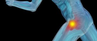
The disease is characterized by the occurrence of pain, limited mobility and crunching in the hip joint, the severity of which directly depends on the degree of neglect of the pathological changes. At the final stages of development, shortening of the affected leg and complete immobility of the hip joint may be observed, which is due to complete fusion of the bone structures that form it.
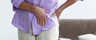
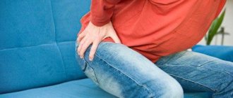
Initially, the disease may occur without pronounced symptoms and cause mild, short-term pain. As a rule, they appear after physical activity, in particular walking, carrying heavy objects, squatting, and bending. But as degenerative-dystrophic changes in the joint progress, the pain intensifies. Over time, they not only become more intense, but also persist longer, and the interval between the start of physical activity and their appearance decreases. At the same time, rest, even a long one, may not bring relief. Subsequently, pain can torment a person even with prolonged immobility of the hip joint, for example, after a night's sleep.
If intra-articular structures impinge on the nerves passing nearby, pain can radiate to the groin, buttocks, thigh and knee. However, they tend to intensify with hypothermia. At the last stage of development of the disease, the pain becomes unbearable. This causes an unconscious desire to spare the leg and put less stress on it, which leads to lameness.
Another symptom of hip arthrosis is a decrease in range of motion. Most often, there is a limitation in the ability to turn the leg in and out, and raise the leg bent at the knee to the chest. Over time, so-called morning stiffness occurs, which disappears after the patient “disperses.” Subsequently, compensatory curvature of the pelvis is possible, which leads to a change in gait. In the future, patients completely lose the ability to make certain movements with the affected leg.
If arthrosis of both hip joints develops simultaneously, the development of the so-called duck gait with the pelvis retracted back and the body tilted forward is observed.
All this may be accompanied by the formation of edema in the hip joint. But if you are overweight, they may go unnoticed.
Often, during movements, especially extension ones, a crunching sensation occurs in the affected joint. It is a consequence of the exposure of the bony surfaces of the femoral head and acetabulum and their friction with each other. In this case, there is a sharp increase in pain.
Also, with arthrosis of the hip joint, painful spasms of the thigh muscles may occur. In extremely advanced degenerative-dystrophic diseases, when the joint space almost completely disappears and the head of the femur begins to flatten, shortening of the affected limb by 1 cm or more is observed.
In general, there are 3 degrees of arthrosis of the hip joint:
- Grade 1 – the joint space of the hip joint is narrowed, and the edges of the bone structures are slightly sharpened, which indicates the beginning of the formation of osteophytes. Clinically, mild pain and some restrictions of movement are observed.
- Grade 2 – the joint space is narrowed by more than 50%, but less than 60%. Significant osteophytes are observed, as well as signs of cysts in the epiphyses of the bones. Patients note significant limitations of movement in the hip joint, the presence of a crunching sound during movements, pain, and atrophy of the hip muscles of varying degrees can be observed.
- Grade 3 - the joint space is reduced by more than 60% or is completely absent, and osteophytes occupy a large surface and are large in size, subchondral cysts are observed. The hip joint is stiff and the pain can become unbearable.
Main signs of problems
At the first manifestations of pathology in the hip joint, you should immediately seek help from a doctor. Symptoms of hip disease may include the following:
- pain in the joint area of varying intensity;
- restriction of movements in the joint, gait disturbance;
- the appearance of swelling in the joint area (with inflammatory, traumatic lesions);
- hyperemic skin over the joint, local or general increase in body temperature (a sign of an inflammatory process);
- difficulties when climbing stairs, when standing up (discomfort, clicking, pain, etc.), often a person needs support;
- the appearance of swelling of the joint area, which is caused by a purulent process in the tissues: all this is accompanied by fever, hyperemia and other inflammatory signs.
Diagnostics
The appearance of pain and other symptoms characteristic of arthrosis of the hip joints is a reason to consult an orthopedist. The doctor will be able to suspect its presence, especially if there have been injuries to the hip or pelvis in the past, based on the data obtained during the survey and examination.
The presence of arthrosis of the hip joint is indicated by pain, the intensity of which increases over several years. Much less often, the rapid development of degenerative-dystrophic changes is observed, when several months pass from the appearance of the first signs to severe constant pain. This is characterized by increased pain while standing or performing physical work. Also typical for arthrosis is the presence of morning stiffness, which persists for up to half an hour, and also occurs after prolonged maintenance of a motionless position. Gradually, there is an increase in mobility restrictions and deformation of the hip joint, which in the later stages of development the orthopedist can notice during the examination.
Nevertheless, all patients are required to be prescribed instrumental research methods, with the help of which it will be possible to confirm the presence of arthrosis of the hip joint and establish its degree, as well as differentiate it from some other diseases accompanied by similar symptoms. As a rule, diagnosis is carried out using:
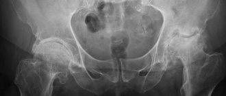
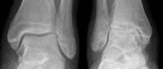
- X-rays - allows you to detect the main signs of arthrosis, in particular narrowing of the joint space and the presence of osteophytes. But recently, CT has become a more informative research method, allowing for more accurate assessment of the condition of the hip joint.
- MRI is a highly informative method for diagnosing various changes in the condition of soft tissue structures, including cartilage tissue, which makes it possible to detect the slightest signs of degeneration of hyaline cartilage.
Patients may also be prescribed laboratory tests, including CBC, OAM, biochemical blood test, etc. They are required to establish concomitant diseases that created the preconditions for the development of secondary arthrosis of the hip joint.
General information about the disease
Degenerative-dystrophic degeneration of the hip joint, called coxarthrosis, develops more often in people aged 40 years and older. Injuries, specific joint diseases, and systematic physical overload lead to earlier development of the disease. According to statistics, a more frequent development of pathology in women is noted, which is due to serious anatomical stress during pregnancy and childbirth.
Coxarthrosis is characterized by a gradual course. At an early stage of the disease, the joint can be completely restored only by conservative methods. In advanced stages, recovery is impossible; the result of treatment is to slow down or stop the destruction of the joint capsule, maintain mobility, and relieve pain.
Treatment of arthrosis of the hip joint without surgery
Treatment of degenerative-dystrophic changes in the hip joint using conservative therapy methods is possible only for arthrosis of degrees 1 and 2. The prescribed measures make it possible to improve the patient’s condition, stop or at least slow down the progression of the pathology and thereby maintain working capacity. But they are not able to lead to complete regression of changes that have already occurred in the joint.
Today, as part of the conservative treatment of arthrosis of the hip joint, the following are prescribed:
- drug therapy;
- exercise therapy;
- physiotherapy.
Patients are also advised to make certain adjustments to their lifestyle. So, if you are overweight, you should take measures to reduce it, that is, increase the level of physical activity and reconsider your diet. If the patient is actively involved in sports and overloads the joint, which causes microtrauma in it, it is recommended to reduce the intensity of training.
Drug therapy
Drug therapy for arthrosis of the hip joint is always complex and includes drugs from different groups aimed at reducing the severity of symptoms of the disease and improving metabolic and other processes in the joint. This:
- NSAIDs are drugs with anti-inflammatory and analgesic effects, produced both in oral forms and in the form of local agents, which allows you to choose the most effective and convenient option for use;
- corticosteroids are drugs that have powerful anti-inflammatory properties and are used in most cases in the form of an injection solution, since when choosing systemic therapy they cause the development of undesirable side effects;
- chondroprotectors - drugs synthesized on the basis of natural components of cartilage tissue used by the body to restore it (prescribed in long courses);
- muscle relaxants - medications indicated for muscle spasms, which causes pain of varying severity;
- B vitamins – help improve nerve conduction, which is required in the development of carpal tunnel syndrome;
- drugs that improve microcirculation - help increase the intensity of blood circulation in the affected area, which leads to an increase in the rate of metabolic processes and helps restore damaged cartilage.
If concomitant diseases are detected, consultation with related specialists and appropriate treatment are indicated.
For very severe, incapacitating pain that cannot be eliminated with prescribed NSAIDs, intra-articular or periarticular blockades can be performed. They involve the injection of a local anesthetic in combination with a corticosteroid directly into the joint cavity, which quickly leads to an improvement in well-being. But procedures of this kind can only be carried out in a medical institution by a qualified specialist, otherwise there is a high risk of complications.
Exercise therapy
Therapeutic physical education plays one of the leading roles in the non-surgical treatment of arthrosis of the hip joint, both idiopathic and post-traumatic forms. But a set of exercises must be selected individually, taking into account the nature of the previous injury, the level of physical development of the patient, and existing concomitant diseases.
You should do physical therapy every day in comfortable conditions without haste. All movements should be performed smoothly without jerking, so as not to cause harm to the already deformed hip joint. This will allow:
- reduce the intensity of pain;
- increase joint mobility;
- reduce the risk of developing muscle atrophy;
- increase the intensity of blood circulation and metabolic processes.
Physiotherapy
To increase the effectiveness of prescribed measures, patients with arthrosis of the hip joint are often recommended to undergo a course of physiotherapeutic procedures. Traditionally, those that have anti-inflammatory, anti-edematous and analgesic effects are selected. This:
- ultrasound therapy;
- electrophoresis;
- magnetic therapy;
- laser therapy;
- shock wave therapy, etc.
In some cases, plasmolifting is indicated, i.e., the administration of the patient’s own blood plasma purified and saturated with platelets. To obtain it, venous blood is collected, which is then centrifuged. As a result, it is divided into red blood cells and plasma, which is used to treat degenerative changes in the hip joint.
Methods for treating coxarthrosis
At different stages of the disease, specialists prescribe different treatments.
Conservative treatment methods are used for the initial stage of arthrosis. This treatment can be carried out at home without going to the hospital.
- Physiotherapy;
- Injections;
- Medications;
- Reduced physical activity;
- Physiotherapy;
- Plasmolifting.
For the second degree of coxarthrosis, more thorough treatment is prescribed:
- Laser therapy;
- Electrophoresis.
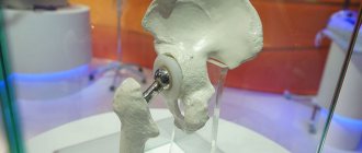
The latest surgical treatment methods are used if the previous ones do not help or if the pathology has developed to the third degree.
- Complete cartilage transplantation (your own or a donor’s).
- Surgery to correct the structure of the joint. The surgeon removes parts of the cartilage with defects and aligns the articular surfaces. Damaged tissues are replaced with implants.
- Complete replacement of a damaged joint with an artificial one.