Arthrosis of the shoulder joint - what is it?
Osteoarthritis of the shoulder joint is a degenerative-dystrophic disease of connective joint tissue, which is chronic in nature and manifests itself with stiffness, accompanied by pain of varying intensity.
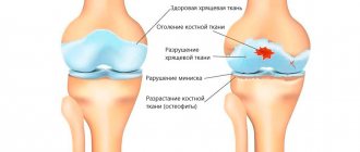
The development of arthrosis of the shoulder joint is characterized by changes occurring in the synovial fluid (located inside the joint). Lack of nutrients leads to degenerative-dystrophic (destructive) changes, due to which cartilage tissue loses its properties and becomes deformed.
Over time, the bone tissue located under the cartilage becomes exposed and, subject to negative effects, becomes deformed (marginal growths form on it - osteophytes, which cause limitation or complete loss of joint mobility).
An important fact is that it is extremely necessary to diagnose osteoarthritis of the shoulder joint in the early stages, when irreversibly destructive processes have not yet occurred in the tissues of the joint. Timely contact with a specialist and initiation of treatment makes it possible to stop the progress of pathological processes, ensuring the ability to maintain the functionality of the joint for many years.
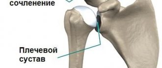
Definition and anatomy of the disease
Definition and anatomy of the disease
The shoulder joint consists of three parts: the clavicle, the scapula and the humerus. The part of the shoulder blade located above the shoulder is called the acromion. The joint connecting the clavicle and acromion is called the acromioclavicular joint. Mobility in this area is relatively small, but the presence of destructive processes in bone and cartilage tissue causes severe pain and inconvenience.
Arthrosis of the acromial clavicular joint is called a degenerative-dystrophic lesion, the progression of which causes destruction of cartilage tissue . As a rule, the pathology is inflammatory in nature and most often occurs as a result of the development of aging processes and external influences.
Due to the destructive effect on the joint, the function of chondrocytes (cartilage cells) is disrupted, which disrupts the production of intercellular substances that provide tissue elasticity and firmness and retain water molecules. Due to these processes, the cartilage becomes thinner and brittle, deformed and, in a neglected state, completely loses its functions.
Possible causes of arthrosis of the shoulder joint
Among the most likely causes of osteoarthritis of the shoulder joint are:
- joint injuries;
- congenital developmental defects (in particular, dysplasia);
- various types of inflammation.
The shoulder is a movable element of the human body skeleton. Due to the possibility of particularly free movement, the risks of injury to this particular joint are much higher. Dislocations are one of the most common causes, which is a prerequisite for the occurrence of osteoarthritis of the shoulder joint.
In addition, the causes of arthrosis of the shoulder joint can be various types of surgical interventions, increased stress, sports and professional characteristics.
Considering arthrosis of the shoulder joint as a secondary phenomenon that develops against the background of inflammation caused by various diseases, it can be caused by:
- infections;
- metabolic/endocrine/autoimmune diseases;
- hereditary predisposition.
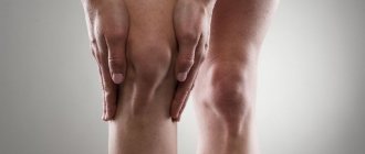
Signs and main symptoms of arthrosis of the shoulder joint
Considering the clinical picture of the disease, it is worth noting that the symptoms of the initial stages of development of pathological processes are weakly expressed. This is often the reason for late diagnosis of osteoarthritis of the shoulder joint.
Symptoms of arthrosis of the shoulder joint include:
- pain (when pressing/abducting a limb);
- accompaniment of movements with a characteristic crunch;
- inactivity;
- local redness and swelling.
Any of the symptoms of osteoarthritis of the shoulder joint requires the attention of a specialist.
As pathological processes develop, symptoms intensify. The earlier the disease is detected, the easier it is to prevent destructive processes, preserving the functionality of the joint as much as possible.
Treatment and rehabilitation
It is impossible to completely cure the disease with conservative methods of therapy. However, this method of helping the patient is effective in terms of controlling the symptoms of the disease. It is possible to relieve the manifestations of acromioclavicular arthrosis and stabilize the progression of osteochondral changes. The basic principles of treatment can be presented as follows:
- effective pain relief - NSAIDs and simple analgesics, intra-articular blockades are used;
- improving blood flow in the joint area - peripheral vasodilators are used;
- anti-inflammatory treatment - hormones are used parenterally for a short course and intra-articular administration;
- chondroprotector therapy – restoration of cartilage tissue;
- preparations for external use – enhance the effectiveness of systemic agents;
- Exercise therapy, massage, acupuncture.
If the entire complex of conservative methods is ineffective and clinical symptoms increase, surgical correction of arthrosis is performed.
The table below presents the main drugs, course of treatment and main dosages for various types of drug delivery to the affected area.
| Drug – type of therapy | Externally | Systemically | Inside the joint |
| NSAIDs | In the form of ointment and gel 2 times a day. Ketoprofen, Ortofen and Nimesulide are commonly used | Intramuscularly for no more than 5 days - Ketorolac, Diclofenac (up to 150 mg per day), then oral tablets in half the dose. For problems with the gastrointestinal tract - Nimesulide 200 mg per day or rectal administration of the drug. General course of therapy - up to 1 month | Not entered |
| Local anesthetics | In combination with NSAIDs (usually lidocaine) | Not applicable | For pain relief, a single dose of 1-2 ml of a 10% solution. It is a diagnostic criterion - pain completely stops when an anesthetic is injected into the joint cavity |
| Hormones | Not applicable | Intravenously in a short course, usually prednisolone at a dose of 90-120 mg. Duration - no more than 5 days | To relieve the inflammatory reaction - hydrocortisone or betamethasone. Usually a single injection is sufficient, which can be repeated after 1 month. |
| Chondroprotectors | In parallel with NSAIDs or in isolation. Chondroitin or glucosamine is used | Long term inside. The effect of therapy for arthrosis of the acromioclavicular joint has not been proven. Average dose - 1000 mg of chondroitin sulfate per day, course - up to 12 months or more | Only in experimental medicine, not used in routine practice |
| Simple analgesics | Not applicable | For rapid pain relief, parenterally or orally. Drugs of choice in the presence of ulcerative defects of the gastric mucosa. Metamizole or Paracetamol is used. The average dose is up to 1500 mg per day for Analgin and 3000 mg for Paracetamol | Not applicable |
In clinical practice, several drugs from different groups are usually combined with different routes of administration. Intra-articular agents are actively used together with oral medications. Chondroprotectors are often prescribed for arthrosis of the acromioclavicular joint. However, no studies have been conducted to prove the effectiveness of drugs from the chondroitin and glucosamine group for diseases of this joint.
Surgical correction is indicated in the presence of large numbers of osteophytes and destruction of bone tissue at the distal end of the acromion and the proximal portion of the clavicle. Usually, grinding of the bone structures is performed to increase the distance between them. A false joint filled with connective tissue is formed. Its function is sufficient for shoulder movements without pain. The operation is performed both openly and using endoscopic techniques.
Degree of development of arthrosis of the shoulder joint
The intensity of the development of destructive processes, as well as the stage of pathological changes occurring inside the joint, makes it possible to distinguish several degrees of development of osteoarthritis of the shoulder joint.
Only the attending physician can accurately determine the existing degree of arthrosis by examining the results of an x-ray. Each degree has a number of characteristic symptoms of arthrosis of the shoulder joint.
Arthrosis of the shoulder joint 1st degree
The initial stage of development of pathological processes. Arthrosis of the shoulder joint of the 1st degree lasts for several years and is manifested by a change in the quality of intra-articular fluid.
The initial stage is characterized by symptoms such as:
- significant loss of joint endurance;
- pain during movements;
- limited mobility accompanied by pain.
Timely treatment of grade 1 osteoarthritis of the shoulder joint has a positive prognosis.
Arthrosis of the shoulder joint 2 degrees
The pain intensifies and can occur even in the absence of movement.
Arthrosis of the shoulder joint of the 2nd degree is more noticeable to the patient and has clear symptoms, the manifestations of which are easily recognizable on an x-ray.
The group of symptoms also includes clicking sounds when making movements, as well as limited mobility, accompanied by sharp pain.
Lack of treatment for stage 2 osteoarthritis of the shoulder joint leads to irreversible consequences, in particular, to a complete loss of joint mobility.
Arthrosis of the shoulder joint 3rd degree
Stage 3 osteoarthritis of the shoulder joint is accompanied by constant pain, pronounced limitation of movements and the inability to move the arm back or raise it up.
X-rays show clear signs of destruction of the articular cartilage. The surfaces of the adjacent bones are severely deformed. Treatment for stage 3 osteoarthritis of the shoulder joint is only surgical intervention, which involves replacing the joint with an endoprosthesis.
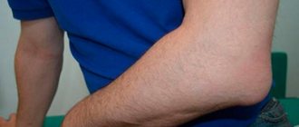
Acromioclavicular joint: movements, functional features
Functional features of the AC node
The collarbone connects to the sternum - firmly and motionlessly. Thanks to this structure, the long bone serves as a support for the hands, which helps to easily perform the following movements:
- Raise your limbs up.
- Perform different movements with your arms above your head.
- Apply the clutch to the upper lock.
- Place your hands behind your back, fastening them together.
At the back, at the level of the waist of the upper limbs, there is a scapula. It consists of a flat, large bone that stabilizes the two largest joints - the shoulder and AC joints. These nodes, together with ligaments and muscles, allow the hands to perform the following complex directed movements:
AK node movements
- Straight arm rotation.
- Retracting straight arms back.
- Raising the upper limbs above the head.
- Performing pronation and supination of the shoulder.
- Bringing the limbs straight in front of the body.
A special disk that divides the AK unit into two parts increases the capabilities and allows the hands to move in three planes. The complex structure is made in such a way that the bones of the node do not fit tightly to each other. This allows the shoulder girdle to perform movements with good amplitude, sufficient for the normal functioning of the arms in the required directions.
Types of arthrosis of the shoulder joint
In medical practice, it is customary to distinguish several types of shoulder arthrosis:
- deforming (chronic form);
- post-traumatic (after bruises/sprains/fractures/ruptured ligaments);
- acromioclavicular (consequence of joint injury);
- humeroscapular (consequence of blows/falls).
The type of pathological process is determined by the attending physician, taking into account the existing symptoms and probable causes of arthrosis of the shoulder joint.
Acromioclavicular joint: shape, structure, anatomy
Acromioclavicular node
The acromion is the coracoid process in the area of the scapula. This bony part, together with the humeral clavicular end, creates one simple, flat, but mobile unit. The hyaline circle divides this joint into 2 chambers.
The base of the joint, consisting of dense bone, is immobilized, since its functional feature is to ensure highly stable and controlled movement of the arm.
- The unique structure of the node helps that the clavicle bone acts as an excellent stabilizer of arm movement.
- It controls the movement of the arms, which allows a person to exercise without causing additional and unwanted stress on the joint.
- This rigidity of the unit allows for precise and active manual movements.
Due to the fact that our limbs are mobile and constantly moving, this helps to ensure 80% of the health of their anterior supporting apparatus along with the articular surface. The rest depends on the genetic predisposition to diseases of the cartilaginous nodes and excessive loads on the joint itself.
Diagnosis of arthrosis of the shoulder joint
Before starting treatment for osteoarthritis of the shoulder joint, it is important to accurately diagnose the type and degree of development of the disease. But what kind of doctor treats arthrosis of the shoulder joint?
You can contact specialists such as rheumatologist, orthopedist, arthrologist for diagnostics and diagnosis. If this is not possible, then it is enough to come to an appointment with a therapist, who will refer you to the right specialist.
To make a diagnosis, a series of tests are performed to identify the presence of diseases that provoke inflammation. In addition, an important step is examination and testing, which makes it possible to confirm/refute symptoms.
Another important diagnostic step is x-ray examination in two projections.
A set of diagnostic techniques allows you to obtain an accurate picture and prescribe competent treatment for osteoarthritis of the shoulder joint.
Acromioclavicular joint: blood supply
Blood supply to the AC
node The blood supply to the AC node is provided by the subclavian vein. It passes through two points of the joint: from below - at a distance of 3 cm inward from the coracoid process, from above - 3 cm down from the edge of the clavicle on the side of the chest. In infants, this vein runs through the middle of the collarbone, and only by the age of 5 will it change location and be located at two points, as in adults.
The subclavian vein is located obliquely relative to the center of the body. During movement, the topography of this vein will not change, since its walls are connected to the ribs and clavicle bone, as well as to the muscles in this area. The outflow of venous blood is carried out through the vascular network, which is located in this area.
Treatment of arthrosis of the shoulder joint
Treatment of arthrosis of the shoulder joint is a long, multifaceted and rather complex process, including both pharmacological agents and behavioral methods.
Depending on the degree of development of pathological processes, both physiotherapeutic methods and the use of medications can be used as treatment. Let us consider the features of treatment of arthrosis of the shoulder joint in more detail.
Physiotherapy as a method of treating arthrosis of the shoulder joint
Physiotherapeutic methods are an excellent treatment for osteoarthritis of the shoulder joint in the initial stages of development of the pathology, as well as as an element of complex treatment of advanced stages.
Among the physiotherapeutic methods of treating osteoarthritis of the shoulder joint, it is customary to distinguish:
- shock wave therapy - exposure of the affected joint to acoustic waves;
- laser therapy – makes it possible to temporarily abandon drug therapy;
- myostimulation – helps restore strength in the affected joint;
- phonophoresis – a combination of ultrasound and the use of a medicinal product (gel/ointment) helps eliminate pain;
- Ozone therapy is an adjuvant that helps reduce pain and speed up recovery.
Exercise therapy for arthrosis of the shoulder joint
Physical therapy for arthrosis of the shoulder joint is an important component of treatment, which involves:
- morning exercises;
- complex of dynamic movements;
- isometric gymnastics.
Each component of exercise therapy for arthrosis of the shoulder joint helps improve the condition of the patient’s body and helps overcome pathological processes.
Gymnastics for arthrosis of the shoulder joint
Physical exercises for the shoulder joint with arthrosis are an extremely important component of complex treatment, since they help slow the progression of the disease.
Gymnastics for arthrosis of the shoulder joint strengthens the muscle frame, thereby reducing the load on the adjacent joint.
It is important to remember that under no circumstances should you overload the joint. All exercises must be carried out under the guidance of a specialist.
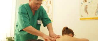
Massage for arthrosis of the shoulder joint
After exercise therapy and gymnastic exercises, it is recommended to have a massage.
Massage for arthrosis of the shoulder joint helps improve blood circulation and restore connective tissue.
It is worth noting that massage for arthrosis of the shoulder joint is possible only in the absence of acute pain, at the stage of reducing inflammatory processes.
During the massage process, it is important to pay attention not only to the inflamed joint, but also to the collar area, as well as the area of the forearms.
Preventive measures
Preventive measures
Axial arthrosis can affect the shoulder joint of a person at any age. In order to minimize the likelihood of this disease occurring, it is necessary:
- Maintain moderation in physical activity;
- Avoid fatty foods and red meat;
- Enrich your diet with fish, dishes containing gelatin and cartilage, fresh vegetables and fruits; introduce foods rich in calcium, as well as vitamins A, B and D;
- Restore normal sleep and rest patterns;
- Avoid critical weight gain;
- Wear orthopedic shoes, use comfortable bedding.
Treatment of arthrosis of the shoulder joint with medications
The key goal of treating arthrosis of the shoulder joint with medications is to relieve pain and eliminate inflammatory processes, as well as improve nutrition of the affected joint and create conditions for successful regeneration of connective tissues.
To achieve the main goal of treating the shoulder joint with medications, drugs from various groups are used. Let's look at some of them in more detail.
IMPORTANT! Only the attending physician can prescribe medications for the treatment of arthrosis of the shoulder joint, based on the formed picture of the disease. Self-medication can lead to irreversible consequences and disability.
NSAIDs in the treatment of arthrosis of the shoulder joint
Nonsteroidal anti-inflammatory drugs (NSAIDs) have excellent anti-inflammatory and analgesic effects. They are prescribed in limited courses, the duration of which is determined by the degree of development of the disease and the individual characteristics of the patient.
Among the most effective remedies in this group is o.
Corticosteroids in the treatment of arthrosis of the shoulder joint
Prescribed for severe pain as an intra-articular blockade. The products have a strong effect. The principle of operation is pain relief and prompt removal of inflammatory processes.
Often, drugs such as Hydrocortisone and Diprospan are used as treatment.
Chondroprotectors in the treatment of arthrosis of the shoulder joint
Prescribed to strengthen and restore connective tissues. They are effective only in stages 1 and 2 of the disease.
Treatment with chondroprotectors is a fairly lengthy process that requires constant attention.
Drugs of this type include: “Artracam”, “Glucosamine”.
Antispasmodics in the treatment of arthrosis of the shoulder joint
Provide relief from muscle tension and pain in the area of the affected joint. They are a mandatory stage of treatment.
“No-shpa” or its analogue “Drotaverine” is often used.
Ointments for arthrosis of the shoulder joint
Preparations in the form of ointment/gel/cream are most applicable for osteoarthritis of the shoulder joint. They are easy to use and have a fairly small number of side effects.
Today there are a large number of drugs of various groups. The most prescribed are “Kapsikam”, “Finalgel”, “Efkamon”, “Sabelnik” and “Viprosal”.
Diagnosis of the disease
The first stage of a patient’s diagnostic examination allows a preliminary conclusion to be made based on the patient’s medical history and examination. All complaints and observations, facts from the medical history, as well as information about previous injuries and operations are taken into account. External examination and palpation of painful areas are necessary to determine the stage of development of the pathology.
The second stage of diagnosis is important to confirm or refute the suspected diagnosis. The following procedures are used for this:
- Collection and examination of blood and urine tests for possible pathological changes that exclude the suspected diagnosis;
- X-ray to determine the presence of damage to both bone and cartilage tissue, as well as the formation of osteophytes;
- Computed tomography and magnetic resonance imaging to examine soft tissues for possible inflammation and destruction;
- Arthroscopy to examine the affected joint from the inside using a microcamera;
- Ultrasound to determine possible changes in hyaline cartilage thickness.
In some cases, a “diagnostic blockade” is used to confirm the diagnosis. During its use, the doctor injects a small amount of analgesic into the joint cavity. The disappearance of pain after injection usually indicates the presence of inflammatory processes.
Dietary nutrition as part of complex treatment of arthrosis of the shoulder joint
Medicines for arthrosis of the shoulder joint, physiotherapy and various types of exercises are not all that are included in the complex treatment of the disease. Another important component is nutrition.
Diet and adherence to the principles of proper nutrition are an integral part of the complex treatment of arthrosis of any type.
The principles of nutrition for shoulder arthrosis include:
- Limiting or avoiding foods that negatively affect joint function (milk, fatty meat, potatoes, eggplants).
- Inclusion in the diet of components that have a beneficial effect on the elasticity of joints (olive/linseed oil).
- Fill your daily diet with antioxidants found in foods such as olives, bananas and cucumbers.
- Complete cessation of bad habits (drinking alcohol, smoking).
- Minimizing fried, pickled, and highly salty and sweet foods.
Endoprosthetics - a modern alternative to surgery
At the second and even third stage of arthrosis, progressive doctors recommend their patients intra-articular injection of a synovial fluid substitute, for example the drug Noltrex. The procedure is carried out in a dressing room, under local anesthesia, for 20-30 minutes. The full course involves several administrations at weekly intervals.
After a short period of time, the person forgets about the pain, mobility is restored, and the result lasts for 1-2 years. Endoprosthetics of synovial fluid is a relatively new treatment for arthrosis, however, due to its long-term effect and the absence of side effects, it is increasingly preferred instead of drugs based on hyaluronic acid.
Treatment with Noltrex is focused on long-term effects rather than temporary relief of symptoms.
The second degree of arthrosis is a certain intermediate stage. The period when it is not too late to start treatment, recover and return to a healthy lifestyle without pain and restrictions. The main thing is not to miss the first signs and take action in time, since otherwise the consequences for the joints can be unpredictable.