If the skeleton doesn't hold
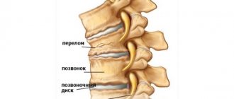
How to achieve a stable position in the truest sense of the word? How to maintain balance in any life situation? What to do if your main internal rod is broken? Read more about spinal fractures.
Fact. As collective analytical information from evidence-based medicine shows, men injure their spine much more often than women: in total, about 82% of the total number of victims.
The classification of fractures varies depending on the nature of the injury and the number of broken vertebrae. They are:
- isolated (single)/multiple (fractures of several vertebrae);
- splintered;
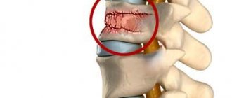
- compression;
- fracture-dislocations;
- compression splintered;
In addition, there are fractures complicated with damage to the spinal cord and uncomplicated.
Content
- 1 Spinal fractures in the lumbar region 1.1 Signs of a spinal fracture
- 2.1 History and complaints
- 3.1 Compression fractures
Recovery after a fracture
Effective rehabilitation is the key to full recovery after a spinal fracture. It is necessary to eliminate the neurological consequences of impaired innervation. Particular attention must be paid to the performance of the muscular system and the circulatory system. A long period of immobilization has a negative impact on the patient's condition. Atrophy of the tendon, ligament and muscle apparatus occurs. All this can lead to the formation of persistent ankylosis and contractures, which is a serious obstacle to full recovery and return to a normal lifestyle.
The recovery process after a compression fracture of the spine takes from 1.5 to 2 years. Throughout the entire rehabilitation period, the patient must strictly follow all recommendations of the attending physician. During the year, it is recommended to undergo periodic courses of physical therapy, massage, physiotherapy, kinesitherapy, etc.
After discharge, the patient is given a referral to the nearest chiropractic clinic. At the appointment, the vertebrologist will study the medical history and examination results. Only after this will he develop an individual plan for rehabilitation and restoration of the functions of the spinal column. As a rule, strict implementation of these recommendations and instructions leads to complete recovery.
Fractures of the spine in the lumbar region[edit | edit code]
Sections of the Spine
A description of all lumbar vertebral fractures is beyond the scope of this book, but a sports physician may have to treat minor fractures. These include compression fractures of the vertebral body, fractures of the spinous and transverse processes, damage to the endplate in adolescents, and stress fractures of the pedicles of the vertebral arch or sacrum.
A compression fracture usually occurs due to excessive flexion; a spinous and transverse process fracture occurs as a result of a direct blow or rotation. In adolescents, an avulsion fracture of the endplate is possible due to more powerful and durable spinal ligaments. There are reports of stress fractures of the pedicles of the lower lumbar vertebrae, usually in power pitchers in cricket. Stress fractures of the sacrum are rare and in most cases are the result of overuse in runners.
Signs of a spinal fracture[edit | edit code]
- Complaints or medical history indicating acute severe injury.
- Gradually increasing unilateral pain in the lower back or buttocks.
- Persistent back pain after exercise therapy.
- Back pain or paravertebral pain with local tenderness on palpation.
- Fracture detection using radiation methods.
Complications of vertebral fractures
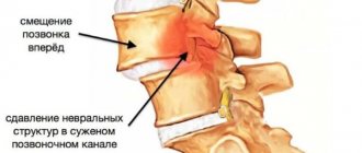
Compression-type vertebral fractures, which provoke a reduction in height by half, often lead to complications in the future in the form of excessive mobility and instability, which, in turn, manifests itself:
- Constant pain symptoms;
- Rapidly progressive degenerative changes;
- Nerve damage.
Fractures in older people can be accompanied by a hump-shaped spinal deformity, as well as constant pain. As for the most severe complications, they consist of compression or rupture of nerve roots or spinal cord. The first occurs both during the injury and as its consequences after some time. As for the second, it occurs at the moment of fracture. The above is fraught with compression of blood vessels and disruption of the blood supply to the spinal cord.
Symptoms and clinical picture[edit | edit code]
History and complaints[edit | edit code]
A fracture of the lumbar vertebrae should be considered if an athlete who received a strong blow or hit something at high speed complains of back pain. Pain that worsens when an athlete sits down and decreases when they lie on their back may be a sign of a compression fracture. It is necessary to question the athlete in detail about neurological disorders and dysfunction of the colon and bladder. It is important to establish the mechanism of injury and the force of impact. Suspicion of a fracture is caused by falls from great heights, strong collisions, and diving. Chronic low back pain that is not amenable to conservative treatment may be a manifestation of a stress fracture of the pedicles of the vertebral arch or sacrum. Athletes should be asked about their nutritional and menstrual habits to identify risk factors for osteoporosis. Increasing pain when extending the lumbar spine may indicate a stress fracture of the posterior half-ring of the vertebra. For chronic pain, it is important to rule out recent changes in the frequency and intensity of training or competition. The doctor should also ask the athlete to indicate the location of discomfort as accurately as possible, since with stress fractures of the sacrum, pain is possible not in the lower back, but in the buttocks.
Physical examination[edit | edit code]
In case of acute pain, the examination should not create additional stress on the back, and if it is necessary to turn the patient, roll him “like a log,” turning the shoulders and pelvis at the same time. The spine is palpated to identify the symptom of a step, hematoma, pain in the midline or on the sides of it. A complete neurological examination should then be performed, including assessment of movement, sensation and reflexes. Perineal sensitivity and anal reflex are checked.
For chronic pain, it is necessary to evaluate the range of active and passive movements in the lumbar region. Increased pain during extension should alert you to a fracture of the posterior half-ring of the vertebrae. It is also necessary to palpate the sacrum, as stress fractures of the sacrum are usually painful to palpation. Two tests can also help identify a stress fracture of the sacrum - maintaining balance on one leg and jumping on one leg.
Radiation diagnostics[edit | edit code]
X-rays of the spine are taken in direct, lateral and oblique projections. Using these images, the shape of the spine, soft tissue swelling, the distance between the spinous processes, the height of the vertebral bodies, and the presence of scoliosis or instability are determined. A compression fracture looks like a violation of the integrity of the anterosuperior part of the vertebral body. It is necessary to compare the anterior and posterior heights of the damaged vertebral body with the heights of neighboring vertebrae. Examine the distance between the spinous processes as carefully as possible. If it is enlarged, it means that an unstable fracture is possible. With simple compression fractures, the height of the vertebral body changes very little and there is neither divergence of the spinous processes nor rotation of fragments (in the photograph in a direct projection).
Fractures of the spinous processes are better visible on the lateral view. It is necessary to measure the distance between the spinous processes as accurately as possible and compare it with the distance between adjacent processes. Although the broken part of the process may be greatly displaced, the remaining part should remain in its normal position in relation to the adjacent vertebrae.
It is better to look for transverse process fractures on a direct photograph. A single transverse process fracture is considered minor, but the presence of multiple transverse process fractures should suggest severe injury.
X-rays usually do not show stress fractures of the pedicles or sacrum. Sometimes these fractures can be detected by signs such as callus or connective tissue adhesions.
Special methods[edit | edit code]
Specifying radiology diagnostics is indicated for compression fractures, multiple fractures of the spinous or transverse processes, and if symptoms persist despite conservative treatment. CT allows you to differentiate between a compression fracture and a more complex fracture of the vertebral body, such as a comminuted or Chance fracture (a horizontal fracture of the body, arch and pedicles of the vertebral arch). In addition, CT allows one to exclude severe concomitant fractures in the case of multiple fractures of the spinous or transverse processes. In athletes with stress fractures of the crura or sacrum, connective tissue adhesions may be visible along the fracture lines. Multiple fractures of the spinous or transverse processes or any neurological impairment are indications for MRI. It provides excellent visualization of the neural structures and ligaments of the posterior spinal column.
If X-rays and CT scans reveal nothing, single photon emission tomography or MRI may reveal stress fractures of the lumbar pedicles or sacrum. Athletes with compression or stress fractures of the lumbar vertebrae undergo bone densitometry to detect osteoporosis.
Rehabilitation measures
Therapy and rehabilitation play the most important role for the health of a victim after a fracture of the lumbar spine. Swimming and special physical therapy are the main recovery methods.
In the absence of adequate therapy or its incorrect provision can lead to the development of the following complications:
- Changes in physiological curves. Kyphoscoliosis is one of the complications that can develop as a result of lumbar trauma.
- Spinal instability. After the first fracture, the risk of re-injury increases significantly.
- Damage to the spinal cord, which can result in paralysis of the lower limbs.
- Involvement of the lower spinal cord - cauda equina. The complication is fraught with urinary and fecal incontinence.
- Hematomas and bleeding resulting from damage to the venous and arterial vessels of the spine.
- Development of a chronic form of pain syndrome. Quite often, a complication occurs as a result of inadequate rehabilitation.
To exclude the development of severe consequences of a transverse process fracture, it is necessary to take a responsible approach to treatment and the recovery period. Upon completion of therapy, the patient must undergo a rehabilitation course. During this period, it is necessary to engage in physical therapy and swimming, attend massage sessions and physiotherapy. In addition to the above measures, it is recommended to wear a belt-bandage on the lower back. The product has a positive effect on the area of injury, muscles and spinal column, and helps maintain the correct position of the back. The period of wearing the bandage depends on the nature of the injury, the duration of healing and rehabilitation. As a rule, the product is recommended to be worn for six months to a year.
Patients diagnosed with a fracture of the spinous process of the lumbar spine are divided into two groups:
- trauma that is not accompanied by disturbances in the functioning of the nervous system;
- with mild or moderate impairment of nervous activity.
During the rehabilitation process, all actions of specialists are aimed at completely restoring the mobility of the spinal column after a period of prolonged immobility and compensating for the consequences of compression of nerve endings.
Rehabilitation measures after a fracture of the transverse process of the spine:
- massage;
- physiotherapy procedures;
- physiotherapy;
- wearing a special corset.
A set of effective measures is selected individually for each patient, taking into account the severity of the fracture, age and health status of the victim. Full recovery will take at least 3 months.
Exercise therapy
Therapeutic gymnastics is used to develop back muscles. The exercise sets consist of moderate strength loads and bending. Exercise therapy is designed to develop a muscle corset for the normal maintenance of the spinal column.
It is recommended to begin performing a set of physical therapy exercises from the first days of treatment, as soon as the contraindications caused by the general manifestations of the injury are eliminated.
Exercise therapy is of great importance in the rehabilitation process after therapy. This is due to the fact that forced maintenance of a lying position and a small degree of physical activity for a long time lead to muscle atrophy. Subsequently, this will negatively affect the support of the spine by the muscular corset.
At first, patients are prescribed breathing exercises. It is performed in a lying position under the supervision of an experienced specialist. He will be able to accurately calculate the required load, which will most effectively stimulate the muscles and work out the joints without aggravating the condition of the damaged spine.
Important! The victim must perform a set of exercises at least twice a day.
The program is developed for each victim individually. However, there are exercises that are prescribed for all types of lumbar fractures. In the first days after the injury, a complex of physical therapy is performed in a lying position, then the axial load on the spine is gradually increased.
Immediately after an injury, the following exercises can be performed (lying on your back):
- flexion and extension of the hands and feet;
- abduction to the sides and alternate bending of the legs at the knee;
- a week later, perform turns on the stomach using hands from the back;
- raising the legs with the knees straightened less than 45 degrees.
After a few weeks, more complex exercises are included in the complex:
- crossing legs raised at 45 degrees (“scissors”);
- in the prone position - alternate lifting of the shoulders and legs;
- slow bends to the sides, sliding straight arms along the body;
- walking on all fours;
- in the same position - simultaneous lifting of the opposite legs and arms.
A month after the injury, the next stage of rehabilitation begins, which consists of a gradual increase in the axial load. The patient is allowed to take a vertical position, that is, stand on his feet. For moderate fractures, the patient is allowed to get out of bed only after 2 months. While rising, the patient is prohibited from sitting down. At first, you are allowed to stand solely on your knees, leaning on the headboard of the bed with your hands. After a certain time, they are allowed to release one hand at a time. You can sit down 3 months after the injury.
The patient is allowed to stand up only if:
- pain syndrome in the injured area of the spine;
- discomfort when tapping damaged vertebrae;
- protrusion at the fracture site.
When the patient is able to assume and maintain the position while standing without support, the final stage of recovery begins. It includes the following exercises:
- walking in place at first with support, and then without it;
- slow torso turns and bends;
- raising your arms up, simultaneously rising on your toes, taking the starting position.
All exercises are performed exclusively under the supervision of a rehabilitation specialist, so as not to aggravate the victim’s condition.
Physiotherapy
Physiotherapy is prescribed to improve blood flow, stimulate nerve endings and eliminate residual pain. Courses of procedures help speed up the recovery process of injured vertebrae.
The following methods are considered the most effective:
- Magnetotherapy. Impact of a magnetic field for therapeutic purposes on damaged vertebrae. Allows you to reduce the manifestations of pain, eliminate swelling and muscle spasms, restore damaged nerve endings, cartilage tissue and blood vessels, and strengthen intervertebral discs.
- Electrophoresis. A method of administering medications directly to the affected area of the spine through the skin using an electric current. During the procedure, a gauze swab pre-soaked in a medication solution is applied to the affected area. Cover the top with a protective gasket and attach the electrode. The drug, accumulating in the tissues, is slowly released, providing an analgesic, trophic and anti-exudative effect. Used to relieve pain, stimulates better penetration of phosphorus and calcium preparations directly into the affected area.
- Reflexology. Activation of points responsible for the functioning of the structures of the spine and the musculoskeletal system as a whole. Prescribed in combination with massage. Improves the course of metabolic processes, relieves swelling of tissues and muscle spasms, and promotes activation of blood flow.
- UHF (ultra high frequency therapy). Treatment with heat generated by ultra-high electromagnetic frequencies. Promotes rapid healing of fractures, reduces swelling, stimulates central and peripheral circulation, reduces pain and eliminates inflammatory processes.
Ultrasound therapy, ultraviolet irradiation and Sollux methods can also be used. The procedures are carried out in courses of 10 to 12 sessions, each lasting about 10–15 minutes. It is worth remembering that each of them has certain limitations and contraindications, which the doctor must take into account when choosing a method of physiotherapy.
Massage
Therapeutic massage is one of the important rehabilitation measures. At the first stage, the doctor’s task is to improve blood circulation and metabolic processes in the area of the spinal lesion. The first sessions are prescribed when the patient is still in hospital, in particular during skeletal traction.
Massage for fractures of the vertebral processes of the lumbar region is performed manually. This helps relieve swelling and compensate for prolonged immobility of the muscular frame of the back. The massage is performed in a hospital setting by a specialist with sufficient qualifications. One course consists of 10 procedures.
The intensity of the impact gradually increases, starting to engage the back muscles. This allows you to restore lost mobility of the vertebrae and significantly speed up the recovery process. But to prevent massage from aggravating the patient’s condition, it should be performed only by a highly qualified specialist.
Corset
If the vertebrae are damaged, a corset is prescribed to redistribute the load and prevent deformation of the spinal column.
There are two types of products, depending on the degree of rigidity:
- Semi-rigid. As a rule, they are used as a preventive measure, since they partially support the back.
- Tough. The product has special ribs made of light metal alloys or other materials. Thanks to this, it is able to fully support the back.
The device stably fixes the vertebrae in one position. The positive effects of the corset are as follows:
- the weight of the back falls completely on the product, due to which the muscles receive relief;
- pain is eliminated by eliminating muscle spasms and pinched nerve fibers;
- blood flow increases, which stimulates the supply of tissues with oxygen and nutrients, and also ensures good warming of the back.
It is recommended to wear the corset for at least 6 hours a day, while walking and during sedentary work. The mode and duration of wearing the device must be agreed upon with a doctor.
Treatment[edit | edit code]
First aid for spinal injury
Compression fractures[edit | edit code]
The basis for the treatment of mild fractures of the lumbar vertebrae is conservative methods. Compression fractures without displacement or with slight displacement require wearing (to unload the spine) a lumbosacral corset or thoracolumbosacral orthosis for 6-12 weeks. X-rays should be taken regularly at this time, as further flattening of the vertebral body or instability may occur. At the end of immobilization and approximately 4-6 weeks after removal of the corset or fixator, X-rays are taken in a standing position with the body flexed and extended for a final assessment of the shape of the spine. Once recovery is clinically and radiographically confirmed, the athlete can proceed to exercise therapy aimed at restoring range of motion and physical performance. After the range of motion is restored, they move on to strength exercises.
Fractures of the spinous and transverse processes[edit | edit code]
Spinous and transverse process fractures are treated by limiting physical activity and wearing a lumbosacral brace to reduce pain. Most of these fractures heal in about 6 weeks. As soon as the pain has subsided and the athlete feels well, exercise therapy begins, aimed at restoring range of motion and physical performance. As they adapt to the loads, they move on to strength exercises.
Stress fractures of the sacrum and pedicles of the vertebral arch[edit | edit code]
Stress fractures of the sacrum are treated conservatively by limiting physical activity and offloading the sacrum. As soon as the symptoms subside, they begin to gradually activate the patient, resume the load on the sacrum and restore physical activity in full.
Stress fractures of the pedicles of the vertebral arch, identified in time, before nonunion occurs, are treated with a thoracolumbosacral orthosis for 6-12 weeks. X-rays are taken regularly to evaluate the shape and stability of the spine. A CT scan may be needed to confirm fracture healing before removing the orthosis. Fractures of the pedicles detected late, when signs of nonunion have appeared, are treated surgically by spinal fusion and instrumental fixation.
Damage to the annular apophysis[edit | edit code]
The literature describes only isolated cases of damage to the annular apophysis in adolescents. There is no generally accepted treatment strategy. Suffice it to say that, according to clinical observations, conservative treatment of asymptomatic injuries has given good results. In patients with radicular symptoms or intermittent claudication (pain that radiates to the legs during physical activity, such as walking), surgical decompression of the spinal cord when the endplate protrudes backwards has yielded good results.
Diagnosis of vertebral fractures
Before prescribing treatment for vertebral fractures, our specialists conduct comprehensive diagnostic studies to determine the exact location of the injury, its type and consequences. For this, the patient is prescribed:
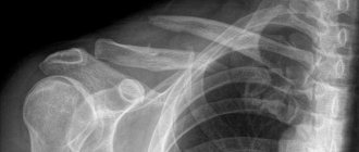
- X-ray studies of the spinal column in lateral and anteroposterior projections;
- Magnetic resonance imaging of the spine, which allows identifying damage to the nerve roots and spinal cord;
- Computed tomography of the spinal column, which makes it possible to see not only fractures of bone structures, but also damage to soft tissues.
Read also[edit | edit code]
- Massage for a spinal fracture
- Fractures of the spine in the cervical region
- Abs workout for back pain
- Sports spinal injury - treatment
- Lumbar sprain
- Backache
- Lower back pain
- Herniated disc
- Spondylolysis
- Spondylolisthesis (treatment)
- Treatment for back pain
- Rehabilitation for back pain
- Massage for osteochondrosis
- Massage for radiculitis
- Massage for intercostal neuralgia
First aid to the victim
If first aid is not provided correctly, the situation will worsen. But the right actions will prevent the development of complications. Immobilization and pain relief with analgesics are the first steps to take. The entire spinal column is fixed. This is being done because immobilizing a certain area is problematic. For these purposes, use any rigid base of the required size. When using a soft stretcher, the victim is placed on his stomach. This transportation option is used only in extreme cases, because it does not allow breathing to be controlled.
Absolutely forbidden:
- put the victim on his feet or sit down;
- pull on the lower or upper limbs;
- touch the fracture site;
- transport in a sitting position;
- get up;
- try to straighten the vertebrae;
- Give medications if the swallowing reflex is difficult.
Important! Perform any movement slowly and with extreme caution. People's movements must be synchronous. Needs support The affected area needs support.
The sequence of first aid actions in the presence of symptoms of spinal injury:
- The victim is placed on his back on a hard board, fixing the spine completely. Pain is relieved with a maximum dose of analgesic. Call an ambulance to ensure the victim is transported to the hospital.
- If the cervical spine is injured, build an improvised cardboard collar that limits mobility.
- In the absence of a solid carrying device, the victim is placed on his stomach on a soft stretcher and secured.
- When a neck is broken, it is necessary to control the patient's breathing. In this case, transport in a prone position is contraindicated.
Even if the injury occurs in the lower back, the neck must be secured with a special collar, since turning the head sets the entire spinal column in motion and can lead to displacement of fragments. Health care workers should be told what analgesic the victim took to rule out the possibility of an overdose. After providing first aid, the victim is transported to the neurosurgery hospital.
Prevention
The best prevention of pathology is regular amateur sports. Simple daily exercise will warm up all the muscles and keep the body in good shape. This will help avoid the development of dangerous consequences of a fracture of the transverse process of the spine.
When climbing to great heights, it is recommended to avoid sudden changes in voltage and be sure to use insurance.
Rules that will help you avoid fractures of the cervical vertebrae:
- strengthen the muscular frame of the back, which will significantly reduce the risk of injury in dangerous situations;
- comply with safety regulations at work and driving a vehicle;
- do not dive in unknown bodies of water;
- eat foods that contain the required amount of vitamins and minerals.
Using sports equipment during training will help prevent fractures of the cervical vertebrae. It is necessary to avoid sudden movements and strengthen the neck muscles. You should turn your head smoothly, without overstraining your spine.
Preventive measures after surgery include monitoring exercise, physical therapy, and a diet high in vitamin D and calcium.
In case of partial or complete paralysis, prevention of bedsores is necessary. It consists of using orthopedic mattresses, using skin care products against bedsores, and frequently changing body positions. Immobilized areas of the body must be kneaded and treated with powders and special ointments.