In this article we will look at why the kneecap flies out.
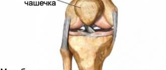
The patella, or kneecap, is a rounded bone that protects the joint from various damages. It is held in place by strong connective tissue formations - ligaments, which create a stable position for it. When a kneecap pops out, a ligament tear or sprain may occur, and the person may experience severe pain during the injury. Afterwards, it can fall back into place on its own, but in most cases it pops out again with further bruises or falls. This injury is characterized by severe pain; it can occur with sudden and incorrect turns of the leg, requiring immediate medical intervention.
Causes
When the kneecap pops out, there are a variety of causes for the injury. Why is this happening? The occurrence of injury is influenced by the patient's weight: if it is too heavy, it places additional stress on the joint, which creates additional risk.
The main reasons are: violation of the articular surface; incorrect immobilization; primary dislocation, which violates tissue integrity; ignoring rest mode during recovery; untimely therapy or advanced disease.
Provoking factors
Among other things, if the kneecap pops out, this problem can arise due to various factors.
These include: high joint mobility; muscle imbalance; femoral muscle atrophy; high position of the kneecap; knee deformity; ligament weakness; increased loads; bowed legs; age. When a person has weak muscles, they cannot hold the cup in its normal position. The same applies to ligaments. Excessive knee mobility also often causes problems. Age also affects the condition of the cup itself, muscles and bones. Cartilage and joints wear out over time, and various disorders appear - osteochondrosis, osteoporosis. All this increases the likelihood of a problem occurring. With strong physical exertion, the cup usually does not pop out immediately - after prolonged exposure and lack of rest for the limbs.
Special types of dislocations
Habitual dislocation
Localized mainly in the patella. It is characterized by repeated regular slipping of the kneecap from its usual location. Delivers severe pain. May provoke arthrosis.
It usually occurs in the presence of several provoking factors.
First of all, this:
- reduction in the thickness of the sliding paths at the location of the femur;
- pathologically high localization of the patella.
It is more often diagnosed in children and female patients.
Subluxation
A distinctive feature of such an injury is the peculiarity of the localization of the pathogenic process: it is not located directly in the joint, but, as a rule, affects the kneecap. There are many reasons that can lead to subluxation. All of them can be classified into 3 main categories.
- Rupture of the ligaments responsible for supporting the patella, or excessive stretching of them.
- Low development of the thigh muscles.
- Anatomical pathologies of leg formation.
Against the background of any of the above reasons, the patella takes on an unstable position. In this state, it is highly susceptible to damage as a result of various types of injuries and falls, careless bending of the legs and even minor physical exertion.
Quite often, subluxation occurs as a result of injury to the joint or as a result of surgery that disrupts the position of the patella.
Congenital dislocation
A relatively rare orthopedic pathology. According to average statistical data, it occurs in every hundred thousand newborns in the world. In the Russian Federation, the disease is diagnosed annually in approximately fifty patients. Pathology develops during the second half of pregnancy. It occurs more often in female newborns than in males.
To eliminate the violation, various means are used: splints, splints, etc. In severe cases, surgery is prescribed. Modern advances make it possible to perform surgical intervention after the patient reaches 3 months of age. In the absence of timely and adequate treatment, the patient will not be able to stand independently or move fully.
Symptoms
What are the signs that indicate that the kneecap is popping out? Most often, acute pain appears immediately, which intensifies during movement. Mostly the joint is slightly bent, its volume is increased, and there is a feeling that it is beginning to fall out. The knee is very swollen. What other symptoms indicate a problem with the patella? The main signs that the kneecap has flown out and is back in place: hemorrhage; edema; a hole on the bottom or top of the knee; a shaky feeling when walking. If the knee pops out, the pain radiates upward to the thigh, and the knee itself also hurts. There may be signs of bruising. The leg does not move at all, the knee quickly swells.
Features of the pathology
The knee part has a complex anatomical structure, which ensures its constant activity.
A person throughout almost his entire life (with the exception of infancy) walks, runs, squats, jumps and even falls. All these movements place high stress on the knee joint.
Many people suffer from excess weight and vitamin deficiency in the body. Such phenomena negatively affect the joint part of the leg, so under the influence of load the knee often pops out.
The peculiarity of the disease is that the joint can fly out and come back on its own. To do this, the person needs to remain still and apply something cold to the leg. But even after this, you will need to visit a doctor, since the damage leads to adverse consequences.
Degrees of impairment
There are three degrees of violation:
- The first is characterized by non-constant pain, a very mobile cup, and the patella is able to return to the correct position on its own.
- In the second degree, significant deformation occurs and severe pain is felt.
- The third is characterized by severe and sharp pain, increased deformation and limited movement.
Externally, you can see changes in the shape of the knee as the patella has moved. But this is not always noticeable, but only in the second and third degrees of pathology.
So, the kneecap flies out. What to do?
Carrying out diagnostics
The specialist examines the limbs: both sick and healthy. This is important in order to make an accurate diagnosis. Magnetic resonance imaging and ultrasound diagnostics are also necessary.
The doctor necessarily prescribes an x-ray of the legs. This eliminates additional injuries (fractures, cracks).
An interesting note: in the presence of partial ruptures, the patella moves slightly downward, while in the case of complete ruptures, the patella moves strongly upward. In addition to such diagnostic methods, the specialist must pay attention to other circumstances: the patient’s age and body weight, joint mobility.
Naturally, the doctor should receive information about the following factors: postural disorders; flat feet; localization of pain; location of the calyx; asymmetrical muscle strength. When all the information is received, treatment is prescribed.
If a person’s kneecap falls out and comes back in, and this happens periodically, the symptom needs a more thorough diagnosis. The joint is examined using MRI and ultrasound; such methods will allow you to visualize the condition of ligaments, soft tissues, tendons and muscles. Based on the results of the study, treatment is prescribed.
Treatment
What actions should I take? It often happens that the kneecap flew out and fell into place, or the patient resets the knee on his own. But this does not eliminate the cause of the pathology. In this case, you should not delay going to the doctor to avoid increasing destabilization, deformation and destruction of the knee. The treatment tactics for a prolapsed knee joint are determined only by a specialist; it depends on the degree of damage to the ligaments, menisci, the presence of marginal fractures in the cup, the condition of the cartilage tissue, and the integrity of the joint surfaces.
When the kneecap flies out, treatment for such a disorder lasts on average about six months. If there is no serious damage, then in such cases conservative methods are usually used, which can help a lot.
Treatment methods
After an injury, it is necessary to provide first aid to the victim. Setting a joint yourself can be dangerous, but there is an algorithm of actions that need to be performed before the doctor arrives. The patient should remain in a lying or sitting position without attempting to use the injured limb.
First aid is as follows:
- the leg should be immobilized in the most comfortable position;
- ice or cold compresses are applied to the knee area;
- if the pain is too severe, you can take painkillers.
Immobilization of the limb is the most important condition.
When the patella dislocates, its ligaments lengthen. The position of the leg, in which the pain is not felt too sharply, means that the ligaments have shortened a little and relaxed. If you move carelessly or try to set the kneecap yourself, they can completely rupture.
Conservative methods
If the patella is displaced for the first time, and the examination does not reveal any obvious violations of the integrity of the ligaments, the patient is recommended to wear elastic bandages for several weeks. Excessive loads should be avoided, but leaning on your leg when walking is not prohibited.
Treatment will consist of several main points:
- use of local anesthetics and anti-inflammatory drugs;
- physical therapy to increase the strength and elasticity of muscles and ligaments;
- UHF;
- physiotherapy to improve blood circulation;
- massage from a competent specialist.
Plaster application is also a conservative treatment method. This is necessary if the injury is accompanied by acute pain and partial rupture of the ligament fibers occurs.
The plaster cast is worn for 2-3 weeks, after which it is changed to an elastic bandage.
Surgery
Surgical intervention is necessary only if the integrity of the ligaments or tendons of the muscles was damaged at the time of the injury. Partial rupture of the fibers is treated under plaster, but if the ligament is completely separated, its ends must be connected by suturing. The operation is performed using arthroscopy - through two small punctures near the knee joint. Healing occurs under a cast or tight elastic bandage.
The rehabilitation period after surgery can last up to 6 months.
Conservative methods
When choosing a treatment regimen, they first resort to conservative methods.
To reduce pain in an acute condition, you need to apply ice to the joint, this can be done as first aid if the injury is acute.
You should immediately contact a specialist to clarify the extent and cause of the disorder; it is especially important to exclude rupture of ligaments and other defects in the integrity of tissues around the joints. This determines further therapy.
At first, you first need to completely, then partially, reduce the usual load on the limb. This is achieved by wearing orthoses, using elastic bandages, bandages or orthopedic devices.
If a person has hemarthrosis, a joint puncture is performed with further aspiration.
Anti-inflammatory non-steroidal drugs - Ibuprofen, Voltaren - will help relieve the inflammatory process.
Severe pain syndrome can be relieved with analgesics.
As soon as the condition improves at least a little, you need to undergo physiotherapy and massage. A complex of physical therapy (primarily static exercises) is required, due to the restoration of muscle balance. This mainly applies to the extensor muscles.
When the knee joint protrudes, physical activity is mandatory. To reduce re-dislocation, you need to train your muscles.
Dislocation of the knee joint (Dislocation of the knee)
Anterior knee dislocation
It is considered the most common dislocation of the LBD.
Accompanied by rupture of the cruciate ligaments and the posterior sections of the articular capsule. Often there is also a rupture of the lateral and median ligaments. Possible compression or damage to the popliteal vessels, peroneal and tibial nerves. If the blood vessels are damaged, the lower leg is pale, cold, cyanotic, the pulse is weak or undetectable. If the nerves are damaged, sensory disturbances and paralysis may occur. The patient notes intense pain in the affected area. The knee is swollen, sharply deformed, and upon examination, hematomas and hemorrhages are revealed. Active movements are impossible; when passive movements are attempted, spring resistance is detected. Support is impossible. With complete dislocations of the leg in a state of extension or slight flexion, shortening of the limb is detected.
With incomplete dislocations, the limb is in a position of slight flexion and there is no shortening. A significant increase in the knee joint in the anteroposterior direction is revealed. The tuberosity and condyles of the tibia protrude along the anterior surface, and the condyles of the femur protrude along the posterior surface. The patella takes an oblique position and “lies” on the articular surface of the LBP.
Posterior knee dislocation
Accompanied by damage to the cruciate ligaments, often the lateral and median ligaments are also torn. It is possible that the patellar ligament may be torn off at its attachment point (in the area of the tibial tuberosity). The patient complains of unbearable pain. In the area of damage, significant swelling, cyanosis, areas of hemorrhage, and severe deformation are determined. There is no movement in the knee, the supporting function of the limb is lost.
With a complete dislocation, the leg is in an extension position, shortened. With incomplete dislocation, the lower limb is bent and there is no shortening. The condyles of the femur stand in front, and the articular surface of the tibia stands behind. The patella is located obliquely and lies on the condyles of the femur. Damage to nerves and blood vessels, manifested by corresponding symptoms, is often observed.
Lateral knee dislocations
Internal and external knee dislocations are rare and are usually incomplete. Accompanied by rupture of the lateral and median ligaments. The knee is deformed, swollen, and sharply painful. There is an inability to support and move. The leg is in a flexed position, the foot is rotated. The patella shifts to the side opposite to the dislocation and takes an oblique-transverse position.
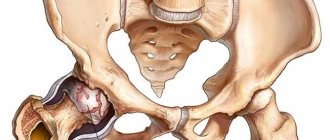
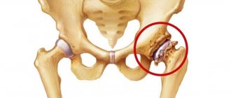
Patella dislocation
The damage is accompanied by severe pain. The knee joint is swollen and slightly bent. Deformation is detected in the anterior sections. The most common option is in which the patella is displaced outward (external dislocation), inward displacement and wedging between other bones are less common. The volume of the knee is increased, as a rule, hemarthrosis is determined. There are no active movements, passive ones are significantly limited. Palpation - the patella is displaced, palpation of the damaged area is sharply painful.
Congenital dislocations of the knee joint
This type of congenital joint abnormality is detected at birth or at an early age. Patellar dislocation is more common in boys and is combined with underdevelopment of the patella and lateral condyle; other anomalies in the development of the knee joint may also be detected. The main manifestations are unsteadiness of gait, instability and rapid fatigue of the limb.
Congenital dislocation of the lower leg often occurs on both sides, and at the same time underdevelopment of the knee and ankle joints is detected. Hypoplasia or aplasia of the tibia is possible. Without surgical treatment, the pathology worsens with age; congenital dislocations of the patella and tibia become the cause of the development of severe arthrosis.
Surgical intervention
If there is no effect from conservative methods or the degree of damage to the joints near the tissues or articular structures is too pronounced, if the process is advanced, a specialist may advise treatment using surgical methods.
Currently, there are many ways of such intervention for problems with the patella. However, treatment using most techniques quite often causes relapses of the pathology and does not exclude the occurrence of secondary intra-articular changes.
If the reason why the patella is constantly moving is due to too much tension on the external ligament compared to the internal one, treatment consists of cutting the ligament using an arthroscope. Such an intervention is well tolerated by patients, does not require long-term recovery, and is minimally invasive. If the kneecap has popped out and there has been lateral displacement, a lateral incision is made. In a number of institutions, a thermal cautery is used to prevent bleeding into the joint with the formation of hemarthrosis.
How is it diagnosed?
Diagnosis of knee dislocation and its type is carried out using a set of measures:
- History taking, visual examination, palpation.
- X-ray to determine the nature of the displacement and exclude bone damage, differential diagnosis of knee dislocation from meniscus damage.
- MRI or computed tomogram.
- Arteriogram – performed for symptoms of damage to the neurovascular bundle to clarify the presence of vascular injury.
- Ultrasound of the joint, Doppler sonography - to analyze blood flow.
- Neurological test of skin sensitivity.
- Checking the presence of a peripheral pulse to determine vascular patency.
Diagnosis should begin as quickly as possible, as soon as the first signs of a knee joint injury appear, because the complex of therapeutic measures and the timing of knee recovery depend on this.
Prevention and recommendations from doctors
To prevent problems that cause the kneecap to pop out, you need to immediately consult a doctor in case of an acute dislocation and take action. Experts recommend doing this even when the patella itself has returned to its place. During treatment, you should listen to the doctor in everything and not violate the limited load. After the acute condition has resolved, it is very important to continue to train and exercise, since the knee can only be stabilized if the muscle tone of the lower leg and thigh is normal. In this case, you need to gradually increase the load, having previously discussed this with your doctor. He will recommend the most optimal rehabilitation measures and special exercises for each patient.