Rheumatism: symptoms, treatment, phases, prevention, causes
Rheumatism is a connective tissue disease associated with inflammation of organs and body systems. Rheumatism is often associated with joint pain and rheumatoid arthritis, but this is only part of the symptoms of the disease. Rheumatic fever affects the heart muscle tissue, skin, blood vessels, brain and nervous system. Joint pain suppresses most symptoms, so a person does not feel other disturbances in the body’s functioning.
Damage to skin and blood vessels
Skin pathologies associated with rheumatism are expressed by redness and irritation in the form of round spots. Subcutaneous nodules may appear on the arms and legs, which do not cause pain and may disappear even without treatment.
Signs of rheumatism:
- temperature increase;
- weakness, dizziness, shortness of breath;
- joint pain in the arms and legs, swelling;
- lower back pain;
- motor limitation;
- pain and compression in the heart;
- round red spots on the skin;
- formations under the skin in the joint area;
- speech disorders.
2. Reasons
The main reason for the development of extra-articular rheumatism is systematic microtrauma, static and monotonously repeated dynamic loads, which is inevitable in some sports, professional activities of musicians, dancers, typists, etc. Risk factors are hormonal changes and imbalances, excess body weight, metabolic disorders, liver and biliary tract diseases, work in conditions of low temperatures and high humidity, infections, diabetes mellitus, and previous myocardial infarction. The influence of hereditary factors and congenital individual structural features of the joint structures of the musculoskeletal system is being studied.
Most often, several such factors are detected simultaneously in the patient’s medical history, i.e. extra-articular rheumatism can be considered a polyetiological disease. It should be noted that the pathogenesis associated with a number of the listed reasons has so far been little studied and remains unclear, but their significance is reliably confirmed by statistics.
Visit our Rheumatology page
Causes
Rheumatic fever appears as a result of complications after a streptococcal infection - sore throat, otitis media, scarlet fever, etc. Tangible symptoms of rheumatism appear after 2 weeks. The disease is more often detected in children aged 7 to 15 years with a hereditary predisposition.
The development of rheumatism is promoted by:
- improper or incomplete treatment of streptococcal infection;
- lack of vitamins and beneficial microelements;
- autoimmune diseases;
- hypothermia of the body.
1.General information
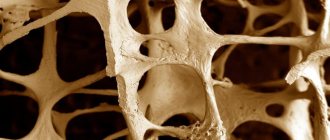
Rheumatism as such is a serious chronic disease, inflammation of connective tissue structures in the body (the literal translation of the diagnosis from Greek means “spreading”), which affects the joints, myocardium and other organs.
Extra-articular rheumatism, as the name suggests, covers mainly periarticular soft tissues (tendons, bursae, aponeurosis plates, ligaments), as well as more distant muscles, neurovascular plexuses, subcutaneous tissue, etc.). Some of these diseases have not been sufficiently studied due to the polymorphism of the clinical picture and the diffuse nature of the lesions. However, another group of diseases, also covered by the collective term “extra-articular rheumatism”, in special epidemiological studies turns out to be very common (up to 8% of people in the general population) and includes tendinitis, fibrositis, ligamentitis, periarthritis, etc., as well as mixed forms of these numerous rheumatic diseases. inflammations that involve the soft tissues associated with the joints. Among the cases, women in the age range from 35 to 55 years, engaged in manual labor, predominate.
A must read! Help with treatment and hospitalization!
Treatment
Rheumatism is diagnosed on the basis of clinical and laboratory indicators: assessment of the body's functional capabilities, analysis, examination using instruments and instruments. Depending on the symptoms, the doctor determines diagnostic methods:
- blood test: general, C-reactive protein, level of antibodies to streptococcus;
- goniomery - a technology for assessing joint mobility by a doctor;
- studying the state and functioning of the immune system;
- microscopic examination of synovial fluid from inflamed joints;
- biopsy of the synovium;
- MRI and ultrasound of joints;
- ECG and ultrasound of the heart;
- echocardiography;
- chest x-ray;
- thermography using thermal vision.
During the active phase of the disease, it is recommended to remain in bed until the symptoms subside. During the inactive phase, it is necessary to reduce physical activity and not put stress on the joints. To avoid stimulating the immune system to activate during illness, the doctor prescribes a diet that excludes the consumption of allergenic foods.
4.Treatment
The arsenal of remedies effective for extra-articular rheumatism is very wide. It is based on various ointments, creams, gels, rubs, etc., causing an increased local blood flow. Physiotherapy, incl. electrotherapy, special types of massage, acupuncture, etc. In some cases, the prescription of hormonal drugs is indicated, in others, non-steroidal anti-inflammatory drugs are used. Treatment usually brings good results if the patient has sufficient patience, compliance (“therapeutic alliance” with the doctor, mutual understanding) and willingness to follow medical prescriptions.
Prevention
The development of rheumatism can be prevented at an early stage, so if symptoms appear, you should contact a rheumatologist, cardiologist or therapist. If you have a streptococcal infection, you should take treatment seriously so as not to provoke complications. To keep connective tissues healthy, do not overcool the body, avoid contact with people with sore throat and strengthen the immune system according to the doctor’s recommendations. Methods of drug prevention can be clarified with your doctor.
3. Symptoms and diagnosis
In the clinic of extra-articular rheumatism, inflammatory symptoms or degenerative changes in tissues may predominate.
In the early stages of the process, symptoms may be absent; at later stages, the most common manifestations are pain and limited mobility in the affected joint.
Tendinitis and tendovaginitis (tenosynovitis) are degeneration of tendon and joint capsule tissues, usually accompanied by minor inflammation.
Ligamentitis is inflammation of the ligaments outside the joint.
Bursitis is an inflammatory process of the mucous “bag” (serous bursa).
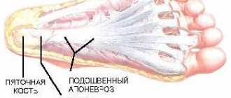
Fibrositis (including fasciitis and aponeurositis) – rheumatic inflammation of various ligamentous structures, etc.
Palpation reveals point tenderness in areas of predominant inflammation. In 30-40% of cases, extra-articular rheumatism is accompanied by the deposition of calcium salts in areas of degenerative-inflammatory necrosis, as well as certain changes in the periosteum.
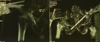
The most important in diagnosis, in addition to collecting anamnesis and direct examination, are radiography, tomographic imaging, and arthroscopy.
About our clinic Chistye Prudy metro station Medintercom page!
Symptoms and varieties
Its nature lies in an infectious-allergic basis. This disease affects connective tissues and blood vessels. It mainly occurs as a consequence of a complication of streptococcal infection. Many people say that older people are most susceptible to this disease, but this is not true.
There are 5 options for placing this disease:
- rheumoerythrema is a skin disease of the epidermis,
- rheumatic carditis - affects the lining of the heart,
- rheumochorea – rheumatic vasculitis of small vessels of the brain,
- rheumopolyarthritis is a disease of the joints,
- rheumopleuritis - inflammation of the bronchi, pleura, lungs.
The main symptoms of this disease are:
- swelling of the joints,
- pain and stiffness of muscles and joints,
- redness of the joints.
If you have even the slightest suspicion of this disease, then treatment should be carried out immediately. After all, delay can cause severe heart disease. To solve this problem, you can use folk remedies to treat rheumatism at home . But it is worth remembering that before starting any manipulations, you should consult a doctor.
How to treat rheumatism at home
Initially, the rheumatologist will make every effort to eradicate the streptococcal infection, since it could be the cause of the disease. Next, after antibiotic therapy, the doctor will definitely prescribe hormonal therapy. Only after this is it worth taking immunomodulatory drugs. Treatment of rheumatism in an adult patient is carried out in several stages:
- inpatient therapy – 1.5-3 months,
- staying in a special sanatorium with cardiorheumatological specialization,
- Frequent visits to the clinic for medical check-ups.
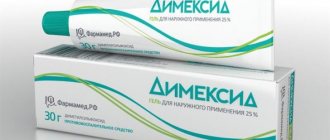
But, in addition to traditional medicine, there are also traditional methods of treating rheumatism . Among a large number of different herbs, infusions, decoctions and other raw materials, the most effective ones are distinguished, such as turmeric, propolis, pine needles, etc. Thus, turmeric has a therapeutic effect on this disease. It actively fights chronic pain and eliminates inflammatory processes.
Propolis is a fairly common substance for the treatment of various diseases. They especially note the ease of use of drugs based on it. It is so affordable that you can do it at home.
Propolis
Take propolis and make a cake out of it. Apply it at night on sore spots. To keep warm, it is recommended to tie it with a warm scarf and then cover it with a scarf.
There is another way. Cool the propolis and grind it on a grater. Next, pour in wine vinegar. Place in a cool, dark place for 10 days. Shake the contents periodically. After the allotted time, the tincture should be placed in the refrigerator for 10 hours and then strained. It is used to prepare compresses. To do this, you need to roll the gauze in several layers and moisten it well. Then they wrap the affected joints, cover them with additional warm cloth and leave them overnight. It will help increase blood circulation and increase the regeneration of damaged tissues.
An ointment for joint rheumatism is prepared from propolis . Take 100 g of Vaseline and heat it. Next, grind 10 g of propolis. Cool the heated Vaseline to 50 degrees and combine with propolis. Place the resulting raw material on the fire and cook for 10 minutes under a closed lid. After preparation, it should be filtered and applied to diseased areas of the skin. It is recommended to do this twice a day.
pine needles
They prepare a pine tincture, which can help even in the most severe cases. For it you need to collect a full liter jar of fresh May spruce needles. Next, fill with alcohol to the top and close tightly with a lid. Let stand in a dark and warm place for 3 weeks. Shake occasionally. Strain and take this way - 8 drops per piece of sugar 30 minutes before meals. Duration – 4-6 months.
Salt
Traditional treatment for rheumatism of the joints notes the particular effectiveness of salt. It helps relieve joint pain in the shortest possible time. To do this, dissolve a tablespoon of salt in a glass of water. The water should be warm. Soak gauze in this solution and apply to the sore spot. Place cellophane and a warm scarf on top. Leave for several hours.
You can not use water, but take fabric bags. Heat salt in a frying pan and pour into bags. Then apply them to sore spots.
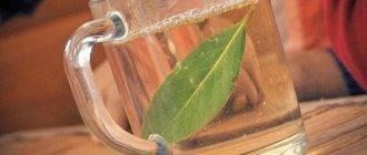
Birch leaves
Take cotton pants. Prepare fresh birch leaves and place them in your pants. Get dressed and go to bed, wrapped in a warm blanket. A strong effect will be achieved when you sweat.
You can also be treated in another way using birch leaves. For it you will need an old bathtub or tub. Fill it with leaves. Place in the sun and wait a few hours. Then get in there and enjoy.
Rheumatic diseases of periarticular soft tissues
Pathological changes first affect the tendons that are subject to the greatest load and mechanical stress. This leads to the appearance of fibril defects, foci of necrosis, the development of post-inflammatory sclerosis, hyalinosis and calcification. Primary changes are localized in the places of fixation of tendons to bone tissue (entheses) and are called enthesopathy. In the future, the process may involve tendon sheaths (tenosynovitis), synovial membranes (bursitis), fibrous capsules (capsulitis), joint ligaments (ligamentitis), etc.
Common symptoms of extra-articular rheumatism include pain and limited joint mobility. Pain is associated with certain active movements in the joint; local painful areas are determined in the areas of tendon fixation. With tendovaginitis and bursitis, swelling is clearly detected along the tendons or in the projection of the synovial membrane.
Humeroscapular periarthritis
It predominantly develops in women over 40-45 years of age. Humeroscapular periarthritis is caused by dystrophic changes in the tendons of the supraspinatus muscle, the rotator muscles of the shoulder (subscapularis, infraspinatus, teres minor and major), tendons of the head of the biceps muscle (biceps) and the subacromial bursa. Involvement of the supraspinatus tendon can result in simple tendonitis, calcific tendonitis, or a tear (or rupture) of the tendon.
Simple tendinitis is characterized by pain in the supraspinatus muscle during active abduction of the arm (Dauborn's sign), with the greatest pain observed when the amplitude of abduction of the limb is 70-90°. A sharp increase in pain is associated with temporary compression of the tendon between the epiphysis of the humerus and the acromion. The calcific form of tendinitis is diagnosed after radiographs of the shoulder joint are taken. Painful symptoms are more pronounced, and the motor function of the joint is more significantly impaired.
A tear or complete rupture of the tendon that anchors the supraspinatus muscle is usually caused by heavy lifting or an unfortunate fall on the arm. It differs from other forms of glenohumeral periarthritis by the typical symptom of a “falling arm,” i.e., the inability to keep the arm in an abducted position. This condition requires arthrography of the shoulder joint and, if a tendon rupture is detected, surgical intervention.
With tendinitis of the head of the biceps, persistent pain and palpation tenderness are noted when trying to strain the biceps muscle. The clinical picture of subacromial bursitis usually develops secondarily, following damage to the supraspinatus muscle or biceps. It is characterized by pain, limitation of rotation and abduction of the limb (symptom of a blocked shoulder). It can occur in the form of calcific bursitis with the deposition of calcium salts in the subacromial bursa.
Periarthritis of the elbow joint
Variants of damage to the periarticular tissues of the elbow joint include enthesopathies in the area of the epicondyles of the humerus and ulnar bursitis. Enthesopathies of the tendons that attach to the epicondyle of the shoulder constitute the pathogenetic basis of the syndrome called “tennis elbow.” Pain is noted in the area of the external and medial epicondyles of the humerus, which intensify at the slightest tension of the extensors and flexors of the hand and fingers.
In the case of olecranon bursitis, a voting protrusion in the projection of the olecranon is determined by palpation.
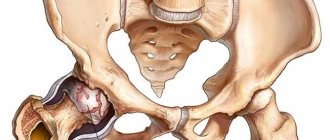
Periarthritis of the hip joint
It develops when the tendons of the gluteus minimus and medius muscles are damaged, as well as the joint capsules in the area of the greater trochanter of the femur. For the clinic of hip periarthrosis, the occurrence of pain in the upper outer thigh when walking and absence at rest is typical. Palpation of the soft tissues in the area of the greater trochanter is painful, and x-rays reveal tendon calcification and osteophytes along the contour of the apophysis of the femur.
Periarthritis of the knee joint
Caused by damage to the tendon apparatus, which provides fixation of the semitendinosus, sartorius, gracilis, semimembranosus muscles to the medial condyle of the tibia. Pain accompanies both active and passive movements (extension, flexion, rotation of the leg), sometimes local hyperthermia and swelling of soft tissue structures are noted.