Swelling of the knees is a serious signal of the development of a complex pathology. There are no interconnected systems in the human body. Their close connection is most obvious during illness. Local damage to the knee is manifested by special signs, of which the first and most important is swelling. They are not considered a separate disease, they are only symptoms that accompany various injuries and diseases, which is important to recognize, diagnose and begin treatment in time. One of the most serious complications is bone marrow edema of the tibia of the knee joint.
The medulla oblongata plays an indispensable role in immunity and blood formation. It is located in the tubular and sternum bones, in the costal and vertebral bone structures. Deviations from the norm in its functions are reflected in the general well-being of a person.
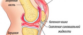
Bone marrow edema of the knee joint
Typically, swelling of the medulla oblongata is the body’s response to injury, infection, and dysfunction of the blood flow. The main causes of swelling in the knees are injuries. The damage changes the structure of the joints, the tissues of the joints are injured, and their blood supply is not regulated. A liquid substance accumulates in the intercellular space, interfering with the functions of the articular joint.
Pathogenesis
It is a characteristic manifestation of traumatic contusions, hidden subcortical and stress fractures. Bone marrow edema is one of the earliest nonspecific MR symptoms of a large number of diseases of the osteoarticular system. The pathomorphological basis of edema is an increase in the content of extracellular (interstitial) fluid in the bone marrow, as well as a local increase in blood flow and perfusion in it.
Epidemiology
The prevalence of bone marrow edema is directly related to etiological factors.
Causes
Factors contributing to the formation of edema:
- the presence of an infectious agent,
- knee injuries, dislocations and subluxations with displacement,
- ruptures of tendons and ligaments,
- hemorrhages, impaired blood supply,
- diseases of the musculoskeletal system ─ osteomyelitis, osteoarthritis,
- oncological neoplasms.
Radiation diagnostics
MRI semiotics
T1-weighted image: area of lower MR signal intensity relative to the normal image of fatty bone marrow.
T2-WI, PD, FS, STIR: area of higher MR signal intensity relative to the normal image of fatty bone marrow.
Contrast-enhanced T1-weighted image: increased signal from edema compared to native T1-weighted image.
The most informative are pulse sequences with signal suppression from adipose tissue (STIR, SPIR, FS), which improve visualization of the edema zone, which will have a hyperintense signal.
The structure of the signal from edema is heterogeneous, the contours of the zone of signal changes are unclear, the boundaries of the edema are relatively sharp, the shape of the zone of the changed signal is incorrect, the localization is at the site of the main process: inflammation, tumor, injury.
The MRI picture of edema in various diseases and skeletal trauma is nonspecific, but it is a highly sensitive symptom
Rice. 1. a – PD cor. scan of the knee joint: against the background of medium-intensity normal bone marrow, areas of edema are identified in both femoral condyles and the lateral tibial condyle (indicated by green arrows). b – T1-VI cor. scan of the knee joint: a hypointense area of bone marrow edema of the medial condyle (green arrow) and an even more hypointense area of fibrous bone remodeling in the area of the fracture of the medial tibial condyle (red arrow).
Rice. 2. MRI of the left hip joint: a – T1-VI cor. scan, b – T2-FS cor. scan 1 – fracture line 2 – area of bone marrow edema.
Causes of aseptic necrosis of the femur
The pathogenesis of aseptic necrosis of the femur is always based on several factors. The process always occurs due to a malnutrition of bone and surrounding tissues due to circulatory disorders. Local ischemia develops after compression of blood vessels, as a result of thrombus formation or any disease of the vascular system.
Of the many pathogenetic factors in the development of aseptic necrosis, special attention is paid to:
- Mechanical impacts - injuries, bruises, surgical interventions that could affect the integrity of the blood supply vessels. The negative result of mechanical damage may appear after several years.
- Unreasonable use of certain medications - hormonal, chemotherapy, glucocorticoid and non-steroidal anti-inflammatory drugs with long-term use affect the composition and quality of the blood, which increases the likelihood of thrombosis.
- Radiation exposure due to specialized therapy, radiation sickness.
- Atherosclerosis.
- Alcohol intoxication - frequent consumption of high doses of alcohol negatively affects the overall metabolism and has a destructive effect on the vascular walls. At the same time, liver dysfunction occurs and cholesterol levels increase. Under the influence of ethanol and cholesterol, the walls of blood vessels lose their elasticity and thicken, which means that the blood supply to the femurs is impaired.
- Diseases of the hematopoietic system.
- Pathologies of internal organs - pancreatitis, Cushing's syndrome, renal and liver failure.
- Autoimmune diseases - multiple sclerosis, systemic lupus erythematosus.
- Diseases of the spinal column.
List of used literature and sources
- Bryukhanov, A.V. Magnetic resonance imaging in osteology: monograph / A.V. Bryukhanov, A.Yu. Vasiliev. – M.: OJSC “Publishing House “Medicine”, 2006. – 200 p.
- Radiation diagnostics of osteomyelitis [Electronic resource] / V.V. Kovalinin [et al.] // Russian electronic journal of radiation diagnostics: electronic journal. – 2014. – T. 4, No. 3. – P. 66-76. – URL: https://www.rejr.ru/nomer/vol-4-3-2014.html (03/29/2018).
- X-ray diagnosis of tuberculosis of bones and joints [Electronic resource] // Second opinion: website – (2013-2017). – URL: https://secondopinions.ru/rentgenovskaya-diagnostika-tuberkuleza-kostey-i-sustavov (03.29.2018).
- X-ray diagnosis of gonarthrosis [Electronic resource] // Second opinion: website – (2013-2017). – URL: https://secondopinions.ru/poleznye-materialy/rentgen/rentgen-sustavov/rentgenovskaya-diagnostika-gonartroza (03/29/2018).
Clinical symptoms of knee swelling
Bursitis and synovitis have similar symptoms at the initial stage of development. The main manifestation is swelling of the soft tissues in the knee area. The knee increases in volume, the skin turns red. On palpation, an oval formation can be felt. As for the patient’s sensations, he feels pain, especially after maintaining a sitting position for a long time. The temperature may rise, the patient limps his leg, it becomes difficult to bend and straighten the knee, and increased fatigue is noted. There is stiffness when walking.
Concussion or contusion of the brain
Contusion (lat. contusio - bruise), or brain contusion. This is damage in the form of macrostructural destruction of the brain substance, often with a hemorrhagic component, occurring at the time of exposure to a traumatic agent. There are mild, moderate and severe bruises.
Mild brain contusion occurs in 10–15% of victims. This may result in a fracture of the skull bones. The duration of loss of consciousness is up to 30 minutes (according to other sources, up to 40 minutes). The duration of congrade and retrograde amnesia is up to one hour. Anterograde amnesia usually does not occur. Upon restoration of clarity of consciousness, general weakness, headache, tinnitus, nausea, vomiting (often repeated), dizziness, impaired concentration, weakened memory, and some slowdown in mental activity are detected. Nystagmus (usually horizontal), anisoreflexia, sometimes mild hemiparesis, and pathological reflexes may be detected. Due to subarachnoid hemorrhage, mild meningeal syndrome may occur, and an admixture of blood may be detected in the cerebrospinal fluid. Brady- or tachycardia and a transient increase in blood pressure by 10–15 mmHg are often observed. Art., other vegetative symptoms.
On CT, 40–50% of victims show areas of low density (areas of edema-ischemia). Histological examination of such lesions reveals edematous brain tissue. There are ruptures of small vessels and pinpoint diapedetic hemorrhages. Regression of these morphological changes occurs within 2–3 weeks.
Symptoms gradually regress within 1–3 weeks after the injury, full restoration of performance occurs after a month, but this does not always happen. Distinguishing between a concussion and a mild bruise is often quite a difficult task.
Moderate brain contusion. It is observed in 10–15% of victims. Loss of consciousness lasts from 30 minutes to one hour (according to other sources - up to 3-4 hours, and depression of consciousness to the level of moderate or deep stupor - up to several hours or days). Amnesia, congrade and retrograde, extends for a period of 1 to 24 hours (according to other sources, for the entire period of stunning consciousness, up to several days). Since the depression of consciousness changes in its depth, sometimes intervals of clarity of consciousness appear, which is why congrade amnesia is as if torn: periods of amnesia are interspersed with short periods of relatively normal memory. For 7–12 days there may be disorientation in time, location, and what is happening; absence or incomplete awareness of one’s condition; disorders of attention, memory, thinking and intelligence; psychotic episodes with perceptual deceptions, delusions, agitation, and inappropriate behavior. There are convulsive seizures, often isolated. Anterograde amnesia may occur - based on the impressions of the first minutes or hours after emerging from a state of traumatic coma.
Upon recovery from stunned consciousness, complaints of severe headache, dizziness, and nausea are revealed. Vomiting is observed, often repeated. Horizontal nystagmus, weakened pupillary response to light, moderate meningeal syndrome and respiratory disorders (tachypnea without rhythm disturbance) are detected; tachy- and/or bradycardia, low-grade fever, and often convergence disorder are also recorded. There is dissociation of tendon reflexes, sometimes moderate hemiparesis, pathological reflexes, sensory disturbances, and speech disorders. During puncture, cerebrospinal fluid flows out under a moderate increase in pressure (if liquorrhea does not occur), it is colored with blood; its rehabilitation occurs within 1.5–2 weeks.
During ophthalmoscopy, in some patients on the 4th day of the disease, dilated and tortuous retinal veins are detected, sometimes blurring of the boundaries of the optic discs. A craniogram reveals skull fractures in 60–65% of patients. CT scan in all cases identifies a focus of brain contusion or several such lesions. Perifocal edema usually does not extend beyond one damaged lobe of the brain. Hydrocephalus develops in 20% of cases.
Histological examination reveals small focal hemorrhages, swelling of brain tissue, subpial hemorrhages (under the pia mater), foci of necrosis of the cerebral cortex and underlying white matter in the area of 1–2 gyri. With depressed fractures of the bones of the calvarium, foci of mechanical damage to the cerebral cortex and adjacent white matter to a depth of 2 cm are observed. Around the focus of destruction, small-focal, often confluent areas of hemorrhage with perifocal edema are detected. Typically, moderate brain contusions do not require surgical treatment unless there is a depressed skull fracture or rapidly increasing hydrocephalus.
Severe brain contusion. Occurs in 10–15% of victims with TBI. Loss of consciousness lasts more than 60 minutes (according to other sources, from several hours to several days, sometimes with the transition of stupor or coma to apallic syndrome or akinetic mutism). Amnesia, retrograde and congrade, extends for more than a day; long-term anterograde amnesia may occur. Coma and recovery from it are often accompanied by motor agitation, which is replaced by immobility and muscle atonia, as well as psychotic states that last up to 1–2 months.
Brainstem symptoms are expressed: floating movements of the eyeballs, separation of the eyeballs along the vertical axis, fixation of upward gaze, anisocoria. The reaction of the pupils to light and corneal reflexes are depressed, swallowing is impaired. Bilateral pathological foot reflexes are detected. Disturbed breathing of the central or peripheral type (tachy- and/or bradypnea). Blood pressure is increased or decreased (it can be normal), in atonic coma it is unstable and requires constant drug support. Muscle tone is changed, hemiparesis and anisoreflexia often develop. Meningeal syndrome is pronounced. Sometimes hormetonia occurs (attacks of tonic spasms of the muscles of the trunk and limbs, repeating approximately every 6-10 minutes) - it occurs spontaneously or in response to painful stimuli. There may be convulsive seizures, and they often recur. During a lumbar puncture, the cerebrospinal fluid flows out under pressure and contains an admixture of blood.
Craniograms reveal fractures of the vault and/or base of the skull in almost all victims. CT always reveals foci of brain contusion, perifocal or diffuse edema of brain tissue, and pathological examination reveals foci of brain destruction over a significant extent, both in surface and depth.
Diffuse axonal brain injury (DABI). Considered as a special form of brain contusion. Clinical signs of DAPM in the foreground include dysfunction of the brain stem: depression of consciousness to the point of deep coma, pronounced impairment of vital functions, requiring urgent drug and hardware correction. The disorder may be accompanied by the formation of intracranial hematomas. Mortality with DAPM reaches 80–90%. Surviving patients develop apallic syndrome.
Return to Contents