Fractures of the diaphysis (body) of the femur
The more severe the fracture of the femoral body, the higher the energy of the damage, the more effort will have to be spent on recovery.
- Indications for surgery
There are practically no indications for conservative treatment of a femoral shaft fracture. Any fracture of the femur is operated on. The only exceptions are small children, in whom good fusion can be achieved within a few weeks, and during the period of further bone growth, even those displacements that remain are gradually compensated. When it comes to teenagers and adults, this is always an operation. It is customary to operate even when the patient’s physical condition is not very good. There is no statement that every hip fracture must be operated on urgently, but the hip must be fixed quickly. If the patient’s somatic status does not allow immediate definitive fixation, an external fixation device is installed. This is an inpatient injury and requires surgery.
- Minimally invasive nail osteosynthesis
No angular deformations or shortening of the bone should be allowed. This will lead to lameness and pathology of the adjacent hip and knee joints. But, at the same time, it is not necessary to perform anatomical tooth-to-tooth reduction for every hip fracture. Preservation of femoral length, femoral axis (i.e., no angular displacement), and absence of rotational displacement are what the surgeon seeks when treating hip fractures.
The gold standard for the treatment of femoral shaft fractures is intramedullary osteosynthesis. Orthopedic traumatologists at the Ilyinskaya Hospital most often use minimally invasive osteosynthesis with a nail. Insertion of a nail through the knee joint is called retrograde insertion, and insertion through the trochanteric region is called antegrade insertion. For normal hip function, the hip axis must be restored. The length of the bone must be restored accurately; shortening of no more than 1 cm is allowed. Axial deformation is no more than 5° and rotation is also no more than 5°. The most important advantage of this surgical approach is that periosteal (coming from the surrounding soft tissue) blood flow is preserved, and trauma to the surrounding soft tissue is minimal. Minimally invasive surgery gives a good cosmetic effect: one small scar in the place where the nail is located and 3 - 5 scars up to 1 cm in size in the places where the screws are inserted (in order to avoid rotational displacements, screws must be passed through the nail). Even with fractures of both legs, with fractures with a large number of fragments, the patient can immediately walk after surgery, using nails as endoprostheses. Limb function is restored very quickly. Severe pain after surgery almost never occurs.
- Indications for plate osteosynthesis
Nails are used in 90% of hip surgeries. However, sometimes surgeons have to use plates. This type of operation sometimes requires a fairly large incision. Although, in most cases, the plate is installed minimally invasively. The plate is used when a femur fracture is combined with a pelvic fracture in the acetabulum area. When fractures of the femoral shaft and femoral neck are combined, a plate is also used. Open growth areas are also an indication for plate installation. There are special nails for children, however, it is believed that it is not necessary to go through the growth zones of adolescents and children with massive nails. An indication for the use of plates is polytrauma when there are severe forms of fat embolism. Plate osteosynthesis is also preferable for combinations of hip and spine fractures. A plate must also be placed when the patient has a combination of a hip fracture and vascular damage. In order for vascular surgeons to begin their work, the bone must first be stabilized. One access is made for both the vessels and the thigh. Orthopedic surgeons quickly place a plate on the thigh, and vascular surgeons immediately begin restoring blood flow. Pregnancy is a contraindication for intramedullary osteosynthesis, since it involves prolonged use of x-rays. In severe forms of obesity, a plate is also placed, since it is difficult to find the point of insertion of the nail.
- Rehabilitation
The more severe the injury, the higher the energy of the damage, the more effort will have to be spent on recovery. Rehabilitation doctors at the Ilyinskaya Hospital use the full range of modern techniques for restoring function after a fracture of the femoral shaft. It includes physical therapy, massage, and various types of hardware physiotherapy. An individual program is drawn up for each patient, taking into account his needs and individual characteristics. Rehabilitation specialists, in collaboration with the operating surgeon and general practitioners, make the recovery process as fast and efficient as possible. Rehabilitation can be carried out both in a hospital setting and on an outpatient basis.
Classification of long bone fractures AO (Association of Osteosynthesis)
With the goal of simplifying clinical diagnosis, strategically assessing the severity and prognosis of injury, and creating a "common language of concepts" among traumatologists, Maurice E. Muller created the AO classification of long bone fractures .
General provisions for the classification of bone fractures according to AO
In general terms, Müller suggests presenting fractures of all tubular bones in the human skeleton in the following form (see diagrams, drawings, tables).
AO/OTA numbering system with anatomical location of fractures of three bone segments
Proximal segment = 1, diaphyseal segment = 2, distal segment = 3
Alphanumeric structure of the AO Muller classification of long bone fractures for adults
Designation of the anatomical location of the fracture according to AO
The anatomical location of the fracture is indicated by two numbers: the first for the bone, the second for its segment (the ulna and radius, as well as the tibia and fibula, are regarded as one bone). The proximal and distal segments of the long bones are defined using a square whose sides are the same length as the widest part of the epiphysis (exceptions 31- and 44-).
Determining the type of fracture in long bone fractures in adults
The exceptions are fractures of the proximal segment of the humerus (11-), proximal segment of the femur (31-), bones (44-), subacetabular fracture (32-)
| Segment | Type | ||
| A | B | C | |
| 1 (proximal) | Proximal extra-articular fracture Articular surfaces are not involved in the fracture | Proximal incomplete intra-articular fracture Part of the articular surface is involved, the rest is partially associated with the metadiaphysis | Proximal complete intra-articular fracture The fracture involves the entire articular surface; metaphyseal fracture completely separates the articular component from the diaphysis |
| 2 (diaphyseal) | Diaphyseal simple fracture One fracture line, cortical contact between fragments after reduction is more than 90% | Diaphyseal wedge fracture A fracture occurs in three or more fragments; the main fragments are in contact after reposition | Diaphyseal compound fracture A fracture involves three or more fragments; the main fragments do not contact after reposition |
| 3 (distal) | Distal extra-articular fracture No involvement of the articular surface by a displaced bone fragment | Distal incomplete intra-articular fracture The fracture involves part of the joint; the uninvolved part has a connection with the metadiaphysis | Distal complete intra-articular fracture The fracture involves the entire articular surface, and there is complete separation of the articular surface from the diaphysis |
Diaphyseal fractures
Stages of diagnosing diaphyseal fractures
| Diaphyseal fracture | ||
| Step | Question | Answer |
| 1 | What bone? | |
| 2 | Fracture of the extreme or middle segments of the bone? | |
| 3 | Type: simple or comminuted fracture (if more than 2 fragments)? | Simple (X2-A) |
| Splintered - go to step 3a | ||
| 3a | Is there contact between the two fragments? | Contact fragments, wedge-shaped (X2-B) |
| Non-contact fragments, complex (X2-C) | ||
| 4 | Group: simple or complex fracture? | Simple spiral (X2-A1), or spiral wedge (X2-B1), or complex spiral (X2-C1) |
| Simple oblique (X2-A2), simple transverse (X2-A3), wedge-shaped bending (X2-B2), wedge-shaped comminuted (X2-B3), complex irregular (X2-C3), complex segmental (X2-C2) | ||
Classification of diaphyseal fractures into three groups
| Type | Group | ||
| 1 | 2 | 3 | |
| A (simple) | Spiral | Simple | Transverse |
| B (wedge-shaped) | Spiral | Bending | Splintered |
| C (complex) | Spiral | Segmental | Wrong |
Segmental fractures
Stages of diagnosis of segmental fractures
| Segmental fracture | ||
| Step | Question | Answer |
| 1 | What bone? | Specific bone (X) |
| 2 | Fracture of the extreme or middle segments of the bone? | End segment |
| 3 | Fracture of the proximal or distal segments? | Proximal (X1) |
| Distal (X3) | ||
| 4a | Type: does the fracture involve a joint? | Extra-articular (XX-A), go to step "6" |
| Intra-articular, go to step "4b" | ||
| 4b | Type: incomplete or complete intra-articular fracture? | If the part is connected to the metaphysis/diaphysis, then - incomplete intra-articular (XX-B) |
| If the part is not connected - complete intra-articular (XX-C) | ||
| 5 | Groups: How many fracture lines intersect on the surface of the bone? | If there's one line, it's a simple one |
| If there are more than 2 lines, this is a comminuted fracture | ||
| 6 | Group: metaphyseal fracture? | Simple extra-articular (XX-A1), or simple intra-articular (XX-C1) |
| Wedge-shaped extra-articular (XX-A2) | ||
| Complex extra-articular (XX-A3), or simple intra-articular (XX-C2), or complex intra-articular (XX-C3) | ||
Classification of segmental fractures into three groups
| Type | Group | ||
| 1 | 2 | 3 | |
| A (extra-articular) | Simple | Wedge-shaped | Difficult |
| B (incomplete intra-articular) | Condyle fracture | Depression of the articular surface | Fracture of the condyle and depression of the articular surface |
| C (full intra-articular) | Simple intra-articular, simple metaphyseal | Simple intra-articular, complex metaphyseal | Complex intra-articular, complex metaphyseal |
Particular provisions of the classification of bone fractures according to AO
Classification of humerus fractures according to AO (1)
| 11-A | 11-B | 11-C | ||||||
| extra-articular unifocal fracture | extra-articular bifocal fracture | intra-articular fracture | ||||||
| 11-A1 | 11-A2 | 11-A3 | 11-B1 | 11-B2 | 11-B3 | 11-C1 | 11-C2 | 11-C3 |
| greater tuberosity | with impacted metaphysis | without impacted metaphysis | hammered with impact | not driven in | with displacement of the articular surface | driven in with slight displacement | driven in with significant displacement | with dislocation |
| Extra-articular unifocal fracture of the greater tubercle of the humerus | Extra-articular unifocal fracture of the humerus with impacted metaphysis | Extra-articular unifocal fracture of the humerus without impacted metaphysis | Extra-articular bifocal impacted humerus fracture with impaction | Extra-articular bifocal non-impacted humerus fracture | Extra-articular bifocal fracture of the humerus with displacement of the articular surface | Intra-articular impacted fracture of the humerus with slight displacement | Intra-articular impacted fracture of the humerus with significant displacement | Intra-articular fracture of the humerus with dislocation |
| 11 - proximal segment of the humerus | ||||||||
| 12-A | 12-B | 12-C | ||||||
| simple fracture | wedge fracture | compound fracture | ||||||
| 12-A1 | 12-A2 | 12-A3 | 12-B1 | 12-B2 | 12-B3 | 12-C1 | 12-C2 | 12-C3 |
| spiral | oblique (>30°) | transverse (<30°) | with spiral wedge | with bending wedge | with splinter wedge | spiral | segmental | wrong |
| Simple spiral fracture of the humerus | Simple oblique fracture of the humerus | Simple transverse fracture of the humerus | Wedge fracture of the humerus with a spiral wedge | Wedge-shaped fracture of the humerus with a bending wedge | Wedge-shaped fracture of the humerus with a comminuted wedge | Compound spiral fracture of the humerus | Compound segmental fracture of the humerus | Complex irregular fracture of the humerus |
| 12 - diaphyseal segment of the humerus | ||||||||
| 13-A | 13-B | 13-C | ||||||
| extra-articular fracture | incomplete intra-articular fracture | complete intra-articular fracture | ||||||
| 13-A1 | 13-A2 | 13-A3 | 13-B1 | 13-B2 | 13-B3 | 13-C1 | 13-C2 | 13-C3 |
| with separation of apophyses | metaphyseal simple | comminuted metaphyseal | sagittal lateral condyle | sagittal medial condyle | frontal | simple, metaphyseal simple | simple, metaphyseal comminuted | comminuted |
| Extra-articular fracture of the humerus with separation of the apophyses | Extra-articular metaphyseal simple fracture of the humerus | Extra-articular metaphyseal comminuted fracture of the humerus | Incomplete intra-articular sagittal fracture of the lateral condyle of the humerus | Incomplete intra-articular sagittal fracture of the medial condyle of the humerus | Incomplete intra-articular frontal fracture of the humerus | Complete intra-articular simple, metaphyseal simple fracture of the humerus | Complete intra-articular simple, metaphyseal comminuted fracture of the humerus | Complete intra-articular comminuted fracture of the humerus |
| 13 - distal segment of the humerus | ||||||||
Classification of forearm fractures: radius and ulna according to AO (2)
| 21-A | 21-B | 21-C | ||||||
| extra-articular fracture | intra-articular fracture | intra-articular fracture of both bones | ||||||
| 21-A1 | 21-A2 | 21-A3 | 21-B1 | 21-B2 | 21-B3 | 21-C1 | 21-C2 | 21-C3 |
| ulna, radius intact | radius, ulna intact | both bones | ulna, radius intact | radius, ulna intact | one bone, extra-articular - another | simple | simple one and multi-fragmented - the other | comminuted |
| Extra-articular fracture of the ulna, radius intact | Extra-articular fracture of the radius, ulna intact | Extra-articular fracture of both forearm bones | Intra-articular fracture of the ulna, radius intact | Intra-articular fracture of the radius, ulna intact | Intra-articular fracture of one forearm bone, extra-articular fracture of the other | Intra-articular simple fracture of both bones | Intra-articular simple fracture of one bone of the forearm and comminuted fracture of the other | Intra-articular comminuted fracture of the forearm bones |
| 21 - proximal segment | ||||||||
| 22-A | 22-B | 22-C | ||||||
| simple fracture | wedge fracture | compound fracture | ||||||
| 22-A1 | 22-A2 | 22-A3 | 22-B1 | 22-B2 | 22-B3 | 22-C1 | 22-C2 | 22-C3 |
| ulna, radius intact | radius, ulna intact | both bones | ulna, radius intact | radius, ulna intact | one bone, simple or wedge-shaped - the other | ulna, simple - radius | radius, simple - ulna | both bones |
| Simple fracture of the ulna, radius intact | Simple fracture of the radius, ulna intact | Simple fracture of both forearm bones | Wedge-shaped fracture of the ulna, radius intact | Wedge-shaped fracture of the radius, ulna intact | Wedge-shaped fracture of one forearm bone, simple or wedge-shaped fracture of the other | Complex fracture of the ulna, simple - radial | Complex fracture of the radius, simple fracture of the ulna | Compound fracture of both forearm bones |
| 22 - diaphyseal segment | ||||||||
| 23-A | 23-B | 23-C | ||||||
| extra-articular fracture | incomplete intra-articular fracture of the radius | complete intra-articular fracture of the radius | ||||||
| 23-A1 | 23-A2 | 23-A3 | 23-B1 | 23-B2 | 23-B3 | 23-C1 | 23-C2 | 23-C3 |
| comminuted ulna, radius intact | ulna, simple and impacted | radial, comminuted | sagittal | frontal, dorsal edge (Barton) | frontal, palmar edge (reverse Barton, Goyrand-Smith II) | simple, metaphyseal simple | simple, metaphyseal comminuted | comminuted |
| Extra-articular comminuted fracture of the ulna, radius intact | Extra-articular fracture of the ulna (simple and impacted) | Extra-articular fracture of the radius (comminuted) | Incomplete intra-articular sagittal fracture of the radius | Incomplete intra-articular frontal fracture of the dorsal edge (Barton) of the radius | Incomplete intra-articular frontal fracture of the volar edge (reverse Barton, Goyrand-Smith II) of the radius | Complete intra-articular simple, metaphyseal simple fracture of the radius | Complete intra-articular simple, metaphyseal comminuted fracture of the radius | Complete intra-articular comminuted fracture of the radius |
| 23 - distal segment | ||||||||
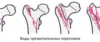
Classification of femur fractures according to AO (3)
| 31-A | 31-B | 31-C | ||||||
| extra-articular trochanteric fracture | extra-articular femoral neck fracture | intra-articular fracture of the femoral head | ||||||
| 31-A1 | 31-A2 | 31-A3 | 31-B1 | 31-B2 | 31-B3 | 31-C1 | 31-C2 | 31-C3 |
| pertrochanteric simple | pertrochanteric comminuted | intertrochanteric simple | subcapital with slight displacement | transcervical | subcapital not impacted with displacement | by type of split | with indentation | with a neck fracture |
| Extra-articular pertrochanteric simple fracture of the trochanteric zone of the femur | Extra-articular pertrochanteric comminuted fracture of the trochanteric zone of the femur | Extra-articular intertrochanteric simple fracture of the trochanteric zone of the femur | Extra-articular subcapital fracture of the femoral neck with slight displacement | Extra-articular transcervical femoral neck fracture | Extra-articular subcapital non-impacted fracture of the femoral neck with displacement | Intra-articular fracture of the femoral head by split type | Intra-articular fracture of the femoral head with depression | Intra-articular fracture of the femoral head with fracture of the neck |
| 31 - proximal segment | ||||||||
| 32-A | 32-B | 32-C | ||||||
| simple fracture | wedge fracture | compound fracture | ||||||
| 32-A1 | 32-A2 | 32-A3 | 32-B1 | 32-B2 | 32-B3 | 32-C1 | 32-C2 | 32-C3 |
| 32-A (1-3) . 1 = subtrochanteric fracture | 32-B(1-3). 1 = subtrochanteric fracture | 32-C(1-3). 1 = subtrochanteric fracture | ||||||
| spiral | oblique (>30°) | transverse (<30°) | spiral wedge | bending wedge | splinter wedge | spiral | segmental | wrong |
| Simple spiral fracture of the femur | Simple oblique femoral fracture | Simple transverse femoral fracture | Wedge fracture of the femur: spiral wedge | Wedge fracture of the femur: bending wedge | Wedge fracture of the femur: comminuted wedge | Compound spiral fracture of the femur | Complex segmental femoral fracture | Complex irregular femoral fracture |
| 32 - diaphyseal segment of the femur | ||||||||
| 33-A | 33-B | 33-C | ||||||
| extra-articular fracture | incomplete intra-articular fracture | complete intra-articular fracture | ||||||
| 33-A1 | 33-A2 | 33-A3 | 33-B1 | 33-B2 | 33-B3 | 33-C1 | 33-C2 | 33-C3 |
| simple | metaphyseal wedge | metaphyseal complex | sagittal lateral condyle | sagittal medial condyle | frontal | simple, metaphyseal simple | simple, metaphyseal comminuted | comminuted |
| Extra-articular simple hip fracture | Extra-articular femoral fracture: metaphyseal wedge | Extra-articular femoral fracture: metaphyseal complex | Incomplete intra-articular sagittal fracture of the lateral femoral condyle | Incomplete intra-articular sagittal fracture of the medial femoral condyle | Incomplete intra-articular frontal femoral fracture | Complete intra-articular simple fracture of the femur, metaphyseal simple | Complete intra-articular simple fracture of the femur, metaphyseal comminuted | Complete intra-articular comminuted fracture of the femur |
| 33 - distal segment of the thigh | ||||||||
Classification of tibia fractures: tibia and fibula according to AO (4)
| 41-A | 41-B | 41-C | ||||||
| extra-articular fracture | incomplete intra-articular fracture | complete intra-articular fracture | ||||||
| 41-A1 | 41-A2 | 41-A3 | 41-B1 | 41-B2 | 41-B3 | 41-C1 | 41-C2 | 41-C3 |
| tear-off | metaphyseal simple | metaphyseal comminuted | with condyle fracture | depressed | with condyle spallation with indentation | simple, metaphyseal simple | simple, metaphyseal comminuted | comminuted |
| Extra-articular fracture, avulsion fracture of the tibia | Extra-articular fracture of the tibia: metaphyseal simple | Extra-articular fracture of the tibia: metaphyseal comminuted | Incomplete intra-articular fracture of the tibia or fibula by splitting off the condyle | Incomplete intra-articular depressed fracture of the tibia or fibula | Incomplete intra-articular fracture of the tibia or fibula with condyle spalling and depression | Complete intra-articular fracture of the tibia: simple, metaphyseal simple | Complete intra-articular fracture of the tibia: simple, metaphyseal comminuted | Complete intra-articular comminuted fracture of the tibia |
| 41 - proximal segment of the tibia | ||||||||
| 42-A | 42-B | 42-C | ||||||
| simple fracture | wedge fracture | compound fracture | ||||||
| 42-A1 | 42-A2 | 42-A3 | 42-B1 | 42-B2 | 42-B3 | 42-C1 | 42-C2 | 42-C3 |
| spiral | oblique (>30°) | transverse (<30°) | spiral wedge | bending wedge | splinter wedge | spiral | segmental | wrong |
| Simple spiral fracture of the tibia | Simple oblique fracture of the tibia | Simple transverse fracture of the tibia | Wedge fracture of the tibia: spiral wedge | Wedge-shaped fracture of the tibia: bending wedge | Wedge-shaped fracture of the tibia: comminuted wedge | Complex spiral fracture of the tibia | Complex segmental fracture of the tibia | Complex irregular fracture of the tibia |
| 42 - diaphyseal segment of the tibia | ||||||||
| 43-A | 43-B | 43-C | ||||||
| extra-articular fracture | incomplete intra-articular fracture | complete intra-articular fracture | ||||||
| 43-A1 | 43-A2 | 43-A3 | 43-B1 | 43-B2 | 43-B3 | 43-C1 | 43-C2 | 43-C3 |
| metaphyseal simple | metaphyseal wedge-shaped | difficult | by type of split | by type of split and depression | comminuted and with depression | simple, metaphyseal simple | simple, metaphyseal comminuted | comminuted |
| Extra-articular metaphyseal simple fracture of the tibia | Extra-articular metaphyseal wedge fracture of the tibia | Extra-articular compound fracture of the tibia | Incomplete intra-articular fracture of the tibia, split type | Incomplete intra-articular fracture of the tibia by split and depression type | Incomplete intra-articular comminuted fracture of the tibia with depression | Complete intra-articular simple, metaphyseal simple fracture of the tibia | Complete intra-articular simple, metaphyseal comminuted fracture of the tibia | Complete intra-articular comminuted fracture of the tibia |
| 43 - distal segment of the tibia | ||||||||
Classification of ankle fractures according to AO (44)
| 44-A | 44-B | 44-C | ||||||
| subsyndesmotic fracture | transsyndesmotic fracture of the fibula | suprasyndesmotic fracture | ||||||
| 44-A1 | 44-A2 | 44-A3 | 44-B1 | 44-B2 | 44-B3 | 44-C1 | 44-C2 | 44-C3 |
| isolated | with a fracture of the medial malleolus | with a fracture of the posteromedial edge of the ankle | isolated | with damage to the tibia or deltoid ligament | with damage to the tibia or deltoid ligament and fracture of the posterior edge | diaphyseal fibula, simple | diaphyseal fibula, comminuted | with proximal fibula injury |
| Subsyndesmotic isolated fracture of the ankles | Subsyndesmotic fracture of the tibia with fracture of the medial malleolus | Subsyndesmotic fracture of the tibia with a fracture of the posteromedial edge of the malleolus | Transsyndesmotic isolated fracture of the fibula | Transsyndesmotic fracture of the fibula with damage to the tibia or deltoid ligament | Transsyndesmotic fracture of the fibula with damage to the tibia or deltoid ligament and fracture of the posterior edge | Supra-shaft simple fracture of the fibula | Supra-syndesmotic diaphyseal comminuted fracture of the fibula | Suprasyndesmotic fracture of the tibia with proximal damage to the fibula |
Proximal femur fractures (damage to the neck and trochanters)
In addition to the main causes (direct impact, road accident, fall from a height, etc.), even a banal tripping can cause injuries in older people, especially if they have osteoporosis and muscle weakness. The clinical picture is as follows:
- pain in the groin and hip joint, worsening with movement. With a trochanteric injury, the pain syndrome is so severe that the patient tries not to move at all;
- in case of fractures accompanied by displacement of bones, the injured leg becomes slightly shorter than the healthy one;
- the symptom of a “stuck heel” is the main sign: the patient cannot lift his straightened leg;
- with trochanteric fractures, swelling and hematoma appear at the site of injury, but if the femoral neck is damaged, they may be completely absent.
The diagnosis is made based on X-ray data, and for intra-articular injuries, an MRI of the hip joint is often additionally prescribed.
A.P. SKVORTSOV, P.S. ANDREEV, I.V. YASHINA, I.V. TsOY, R.F. KHASANOV
Republican Clinical Hospital of the Ministry of Health of the Republic of Tatarstan, Kazan
Yashina Irina Vladimirovna
traumatologist, department of pediatric traumatology and orthopedics
420064, Kazan, st. Orenburgsky Trakt, 138, tel. (843) 296-31-40, e-mail:
The authors developed and successfully used a monolateral external fixation device for the treatment of proximal femur fractures in children. A peculiarity of the treatment of this type of fractures using external fixation devices is that in the presence of a short proximal bone fragment, difficulties arise in creating stable osteosynthesis, which is achieved by the spatial arrangement of fixing elements (intraosseous rods). The developed design of the external brackets makes it possible to achieve rigid fixation of the proximal bone fragment on the support of the device, thereby creating stable osteosynthesis when performing both orthopedic and traumatological interventions on the femoral segment in children.
Key words:
monolateral apparatus, proximal femoral diaphysis, layout, transosseous osteosynthesis.
AP SKVORTSOV, PS ANDREEV, IV YASHINA, IV TSOY, R.PH. KHASANOV
Republican Clinical Hospital of the Ministry of Health of the Republic of Tatarstan, Kazan
Operative treatment of fractures and diseases of proximal diaphysis of thigh bone in children
The authors developed and successfully applied the composition of
monolateral external fixator for the treatment of proximal femoral fractures in children. The peculiarity of treatment of this type of fracture with the use of external fixation devices is that when there is a short proximal bone fragment difficulties occur in creating a stable osteosynthesis, which is achieved by the spatial arrangement of the fixing elements (intrabone rods). The developed design of outrigger brackets can achieve a rigid fixation of the proximal fragment of bone on the support of the apparatus, therefore, it creates a stable osteosynthesis in the performance of orthopedic and traumatologic procedures for femoral segment in children.
Key words:
monolateral fixator, proximal diaphysis of the thigh, composition, transosseous osteosynthesis.
In the treatment of diaphyseal femoral fractures in children and adolescents, monolateral external fixation devices are currently widely used. Their advantages include low weight and dimensions, speed of application with the possibility of postoperative additional correction of the position of fragments, and convenience for the patient in the postoperative period [1]. However, when treating fractures of the proximal diaphysis of the femur (trans-, subtrochanteric fractures) or when correcting the neck-shaft angle (CHA) with varus or valgus deformity of the femoral neck, one has to face certain difficulties in fixing the proximal femoral bone (FFA). This is explained both by the architectonics of the POBC and by the fact that we are dealing with a short proximal fragment, and for its stable fixation a “spatial scattering of fixation elements” is necessary (cited by G.A. Ilizarov). Orthopedists achieve this by installing two intraosseous rods on the proximal support of the device. In this case, two options for fixing the POBC are possible. The first is to place one rod strictly in the frontal plane near the fracture line (subtrochanteric fracture), and another intraosseous rod along the axis of the femoral neck (Fig. 1).
Picture 1.
Option for fixing the proximal femur with the creation of a “skew” in the frontal plane
The second option for fixing the FB is to place intraosseous rods along the axis of the femoral neck at an angle of 25-30° relative to each other in the frontal plane (Fig. 2).
Figure 2.
Option for fixing the proximal femur along the axis of the femoral neck with the creation of a “skew” of 25-30° relative to each other
The use of intraosseous rods placed in this way has shown to be effective in terms of creating rigidity of fixation and postoperative controllability of the proximal bone fragment [1]. For this purpose, two standard external brackets of the Ilizarov apparatus are used, connected to each other at an angle of 90° [2, 3]. However, the installation of an intraosseous rod on the proposed structure of assembled outrigger brackets is unstable, since there is a possibility of rotation of the outrigger bracket relative to the support, as well as their rotation relative to each other. This is observed when correcting the deformity of the trochanteric group (for example, when correcting the SDU), which is explained by the constant growing compressive forces of the muscles of the trochanteric group during the correction. Such spontaneous rotation leads to loss of distraction mode and premature fusion of fragments in the osteotomy area. Rotation of the brackets installed on the proximal support is also observed in the treatment of fractures of the proximal femur, which usually occurs during reposition of fragments. Thus, the use of known standard devices does not allow (adequately arising during reposition or distraction loads) the fixation of the intraosseous rod to the supports of the external fixation apparatus, according to the spatial location of the femoral neck (taking into account the antetorsion angle and the neck-shaft angle).
Clinical example
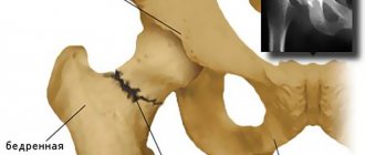
B-naya O-va, 11 years old, ist. disease No. 1767, was admitted to the pediatric orthopedics department on May 14, 2010 with a diagnosis of a closed subtrochanteric fracture of the left femur with displacement (Fig. 3).
Figure 3.
X-ray of patient O-voy, 11 years old, with a diagnosis: Closed subtrochanteric fracture of the left femur with displacement
Under combined anesthesia, the patient underwent PCOS of the left thigh in the operating room of the emergency department on the day of admission. The patient was placed on an orthopedic table, distraction was performed on the table, followed by X-ray control. Two intraosseous rods were inserted into the proximal fragment: one strictly in the frontal plane near the fracture line, the other along the axis of the femoral neck. The latter are mounted using two external rod fixators located on the proximal support.
On the distal support, intraosseous rods are inserted into the bone fragment perpendicular to the axis of the fragment with a rotational obliquity relative to each other (Fig. 4, 5).
Figure 4.
X-ray image of the b-nail after application of the device, direct projection
Figure 5.
X-ray image of the b-nail after application of the device, lateral projection
In this case, the position of the support is also perpendicular to the axis of the distal fragment. With respect to the bone fragments reduced on the orthopedic table, the supports should be located at the same distance from the latter; this is achieved by temporarily installing a threaded rod and correcting the position of the supports by moving them using special nuts with a spherical head. After the visual position of the threaded rod becomes parallel to the axis of the damaged segment, a second threaded rod is installed. X-ray control is done. By moving special nuts along the intraosseous rods, and, if necessary, rotating the bracket around its axis along radial teeth that contact the counter-shaped teeth located on the lower surface of the pedestal, the final reposition of the fragments is carried out.
Using this device, 20 patients with fractures of the upper third of the femur and 7 patients with varus deformity of the femoral neck were operated on. The undoubted advantage of the design used is the ease of installation of intraosseous threaded rods on the proximal support of the device, which not only significantly reduces the operation time, but also due to the achieved “spatial spread of transosseous elements” we obtain stable fixation of the proximal femur.
LITERATURE
1. RF Patent No. 2201168, A61B 17/66, BIPM No. 9, 2010
2. L.N. Solomin. Basics of transosseous osteosynthesis using the G.A. apparatus Ilizarov. - St. Petersburg, 2005. - P. 216 (Fig. 2, 4, 7).
3. Golyakhovsky V., Frenkel V. Guide to transosseous osteosynthesis using the Ilizarov method. — Moscow: BINOM ,
St. Petersburg: Nevsky dialect. - 1999. - P. 72 (Fig. 3-5).
conclusions
1. Ipsilateral fractures of the neck and diaphysis of the femur are a relatively rare injury in the structure of skeletal trauma due to the peculiarities of mechanogenesis; As a rule, they are observed in victims with polytrauma and represent a difficult problem in terms of diagnosis and treatment.
2. Mandatory radiographic examination of the hip joint area in patients with polytrauma with an active search for possible hidden injuries allows us to eliminate diagnostic errors.
3. The use of biomechanically sound osteosynthesis tactics based on a differentiated approach to assessing the characteristics of a fracture of the neck and diaphysis of the femur makes it possible to restore the motor and support function of the damaged limb.
Medical reference books
Femoral shaft fractures
Fractures of the femoral shaft include fractures whose line runs between points 5 centimeters distal to the lesser trochanter and 5 centimeters proximal to the medial epicondyle.
Epidemiology
Most often, fractures of the femoral diaphysis occur in young men (the result of exposure to great force - high-energy trauma) and elderly women (falling on the side, twisting - osteoporotic fractures).
Clinical anatomy
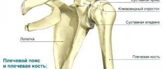
The femur is the largest tubular bone in the human body and is surrounded by a large array of muscle tissue. It must be remembered that the femur has a physiological curvature, bending anteriorly (antecurvation) and outwardly (varus), and the amount of curvature may vary among individuals. Compressive stresses prevail along the medial surface of the femur, and tensile forces due to the action of muscles prevail along the outer surface. The peculiarities of muscle attachment along the diaphysis of the femur determine typical displacements of fragments during fractures at different levels. For fractures in the upper third of the femur, the proximal end is flexed and externally rotated (action of the iliopsoas muscle, which attaches to the lesser trochanter), and also abducted (the gluteus medius and minimus muscles, which act on the greater trochanter). The distal end is shifted along the length by the traction of the biarticular muscles and inwards by the traction of the adductor muscles. In case of fractures in the middle third, the bone fragments are affected mainly by biarticular muscles and the most typical is displacement along the length. Due to the traction of the short adductor muscles, the proximal fragment moves slightly inward and posteriorly. In case of fractures of the femur in the lower third, the distal fragment, under the action of the two heads of the gastrocnemius muscle, which is attached to the condyles, is displaced posteriorly, and the proximal fragment is located anterior to it and somewhat inward. The shorter the distal fragment, the greater its angular displacement - up to a right angle with respect to the bones of the leg, which may cause compression or even disruption of the integrity of the neurovascular bundle in the popliteal fossa with acute disruption of the blood supply to the distal part of the limb. Therefore, the elimination of such displacement should be done urgently. The thigh muscles are divided into three main sections using fascial bridges:
- the anterior section contains the sartorius muscle, the quadriceps femoris and adductor minor muscles, the femoral artery, vein and nerve, the external cutaneous nerve of the thigh; - the internal section includes the gracilis muscle, adductor muscles (longus, brevis and major), external obturator muscle, obturator artery, vein and nerve, deep femoral artery; — the posterior section contains the biceps femoris muscle, the semitendinosus and semimembranosus muscles, a portion of the adductor magnus muscle, the sciatic nerve, the posterior cutaneous nerve of the thigh, and branches of the deep femoral artery. The blood supply to the diaphyseal part of the femur occurs mainly due to the deep femoral artery. One or two bone-feeding vessels penetrate the femur along the linea aspera, providing endosteal circulation. The periosteal vessels also penetrate the femur along the linea aspera and supply the outer third of the circumference of the femur. The remaining two-thirds of the circumference of the femur is supplied by endosteal vessels. As a result of most femoral fractures, the endosteal blood supply to the femur is disrupted and the primary bone healing process is provided by the periosteal vessels. Endosteal circulation is restored later. Reaming the medullary canal when installing an intramedullary nail can further disrupt the endosteal circulation, which requires 3-4 weeks to recover. To ensure the normal healing process of a femur fracture, damage to the periosteum should be avoided, especially along the posterior surface, where the vessels feeding it penetrate into the femur along the line aspera.
Mechanogenesis of damage
Diaphyseal fractures of the femur most often occur from direct trauma as a result of high-energy exposure (road traffic accidents, falls from a significant height, gunshot wounds). Less common is indirect injury - twisting of the hip (for example, during a wrestling competition). The percentage of pathological fractures is quite large, which most often occur at the level of the metaphyseal-diaphyseal junction. Any fracture of the femur resulting from inappropriate trauma should be considered pathological. Stress fractures, which occur in military personnel or runners, are rare; Typically, patients note an increase in the intensity of the training process on the eve of the onset of hip pain.
Clinical picture
Fractures of the femur, even closed ones, are accompanied by extensive damage to soft tissues, and therefore significant pain and blood loss (1000-1200 ml). These factors determine the frequent development of painful shock, and if this does not occur, then such patients should be considered shock-risk and should be given anti-shock therapy with adequate replacement of blood loss, especially during surgical treatment. The diagnosis of a femoral shaft fracture is usually obvious.
Clinical signs:
- passive position of the lower limb in external rotation of the distal fragment, - shortening of the femur compared to a healthy limb (up to 8-10 cm), - soft tissues at the level of the fracture are tense due to large hemorrhage, - due to shortening of the limb, folds of skin appear above patella, - muscle tone decreases, - mobility of fragments is expressed. It is imperative to check the pulsation of the arteries and the sensitivity of the skin on the foot. Fractures of the femur are often accompanied by damage to the ligamentous apparatus and menisci of the knee joint (up to 50% of cases), but assessment of their condition is possible only after stable fixation of the fracture.
X-ray examination
X-ray examination of the femur in two projections, hip and knee joints, plain X-ray of the pelvis. When assessing radiographs, it is necessary to determine the type of fracture, the quality of bone tissue, the presence of bone defects accompanying splintered injuries, air in the soft tissues, and the amount of shortening of the femur. Particular attention should be paid to the proximal femur (pertrochanteric and neck fractures), and the condition of the acetabulum should be assessed.
Classification
Based on location, fractures of the diaphysis are distinguished in the upper, middle and lower thirds of the femur, as well as isthmus, subisthmus and supracondylar. The nature of the fracture is transverse, oblique, splintered, with the presence of a butterfly-shaped fragment.
Classification of femur fractures according to AO/ASIF
32 – femur, diaphyseal segment.
A1 – simple fracture, spiral:
A1.1 – subtrochanteric region;
A1.2 – middle section; A1.3 – distal section. A2 – simple fracture, oblique (> 30°):
A2.1 – subtrochanteric region;
A2.2 – middle section; A2.3 – distal section. A3 – simple fracture, transverse (<30°):
A3.1 – subtrochanteric region;
A3.2 – middle section; A3.3 – distal section. B1 – wedge-shaped fracture, spiral wedge:
B1.1 – subtrochanteric region;
B1.2 – middle section; B1.3 – distal section. B2 – wedge-shaped fracture, wedge from flexion:
B2.1 – subtrochanteric region;
B2.2 – middle section; B2.3 – distal section. B3 – wedge-shaped fracture, fragmented wedge + detailing for all subgroups:
B3.1 – subtrochanteric region;
B3.2 – middle section; B3.3 – distal section. C1 – complex fracture, spiral + detailing for all subgroups:
C1.1 – with two intermediate fragments;
C1.2 – with three intermediate fragments; C1.3 – more than three intermediate fragments. C2 – complex fracture, segmental:
C2.1 – with one intermediate segmental fragment + detailing;
C2.2 – with one intermediate segmental and additional wedge-shaped fragments + detailing; C2.3 – with two intermediate segmental fragments + detailing. C3 – complex fracture, irregular:
C3.1 – with two or three intermediate fragments + detailing; C3.2 – with fragmentation in a limited area (<5 cm) + detailing; C3.3 – with widespread fragmentation (>5 cm) + detailing.
Detailing
B3: 1) spiral wedge; 2) wedge from bending. C1: 1) purely diaphyseal; 2) proximal diaphyseal-metaphyseal; 3) distal diaphyseal-proximal. C2.1: 1) purely diaphyseal; 2) proximal diaphyseal-metaphyseal; 3) distal diaphyseal-proximal; 4) oblique fracture lines; 5) transverse and oblique fracture lines. C2.2: 1) purely diaphyseal; 2) proximal diaphyseal-metaphyseal; 3) distal diaphyseal-proximal; 4) distal wedge; 5) two wedges (proximal and distal). C2.3: 1) purely diaphyseal; 2) proximal diaphyseal-metaphyseal; 3) distal diaphyseal-proximal. C3.1: 1) two main intermediate fragments; 2) three main intermediate fragments. C3.2: 1) proximal part; 2) middle section; 3) distal section. C3.3: 1) purely diaphyseal; 2) proximal diaphyseal-metaphyseal; 3) distal diaphyseal-proximal. General details: 7) bone defect; incomplete separation; 9) complete separation.
Treatment
Conservative treatment
Currently, it is limited to cases where there are contraindications to surgical treatment associated with concomitant pathology. For type A1 and A2 fractures without fragment displacement, fixation with a coxite plaster cast is possible for 8-10 weeks. 10-14 days after applying the bandage, X-ray monitoring of the position of the bone fragments is necessary to avoid secondary displacement. After removing the plaster cast, rehabilitation is 4-6 weeks (walking with crutches, and then with a cane). Depending on the level of the fracture, skeletal traction systems have their own characteristics. For fractures in the upper third, the wire is placed in the supracondylar zone of the femur. The limb is given a position of abduction by 30-40° (sometimes up to 100°-110°) and flexion at an angle of 50°-70°, and sometimes even up to 90° or more, which is due to the typical displacement of the proximal fragment under the influence of muscles. Initial load – 4-5 kg, setting load – 8-12 kg. For fractures of the femur in the middle third of the limb, the average physiological position is given. Elimination of displacement along the length is achieved by increasing weights, displacement in width is eliminated by adjusting loops. For fractures of the femur in the lower third of the limb, the knee joint is placed in a position of significant flexion (sometimes to a right angle), the foot is placed in a plantar flexion position. This position leads to relaxation of the calf muscle, which eliminates the active cause of the displacement. If the length of the fragment allows, the wire is passed through the femoral condyles; it is also possible to pass the wire through the tibial tuberosity. Skeletal traction can be used as preparation for surgery. Its purpose in such cases is to eliminate deformation and painful muscle spasms, and minimize acute bleeding. In such cases, the wires are passed through the tibial tuberosity and the calcaneus (traction by the femoral condyles can lead to inflammation of the soft tissue around the wire, which is undesirable in the segment where surgery is to be performed).
Surgical treatment
In most cases of femoral shaft fractures, this is the standard treatment. Surgery should be performed within the next 24 hours after injury. Early stabilization of a femur fracture is especially important for patients with multiple injuries.
Intramedullary fixation
It is considered the standard technique for treating fractures of the middle third of the femur.
The advantages of intramedullary rods, compared to plate fixation, include less traumatic surgery with less damage to the quadriceps femoris muscle, a relatively low percentage of purulent complications, and less stress in the implant. Closed reduction followed by extrafocal introduction of an intramedullary fixator reduces blood loss and preserves the periosteal blood supply to the femur. Reaming during installation of an intramedullary nail disrupts the endosteal circulation, but serves as a source of additional osteoinductive and osteoconductive material for fracture healing. Antegrade insertion of an intramedullary rod.
It is advisable to use a special radiolucent table with the possibility of skeletal traction and an electron-optical converter.
The patient can be positioned on his back or side. The latter position is more convenient for determining the site of insertion of the intramedullary rod, but is contraindicated if there is damage to the chest and lungs. The pyriform fossa of the femur has an advantage as a point of insertion of the rod, since it corresponds to the medullary canal of the femur. But the starting point on the greater trochanter is easier to identify. Use of the greater trochanter will require the use of a proximally valgus nail. The use of modern locking intramedullary systems does not require careful drilling of the medullary canal with the installation of a large diameter rod that is in intimate contact with the femur along its entire length. The role of reaming the medullary canal during intramedullary fixation is controversial. On the one hand, there are negative manifestations in the form of impaired endosteal circulation, increased intraosseous pressure and the risk of fat embolism, on the other hand, better stability of the implant, promoting bone fusion and increasing its strength. Blocking of intramedullary rods is necessary to prevent dislocation of fragments along the length and rotational displacements. The number of locking screws depends on various factors, including the comminuted nature of the fracture, its location, bone quality, weight, and activity level of the patient. Retrograde insertion of an intramedullary rod
is performed through the knee joint. The main advantage is the ease of determining the point of insertion of the intramedullary nail. Relative indications are: - the presence of other injuries of the same lower limb, such as a fracture of the femoral neck, pertrochanteric fracture, fractures of the acetabulum, patella, tibia; - diaphyseal fracture of the femur on both sides; - the patient is severely overweight; — fracture of the diaphyseal part of the femur after knee replacement; - fracture of the femur and the presence of a stump at the level of the knee joint on the same side.
Contraindications
— Range of motion in the knee joint is less than 60°. — Presence of an open traumatic wound.
External fixation with rod devices Advantages:
- can be completed in a short period of time - within 30-40 minutes;
— the blood supply to the femur fragments is minimally disrupted; — absence of foreign materials at the fracture site; — in cases of open fractures of the femur, extrafocal osteosynthesis with a rod apparatus can reduce the risk of suppuration; — controlled external fixation devices based on rods allow you to correct the position of bone fragments, as well as stimulate the process of fracture consolidation through compression or distraction. Disadvantages:
- infection of the soft tissue around the rods, sometimes leading to osteomyelitis; — limitation of movements in the knee joint associated with the passage of rods through soft tissues; — the need to care for the rod apparatus and constant medical supervision. Fixation with a rod apparatus can be used as temporary immobilization, followed by the use of other methods of surgical treatment, and can also act as a final method of stabilization.
Fixation with metal plates Advantages:
— the ability to achieve anatomical reduction of bone fragments;
— no additional trauma to the proximal and distal femur. Disadvantages:
- the size of the surgical approach, which is accompanied by blood loss, the risk of infection of the postoperative wound, damage to soft tissues, including the quadriceps femoris muscle, with a subsequent decrease in its strength and the possibility of developing myogenic contracture in the knee joint;
— disruption of the vascularization of bone fragments when installing the plate and, in the case of absolute stability of the fragments, “stress shielding” effect of the metal structure on bone tissue, that is, lack of stimulation of bone formation at the level of the fracture through dosed axial load with irritation of the periosteum; — since the metal plate takes on the main load, the percentage of implant fractures with the occurrence of refractures is high. Indications:
— the presence of a wide bone marrow canal that does not allow the use of intramedullary fixation; - previously existing femur fractures, fractures that healed in the wrong position; - other injuries of the same lower limb, such as a fracture of the femoral neck, pertrochanteric fracture, fractures of the acetabulum, patella, tibia; - presence of spinal injury; — adolescence (the use of intramedullary fixation is contraindicated due to existing bone growth zones); - pregnancy. It is possible to use both wide access and minimally invasive implant installation. The latter makes it possible to preserve the vascularization of bone fragments to a greater extent, but requires additional equipment (special operating table, electron-optical converter). Absolute stability of the fragments – absence of mobility at the level of the fracture under physiological load. Achieving absolute stability is necessary for all intra-articular fractures and only in some cases of diaphyseal fractures. The latter include simple fractures with transverse and oblique fracture lines, when static compression is achieved using a preload plate or lag screw (necessarily in addition to a neutralizing plate). The minimax principle is used - maximum stability with minimal fixation, but at least 4 screws are inserted into each main fragment (a biomechanical condition to achieve absolute stability). Relative stability of fragments – controlled mobility between fragments during physiological loading. Indicated in the case of an extra-articular location of the fracture zone, sufficient quality of bone tissue (normal or osteopenia), and in complex fractures with the presence of many fragments. General principles of bridge osteosynthesis: 1) 3-4 screws in each fragment; 2) the length of the fixator exceeds the length of the fracture zone by 2-3 times; 3) the absence of any screws in the fracture zone, as they will interfere with the controlled mobility of the fragments. Given the large length of the plate, it is necessary to use a minimally invasive technique, which involves preserving the periosteum with closed axillary insertion of the implant. Modern plates for such purposes are a submersible rod apparatus due to the possibility of blocking screws in the plate (LCP plates). Locking the screws allows a gap between the plate and the bone, which has a beneficial effect on blood circulation to the bone and healing of the fracture. In patients with polytrauma, intramedullary fixation with reaming of the medullary canal is contraindicated, as well as external osteosynthesis, which requires adequate surgical access and a sufficient amount of time. Extrafocal osteosynthesis is recommended using a rod-based external fixation device as a temporary or even final stabilizer. From 3 to 10% of femoral shaft fractures are accompanied by neck fractures, and in 30% of such cases, neck fractures are not immediately detected. To stabilize bone fragments, it is possible to use intramedullary rods (by ante- or retrograde insertion) or compression plates (in both cases, the neck is fixed with screws). Also used are an intramedullary nail with a femoral screw (Gamma-nail, PFN) or an intramedullary nail with an antirotation mechanism (PFNA). When using Gamma-nail, rotational instability of fragments of the femoral neck is possible; PFN and PFNA exclude such instability. In the first case, due to an additional derotational screw, in the second, due to the spiral shape of the blade fixing the neck. Ipsilateral distal femur fractures may appear as an extension of the fracture line from the shaft to the lower third or as a separate fracture. Fixation of fragments is possible using a dynamic condylar screw (DCH), an LCP plate for the distal femur (LCP DF), an intramedullary rod for fixation of the diaphysis and a plate for the distal femur.
Complications
Damage to the sciatic nerve is rare, since at the level of the femoral diaphysis the sciatic nerve is located in the thickness of the muscles and is not damaged by bone fragments. Most injuries result from traction or compression during surgical reduction. Damage to the femoral artery is possible at the border of the upper and lower third of the thigh, where the femoral artery enters the superior opening of the adductor canal, but more often occurs in the popliteal fossa, when the distal fragment is displaced posteriorly by the traction of the gastrocnemius muscle. Refractures (essentially, are secondary displacement of fragments in the absence of full fusion along the entire length). Patients are vulnerable at the early stage of callus formation and after removal of the metal structure. Nonunion is said to occur when the period after the injury is more than 6 months. This is usually due to impaired blood supply (wide detachment of the periosteum during surgery), infection, or smoking abuse. Evidence of the occurrence of a false joint is the appearance of end plates on bone fragments. Malunion usually includes varus deformity, external rotation, and/or longitudinal malalignment. Fractures of metal structures are most often associated with the use of plates. Heterotopic ossification with the formation of contractures in the knee joint.
Material and methods
In the period from 2002 to 2013, we monitored 179 patients with PSBC aged from 18 to 60 years. The criteria for selecting patients into this group were age under 60 years, sufficiently high physical activity of patients before injury, no history of chronic diseases of the endocrine, cardiovascular and respiratory systems, normal bone mineral density according to densitometry. In 107 (59.78%) victims the injury was of a high-energy nature, in 67 (37.43%) multiple and combined injuries were observed.
Ipsilateral fractures of the femoral neck and diaphysis were observed in 17 patients (9.5%). The age of the victims ranged from 22 to 55 years. Among them were 14 men aged from 22 to 50 years (mean age 43.3 ± 2.1 years) and 3 women aged 23, 40 and 55 years. In addition to ipsilateral fractures of the neck and diaphysis of the femur, 10 victims had other injuries to the musculoskeletal system and internal organs. The subject of the study was methods of reposition and osteosynthesis of cervical and diaphyseal fractures of the femur, timing of surgical intervention, factors determining the choice of fixators, clinical and radiological results of treatment.
Introduction
At the turn of the 20th and 21st centuries, the problem of femoral neck fractures (FCF) ceased to be predominantly geriatric [2]. The increase in the number of victims with proximal femur fractures worldwide is accompanied by an increase in the incidence of PFS among young people. And although HIPs account for only 2 to 6% of all fractures in the hip joint in people under the age of 60 years [2, 4], the general trend towards an increase in their number is due to an increase in injuries, as well as an increase in the upper limit of young age. Based on an individual assessment of physiological age criteria (high functional activity, good bone tissue, absence of chronic diseases), people under 65 years of age are classified as “young”. “Senile” age is considered to be over 75 years of age due to a decrease in the functional activity of patients, the presence of involutive osteoporosis and chronic diseases [4]. In young people, especially those under 50–55 years of age, PPHL rarely occurs as a result of a simple fall on the outer thigh. They are predominantly the result of high-energy exposure—a fall from a height (catatrauma) or a traffic accident [2].
The mechanogenesis of injury determines the presence of multiple and combined injuries in victims and a significant frequency of traumatic shock. According to George J. Haidukewych et al. (2004), among victims with PSBC aged from 15 to 50 years, severe multiple and combined injuries were noted in 43% of cases.
Osteosynthesis is recognized as the main method of treatment for PSCD in young people [7, 9].
The specific features of damage to the hip joint in cases of hip joint fracture at a young age require a differentiated approach to the choice of the method of internal fixation of fragments depending on the location, nature of the destruction of the neck, the degree of displacement of the fragments and the spatial orientation of the fracture plane according to Pauwels [5].
A separate problem is represented by ipsilateral (unilateral) fractures of the femoral neck and diaphysis (IPHF), which are observed in more than 20% of cases in this age group. At the same time, in 19–31% of victims, femoral neck fractures are diagnosed untimely.
In this regard, the issues of timely diagnosis of PSBC and the choice of optimal tactics for their treatment in patients with polytrauma become relevant. Literature data regarding the choice of a fixator, the sequence of stabilization of a diaphyseal or neck fracture, and the effect of forced late osteosynthesis of the neck on the incidence of avascular necrosis of the femoral head are contradictory.
The purpose of the work is to analyze the long-term results of surgical treatment of ipsilateral fractures of the femoral neck and diaphysis.