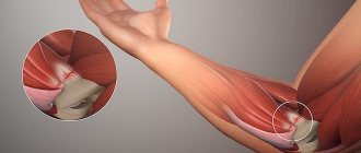
Treatment of tibia fractures is one of the most common tasks in the life of a traumatologist. Fractures of the tibia are most often high-energy, that is, they occur as a result of a fall from a height, traffic accidents, or twisting of the body around a fixed tibia (for example, when the leg is stuck between stones or is fixed by a ski boot and the body is twisted to the side). Low-energy fractures are possible in grandparents with osteoporosis. Most often, both bones of the leg are broken at once. Depending on the location where the bone is broken, fractures of the upper middle and lower third of the tibia can be distinguished; in addition, fractures of the articular ends forming the knee and ankle joints, ankle fractures, and so-called pilon fractures can be distinguished separately, but we will tell you about this in other articles.
Symptoms of a tibia fracture.
Symptoms of a tibia fracture are severe pain, the inability to step on the leg, and most often the inability to give your body any position other than lying down. In case of fractures of the diaphysis of the tibia (upper/middle/lower third), a deformation visible to the eye is often determined; sometimes bone fragments simply stick out under the skin or even perforate it, transforming the fracture into much more severe, open fractures. If you are assisting someone who, in your opinion, may have a broken leg, you should try to carefully fix it if necessary for transportation, since when moving the body, the fracture may no longer be closed due to the fact that the fragments damage the skin and meet the environment. Immediately after the fracture and also within 2-3 days, swelling will increase and a hematoma will appear, since the bone is well supplied with blood and a significant amount of blood will be released from the fracture into the surrounding soft tissue.
Causes
A fibula fracture occurs when a person hits something while falling, running, or jumping. The greatest number of injuries occurs in winter, while the tibia is damaged much less frequently. A fracture at the base of the lateral condyle can also occur if a person is hit in the leg with something, such as a stick or a kick during a fight.
Treatment of shin fractures.
Treatment of tibia fractures in most cases is surgical, since it is often not possible to achieve adequate comparison of bone fragments and avoid secondary deformation in the cast. In addition, long periods of immobilization (12 weeks or more) lead to the formation of contractures in adjacent joints and severe muscle atrophy, which significantly complicates subsequent rehabilitation.
Surgical treatment of tibia fractures most often proceeds according to one of the following scenarios: closed reduction, osteosynthesis with a locking pin, open reduction, osteosynthesis with a plate and screws, as well as treatment of open fractures with external fixation devices. It is possible to use several techniques simultaneously in combination or to sequentially move from one technique to another.
First aid
Correct actions before the ambulance arrives and hospitalization in the trauma department determine the intensity and nature of the recovery process.
If the victim is experiencing severe pain, the use of analgesic drugs will be required:
- Analgin;
- Paracetamol;
- Dolaren;
- Ketonal.
The lower limb must be placed in a stationary position to prevent bone displacement and soft tissue damage. For this purpose, it is necessary to apply an improvised splint to the sore leg. As available materials, you can use boards or sticks, which are laid on both sides and attached with a rope or bandage along the shin and thigh surface.
Clinical example.
Patient G., 56 years old, fell from a stepladder at the dacha, received a closed fracture of both bones of the right leg with displacement, and was hospitalized at K+31 by the insurance company. On the day of admission, a complete preoperative examination of the patient was performed, skeletal traction was applied, and he was hospitalized in the department.
On the same day, osteosynthesis of the tibia fracture was performed with a pin with locking, and the fibula with a plate and screws. Activated on the second day. On the third day he was discharged in satisfactory condition for outpatient follow-up treatment.
6 weeks after surgery, axial load on the limb is allowed. The fracture healed 12 weeks after surgery in a satisfactory position.
The patient is fully activated, walks without additional support without limping, and is not bothered by pain. The attentive reader will be able to notice
Recovery
Rehabilitation after a fracture of the tibia without complications should begin almost immediately after applying a cast. The patient is advised to move his toes and gently turn his foot. Magnetic therapy, laser therapy, electrophoresis, and physical therapy are also prescribed.
Once the patient is able to assume an upright position, it is necessary to begin walking with the help of crutches, but minimal load should be placed on the leg. If pain occurs, the patient is given anesthesia. After the cast is removed, a therapeutic massage and exercise in the pool are prescribed. Throughout the recovery period, the patient must adhere to a specially designed diet high in calcium.
Full recovery occurs in about six months if the person follows all the recommendations of the treating doctor.
Second clinical example.
Patient B., 62 years old, suffering from osteoporosis, suffered a fracture of both bones of the right leg in the lower third in January 2021. After a preoperative examination, osteosynthesis of the fracture with 2 plates was performed on the day of treatment. The patient was discharged 4 days after treatment.
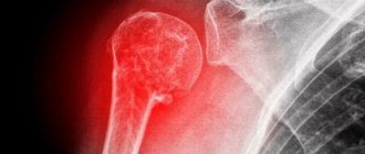
X-ray control after surgery.
X-ray control 3 months after surgery to determine the consolidation of fractures.
X-ray control 6 months after the injury, complete consolidation of the fractures is determined.
But our patient’s misadventures did not end there. Repeated injury, fall at home on the area of the right knee joint. Radiographs at presentation. A comminuted fracture of the upper third of the right tibia is determined.
After a preoperative examination, on the 2nd day after hospitalization, surgery was performed, open reduction, osteosynthesis of the fracture of the upper third of the tibia with a plate and screws. The patient was discharged on the 5th day after hospitalization in satisfactory condition.
The fractures have consolidated, the patient is being treated for severe osteoporosis by an endocrinologist.
Diagnostics
In order to make a diagnosis for which treatment will be prescribed, the doctor conducts a detailed examination, during which:
- Examines the site of injury for the presence of wounds, hematomas, swelling, and deformation of the limb;
- Interviews the victim, clarifying the circumstances of the origin of the injury;
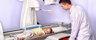
- Conducts an X-ray examination showing the type of fracture, as well as a computed tomography scan, which will show the condition of the ligaments, muscle tissue, blood vessels and joint capsules.
After the diagnosis is made, the victim is sent to the hospital surgical or orthopedic department.
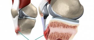
Ligament damage
The medial and lateral collateral ligaments, the anterior and posterior cruciate ligaments (ACL, PCL) take part in the formation of the knee joint. All injuries to these structures can be divided into partial and complete. A preliminary diagnosis can be made by studying biomechanical processes. Pain during supination (turning the leg outward) indicates damage to the medial collateral ligament. If the patient has problems with pronation (turning in on the inside), then the lateral collateral ligament may be injured.
Injuries to the ACL and PCL usually occur after direct impacts, hyperextension (overextension), and rotation of the hip with a fixed shin. Such injuries are typical for athletes (basketball players, wrestlers, track and field athletes). Other reasons that may cause a sprain or rupture of ligaments include carelessness in everyday life, road accidents, falls from a height. When an injury occurs, severe pain occurs.
A rupture of the ACL is accompanied by a click, which is not the case with the PVD. With such injuries, instability of the knee joint and a feeling of displacement when walking are observed. When visually assessing the patient's condition, swelling of the soft tissues in the knee area and hemorrhage inside the joint are observed. The last symptom may be absent if the posterior cruciate ligament is torn. The reason is that damage to adjacent structures - the posterior part of the knee joint capsule - is possible. Because of this, blood enters first into the popliteal fossa, and then into the space between the fascia. On palpation, the patient has a pronounced pain syndrome.
Injury to the medial and lateral collateral ligaments is accompanied by abnormal lateral mobility of the tibia, and the ACL and PCL are characterized by “drawer” displacement. During the acute phase, the patient is prescribed local anesthesia. When the sensation of pronounced pain disappears, instability of the knee joint remains. In order to avoid pathological displacements while walking, the leg should be wrapped with an elastic bandage. Over time, there is a gradual decrease in muscle mass due to atrophy, as well as symptoms of post-traumatic arthrosis.
When performing an X-ray of the knee, a narrowing of the joint space is observed. Using MRI, you can find out about the condition of the ligaments. Diagnosis is best done through a procedure such as arthroscopy. This is a minimally invasive diagnostic method for visualizing structures on a monitor screen. In addition to diagnostics, arthroscopy is also used to restore the integrity of ligaments. Tears are eliminated through conservative treatment. Arthrocentesis is also performed and a cast is applied for four weeks. Afterwards, the doctor may prescribe physical therapy and massage. If a ligament rupture occurs, it is repaired through surgery (stitching, plastic surgery). During the rehabilitation period, a number of physiotherapeutic procedures are prescribed for a speedy recovery.
Rupture of the connective tissue of the quadripses and part of the patella (the ligament itself) can occur with a strong blow, as well as with simultaneous overstrain of the thigh muscles and sharp flexion of the lower leg. When injured, sharp and intense pain manifests itself, and gait is disturbed (the leg bends reflexively). Blood does not enter the joint cavity. Palpation of the damaged area is accompanied by increased pain, there is no displacement and pain in the bones.
Complications and prevention
The most common adverse effects of injury are:
- malunion with leg deformity;
- soft tissue infection;
- stiffness and destructive processes in the ankle and knee joints;
- circulatory disorders due to pathological changes in blood vessels;
- neuropathy of the small or tibial nerves;
- thrombophlebitis of the veins of the lower extremities;
- thromboembolic complications.
Prevention of fractures should be carried out for patients whose bone tissue has increased fragility (osteoporosis, rickets). In order to increase bone strength, individuals at risk should be prescribed calcium supplements with vitamin D; women during menopause may require hormonal therapy with estrogen drugs to prevent osteoporosis.
During periods of ice and difficult weather conditions, older people should take safety precautions to prevent falls and possible injury.
Treatment of tibia fractures should be carried out by qualified specialists after a thorough diagnosis. The choice of treatment method depends on the nature of the damage. Active rehabilitation measures help patients fully restore motor function.
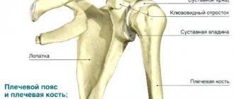
Anatomy of the tibia
The shinbones have a tubular structure, are very strong, and can withstand heavy loads. The upper part of the tibia has a triangle configuration with clear edges and 2 condyles (bulges). They contain articular surfaces that articulate with the femur and form a complex knee joint with the patella.
The articular surface on the lateral condyle articulates with the head of the fibula. The upper part of the tibia has a tuberous surface, which serves for attachment of ligaments and muscles of the limb.
Its lower section takes part in the formation of the ankle joint and forms the medial malleolus on the inside. The thin fibula in its lower section forms the lateral malleolus, which, like the head, protrudes outward and is easily palpated.