Today, a large number of diseases and pathologies of the musculoskeletal system (MSA) have been registered, which occur quite unnoticed, but are difficult to treat and often lead to irreversible consequences. These include spondylosis.
Spondylosis
is a chronic disease of the spine, the progression of which leads to severe thinning and significant wear of the tissues of the intervertebral disc, which causes the appearance of hernias and the formation of osteophytes.
In the absence of timely diagnosis and quality treatment, the disease can lead to vertebral fusion and loss of mobility.
Anatomical structure of the spinal column
The spine is a part of the skeletal system of the human body, consisting of vertebrae connected to each other by a large number of ligaments and cartilage. The dimensions, as well as a number of individual characteristics of the structural elements of the skeleton of the human body are determined by the department in which they are located. So, they distinguish:
- cervical
– 7 cervical vertebrae of relatively small size with openings in the transverse processes through which a channel for blood vessels (arteries and veins) runs;
- chest
– 12 vertebrae of relatively large size, increasing towards the bottom. On the transverse processes there are depressions into which the heads of the ribs enter, which makes it possible to limit the area of the chest of the human body;
- lumbar
– 5 massive vertebrae with a relatively small arch;
- sacral
– 5 vertebrae, which over time transform into a single bone (sacrum).
The key function of the spine is to support and protect the human body. However, in addition, there are also such functions as:
- supporting;
- protective;
- depreciation;
- motor.
Transformations of the structure of the spine during the natural maturation of the body
The spine of a newborn and an adult differs somewhat in a number of features, the most striking among which is the degree of flexibility (in infancy it has excessive flexibility, which does not allow one to maintain posture). In addition, as a person grows older (from birth to 25 years), the spine undergoes a number of consistent changes:
- the first stage (up to 7 years) - during this period, intensive growth is observed, which contributes to an increase in the volume and weight of the vertebral body and connective tissues. There is a gradual replacement of cartilage with bone, the arches fuse with the body and the natural curves of the musculoskeletal system are formed;
- second stage (7-15 years) – the mass of the vertebrae continues to increase, ossification nuclei are formed in their body, which arise in the thickness of the vertebral growth zone;
- the third stage (15-25 years) – thorough formation of the ossification nucleus, which leads to deterioration of flexibility and limited range of movements. The length of the spinal column gradually decreases, and flexibility and elasticity are significantly reduced.
Surgery for spondylosis
Surgical intervention is recommended if conservative treatment is ineffective and there is no improvement in the patient's condition. In addition, urgent surgery for spondylosis is indicated for:
- severe spinal canal stenosis;
- severe compression of the nerve root;
- large intervertebral hernias;
- violations of control over the functioning of the pelvic organs, including the genitals, bladder and rectum;
- paresis of the limbs;
- serious disorders of internal organs caused by the development and progression of spondylosis.
In such situations, depending on the root cause of the development of spondylosis, decompression and/or stabilizing operations of various kinds can be performed:
- microdiscectomy;
- laminectomy;
- endoprosthetics;
- autotransplantation.
Microdiscectomy
Microdiscectomy is the most commonly performed operation for spondylosis, during which a thinned intervertebral disc is removed microsurgically. It involves making a minimal incision to provide access to the spine. Its size does not exceed 3 cm. This allows you to significantly minimize surgical trauma, minimize the risk of developing postoperative complications and significantly facilitate and reduce the duration of rehabilitation.
The essence of the operation is as follows:
- the patient is put under general anesthesia;
- under the control of the image intensifier, a soft tissue incision is made in the projection of the affected spinal motion segment;
- the muscles are carefully moved apart and fixed in the desired position with special instruments, this eliminates the risk of damage and helps reduce pain in the early postoperative period;
- under the control of a special surgical microscope with 8x magnification, the neurosurgeon carefully pushes back the nerve root and, using miniature surgical instruments, removes the intervertebral hernia and, if necessary, the entire disc;
- a thorough sanitation of the surgical field is carried out followed by the installation of endoprostheses or autografts;
- suturing.
Microdiscectomy takes 45–60 minutes. The duration of hospitalization is 7 days, after which the patient is discharged from the hospital. At the same time, he always receives a list of recommendations on how to behave during the rehabilitation period, what medications to take and what physical procedures to undergo.
The outcome of the operation largely depends on the correctness of the recovery period. Therefore, it is recommended not to neglect medical recommendations and undergo rehabilitation under the supervision of a rehabilitation specialist.
Laminectomy
Laminectomy is an operation designed to eliminate compression of the spinal cord and nerve roots caused by various diseases. Elimination of high pressure is carried out through resection of the vertebral arches, spinous processes and intervertebral discs. Thanks to this, they stop compressing the spinal cord and the nerve roots extending from it, which leads to normalization of the transmission of bioelectric impulses and the elimination of neurological symptoms, including pain, sensory disturbances, and paralysis.
Depending on the severity of the situation, laminectomy may remove part or all of the compressive element. This creates additional space for the spinal structures, which ensures a sufficient decompression effect. Often, to maintain normal spinal mobility and physical capabilities of the patient after laminectomy, special stabilizing systems are installed.
Often this operation is combined with other surgical interventions on the spine, in particular the correction of its deformities. But laminectomy is a highly traumatic operation, since it involves excision of important structural elements of the spine.
There are several types of laminectomy. The specific one is selected depending on the reasons for the development of spondylosis:
- hemilaminectomy – removal of the arch of one vertebra on one or both sides while preserving the spinous processes;
- interlaminar - involves incision of the yellow ligament of the spine and removal of the arches of the most affected vertebra and neighboring ones;
- total – removal of the vertebral plates with the spinous processes.
Laminectomy involves making a soft tissue incision and skeletonizing the vertebrae of the affected segments under general anesthesia. The surgeon exposes the structures to be resected: arches, spinous processes, facets.
The most complex and time-consuming process is the skeletalization of the vertebrae of the cervical spine, since the tops of their arches are bifurcated and deeply embedded in the muscles.
During laminectomy, the fascia is often injured, which complicates the recovery period. The incision made is expanded with a retractor. The exposed spinal structures are cut with special forceps and removed from the patient's body. In each case, it is individually determined which fragments must be removed in order to eliminate spondylosis. In some cases, it is enough to remove only the osteophytes and install an endoprosthesis between the vertebral bodies; in other situations, especially with very advanced spondylosis, it is necessary to resort to a total laminectomy.
Endoprosthetics
Because with spondylosis, the intervertebral discs are usually so destroyed that they are completely unable to perform their functions, they are often completely removed. But this requires the introduction of an implant to close the resulting defect. For this purpose, it is preferable to use endoprostheses that help stabilize the operated spinal motion segment, maintain mobility and shock absorption within natural values. Today they are the best alternative to spinal fusion, i.e. complete fusion of the vertebrae.
Modern prostheses completely take over the functions of the removed disc. Their huge advantage is the elimination of overload in neighboring segments and reducing the risk of developing degenerative processes in them.
Today, implants are used for endoprosthetics:
- M6;
- Bryan;
- DCI;
- DePuy et al.
The most advanced are the M6 and Bryan models, which completely replicate the structure and structure of natural intervertebral discs. They are available in different sizes, which allows you to choose the most suitable prosthesis for a particular patient. Such devices are capable of qualitatively softening axial loads and providing mobility in 6 planes. Their compression and elongation indicators are not inferior to natural discs, which guarantees the preservation of normal biomechanics.
The operation is performed under general anesthesia immediately after removal of the intervertebral disc in one way or another. It takes 2–3 hours. Different types of prostheses have different installation techniques, with which the operating neurosurgeon must be thoroughly familiar. Such operations have a high level of security. With them, the risk of complications is no more than 1–2%.
You can usually move after surgery on the 5th day, and complete fusion of the endoprosthesis with the vertebral bodies is observed on average after 3 months. But for proper implantation of an expensive device, it is necessary to strictly follow the doctor’s recommendations and strictly adhere to the rules throughout the entire recovery period.
Autotransplantation
Decompression surgeries often leave behind defects that must be closed to maintain normal spinal height and support function. Sometimes, for this purpose, fragments taken from the patient’s own bones are used and placed into the resulting defect. Most often, part of the ilium is taken for these purposes.
Over time, the autograft takes root and firmly fuses with the remaining bone structures of the spine, i.e., spinal fusion is achieved. As a result, movements in the operated spinal motion segment become impossible, but when only one or two are fixed, patients usually do not notice a difference in the possible range of movements.
Nevertheless, whenever possible, spinal fusion is abandoned in favor of the installation of endoprostheses, since rigid stabilization of the spinal motion segment increases the risk of developing degenerative processes at adjacent levels of the spine. The fusion of two or more vertebrae makes significant changes in the biomechanics of the ridge, so after this often destructive processes begin or worsen in the osteochondral structures of the overlying and underlying segments.
The vertebrae can also be fixed in the desired position with metal structures, which are a set of plates and screws. As a result, spinal fusion is also achieved.
Thus, the prognosis largely depends on at what stage of development of the pathological process it was detected. With early diagnosis and the beginning of adequate treatment, the vast majority of patients manage to maintain the functionality of the spine, normal performance and continue to lead a normal lifestyle.
What is spinal spondylosis
Spondylosis is a degenerative chronic form of spinal damage caused by various factors, including:
- natural aging;
- abnormal load on the musculoskeletal system;
- ODA injuries.
The chronic form leads to deformation of bone surfaces, which often occurs in the absence of severe symptoms, but is accompanied by degenerative changes in the intervertebral discs.
The diagnosis is established on the basis of instrumental research methods of various types. Treatment is predominantly conservative.
Exercises
Sample exercises for lumbar spondylosis (each repeat 5-6 times):
- Lying on your back, arms straight along the body, bend your knees and pull them to your chest.
- Get on all fours, raise your head and slowly bend your lower back, returning to the starting position.
- The position is the same, raise your head and straighten your leg, stretching it back, then do the same with the other leg.
- The position is the same, lower your forearms to the floor, return to the starting position.
- Lie on your back, hands behind your head, bend your legs at the knees and pull them towards your stomach, clasp your knees with your hands and pull your head towards them, return to the starting position.
Causes of spinal spondylosis
Diagnosed cases, as well as the criteria for the clinical picture, show that the key reason for the occurrence of such an unpleasant disease is a metabolic disorder, which is closely related to the irrationality of physical activity or its complete absence. However, in addition to this, there are also a number of other important and significant reasons that are important not to be overlooked during diagnosis. Let's look at them in more detail.
Injuries
Suffered injuries to the musculoskeletal system often lead to bone growths - the formation of osteophytes. This is what often leads to spondylosis.
According to existing data, the integrity of the bone surface after a fracture is restored through the formation of a bone callus, however, there is a high risk that it will not close the defect, but will form a new one.
Special attention should be paid to microtraumas, which occur almost unnoticed, but at the same time entail quite serious consequences that are difficult to correct.
Loads of various durations
One of the key functions of the spine is support. The design of the spinal column and each individual vertebra is formed in such a way that it distributes the entire load, which became possible due to the ability of the disc to stretch and flatten. At the end of the effect, cartilage tissue is normally restored to its natural position and condition.
In a situation where the load is carried out for a long period of time, the area of contact between the vertebrae increases noticeably, which is realized through the proliferation of connective tissue. The longer the negative influence is carried out, the higher the chances that the compensatory reaction will transform into a pathological one.
Activation of inflammation
Local inflammatory reactions that can create optimally favorable conditions for the formation of osteophytes are also identified as one of the most likely causes of spondylosis.
Considering already diagnosed cases, it can be noted that the pathology is observed in the presence of a predominantly generalized process (for example, rheumatoid arthritis or tuberculosis).
It is worth noting that the disease itself does not belong to the category of inflammatory, but it is closely related to impaired blood supply and affects blood vessels or other soft tissues located in relative proximity to the spine.
Metabolic disorder
The development of spondylosis is based primarily on metabolic disorders, which certainly leads to a lack of nutrition in the tissues of the intervertebral disc. All this suggests that even in a situation where the initiation of harmful processes was preceded by injury, the risk of metabolic abnormalities exists and leads to degeneration of the elements of the musculoskeletal system.
Disorders of this type are observed in patients with diabetes, various degrees of obesity and, of course, acromegaly (disorder of the pituitary gland).
Lack of physical activity
The cause of spondylosis, such as low mobility, is often underestimated by specialists, however, it is among the most likely.
Detrimental processes of the degenerative-dystrophic type are directly related to the lack of nutrition of the discs, through which a large number of vessels pass. Prolonged physical activity of a static type leads to increased pressure on the tissue, and the lack of unloading (mobility) causes rapid wear of the disc and the formation of osteophytes.
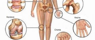
Which structures are subject to negative changes?
Degenerative age-related changes are most often observed in the following structures:
- intervertebral discs;
- facet joints;
- bone apparatus and ligaments.
Over time, as the body ages, gradual wear and tear of all body tissues occurs. The annulus fibrosus and nucleus pulposus of the intervertebral discs also undergo changes. They weaken, the fibrous ring wears out, and the amount of fluid in the nucleus pulposus decreases.
This negatively affects the shock-absorbing properties of the intervertebral discs. The result is a decrease in the height of the intervertebral discs, increasing the risk of hernia formation and deforming spondylosis.
Facet joints, which are present in each vertebral body. Thanks to these structures, flexion, rotation and extension of the spine occur. The shell of these joints is cartilage tissue. This is a connective type of tissue that contains lubricant and also has a sliding surface.
When age-related degenerative changes occur in the facet joints, the cartilage tissue gradually decreases in size and bone growths called osteophytes are formed. These degenerative changes can lead to osteoarthritis and osteoarthrosis, which are characterized by joint hypertrophy.
Bone spurs form near the edges of the intervertebral disc plates. As a result, the following negative changes are observed:
- deterioration of blood microcirculation in vertebral structures;
- compaction of the end plates due to sclerotic processes;
- thickening or compaction of bone structures under the end plates.
Ligaments consist of fibrous tissue fibers, which are the connecting link for the vertebrae. Thanks to them, the spinal column is protected from excessive load. With pathological changes in this area, a loss of ligament strength occurs, and this is fraught with a direct negative effect on the spinal structures.
Stages of spondylosis
Experts around the world diagnose not just spinal spondylosis, but its certain degree, which is determined by a set of criteria. So, today, it is customary to distinguish two large groups of stages of the pathology:
- X-ray;
- functional failure.
Considering the issue from a clinical point of view, radiological stages are considered more informative. They are among the basic information that a specialist uses when making a diagnosis. Let's consider the issue in more detail.
X-ray stages
First
– according to the results of an x-ray examination, a large number of bone growths are clearly visualized, which are limited to the boundaries of the vertebra. It is important to note that the height of the vertebrae and intervertebral discs remains unchanged.
Second
– X-ray images can reveal the presence of osteophytes that go around the intervertebral disc, going beyond its limits. There are cases when bone growths form a new joint.
Third
– osteophytes fuse into a single bracket and block the vertebra, which eliminates the possibility of movement in the selected segment of the spinal column and causes a narrowing of the canal.
Stages of functional failure
The stages under consideration reflect the patient’s ability to work, maintaining his ability to perform the simplest actions.
First
– a slight degree of change in the physiological curves of the spine leads to a slight limitation of mobility.
Second
– pathological changes limit a person’s ability to work, which often forces him to change jobs. This stage of pathology corresponds to the third group of disability.
Third
– the formation of a bone brace provokes a blockage of mobility, which sharply limits a person’s ability to work, which corresponds to the second group of disability. In a situation where bone fusion is pronounced, a person completely loses the ability to self-care and receives the first group of disability.
Causes and features of the development of spondylosis
The pathology is a direct consequence of degenerative changes in the intervertebral discs, i.e. osteochondrosis. Scoliosis, pathological kyphosis or lordosis, as well as the presence of certain systemic diseases increases the risk of its development. Thus, spondylosis can be partly called the final stage in the progression of osteochondrosis. Therefore, it is most typical for people leading a sedentary lifestyle, in particular, office workers and drivers.
Under the influence of various factors, biochemical changes begin to occur in the intervertebral discs, which gradually lead to a decrease in the percentage of water and proteoglycans in them. The consequence of such processes is the destruction of collagen fibers that form the fibrous membrane and a significant reduction in the shock-absorbing capacity of the disc.
At the same time, the tone and elasticity of the ligaments decrease and their fragility increases. As a result, the pressure of the vertebrae on the disc increases, especially strongly if there are concomitant pathologies of the spine, and it begins to flatten. In this case, the spinal roots inevitably suffer, which are compressed by the surrounding tissues. This leads to the development and constant progression of neurological symptoms.
As the load on the vertebral bodies increases and depreciation decreases, they begin to grow. This is how the body tries to compensate for the changes that have occurred and the remaining intervertebral disc. This begins the process of osteophyte formation. They can have a variety of shapes, and sometimes go around the intervertebral disc, taking it into a ring.
If you do not intervene in time, the osteophytes of neighboring vertebrae will fuse with each other and form a powerful bone bracket. As a result, the vertebrae will be firmly connected, which will completely eliminate the possibility of movement in the affected spinal motion segment, and will also lead to:
- injury to tendons and blood vessels;
- circulatory disorders;
- compression of the spinal roots or even the spinal cord itself;
- development of severe neurological symptoms;
- partial or complete paralysis.
Spondylosis can affect absolutely any part of the spine: cervical, thoracic, lumbar and lumbosacral. Most often, spondylosis occurs in the cervical and lumbar spine due to their high mobility.
Degrees of spinal spondylosis
In addition to stages, there are also degrees of development of chronic pathology, each of which is capable of characterizing the general condition of the patient and helps to form an idea of the characteristics of the course of the pathology.
There are 4 degrees of the disease:
- slowly progressive;
- moderately progressive with systematic exacerbation;
- rapidly progressing with rapid formation and growth of bone growths;
- lightning variant with a sharp and acute onset and an extremely unfavorable course.
It is important to pay attention to the fact that the uncomplicated variety is characterized by a slow course, in rare cases – moderately progressive. Other ailments appear as secondary, against the background of more severe pathological conditions.
The essence of the disease
The reasons for the development of spondylosis deformans have not been fully established. Some scientists consider the pathology as a destructive dystrophic lesion of the lumbar region, others - that the disease occurs due to a single injury or multiple repeated microtraumas of the longitudinal ligaments against the background of protrusion of the fibrous rings of the intervertebral discs. The rings lose their ability to withstand the pressure of the pulpy kernels. When exposed to significant loads, they bend and detach the longitudinal ligaments from the areas of their connection with the surfaces of the lumbar vertebral bodies. Factors that provoke the development of spondylosis deformans include:
- elderly and senile age;
- severe acute or chronic diseases of the joints and pathologies of internal organs;
- excess body weight;
- serious sports or professional physical activity;
- spinal injury - severe bruises, fractures, prolonged compression.
Osteophytes.
After the longitudinal ligaments are torn off, a slight hemorrhage occurs, and then a growth of bone tissue gradually forms. Repeated injury causes a new detachment with the formation of an osteophyte. At the same time, the size of the vertebrae does not change for a long time, and there is no decrease in their functional activity. This explains the long asymptomatic course of the disease. What happens as the pathological process progresses:
- bone growths compress the anterior longitudinal ligaments;
- mobility of the anterior spinal column is limited;
- in some cases, the intervertebral foramina narrows.
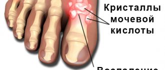
When conducting research, it was found that the development of pathology can be provoked by diseases occurring against the background of metabolic disorders. For example, gout, in which crystals of uric acid salts are deposited in bone and joint tissues, ligaments, and tendons.
The formation of osteophytes changes the anatomical structure of the anterior spinal column. Doctors consider the formation of bone growths to be a protective adaptive reaction of the body. In this way he tries to stabilize the spinal column. The presence of marginal bone growths prevents the deformed vertebrae from moving.
Symptoms of spondylosis
Among the symptoms of spondylosis, there are a large number of different conditions of the human body. It is customary to identify a general symptomatic picture, which suggests dominant or provoked disorders:
- local tenderness
– unpleasant sensations occur when turning the head, often in the first hours after waking up;
- limited mobility
– a feeling of stiffness caused by local pain syndrome is observed in the morning after waking up, however, in some cases (during the formation of a bone brace) it is constantly present;
- muscle overstrain
– the symptom is caused by a high concentration of lactic acid in the muscles, which causes pain and can lead to a sharp thickening and bulging of soft tissues;
- fainting
- a characteristic feature of the advanced stage. Fainting is short-lived and is preceded by a general decrease in well-being, nausea and weakness.
Cervical spondylosis
Cervical spondylosis can be caused by various reasons. There are cases where this type of disease progresses asymptomatically.
Symptoms of cervical spondylosis
When the cervical spine is affected, the following symptomatic signs are observed:
- recurrent or constant headaches
– observed more often after prolonged static stress, may be accompanied by dizziness and tinnitus. An important characteristic is the development of a symptom with an ophthalmological component (fogginess, flickering, etc.);
- pain in the back of the head
– provoked by turning the head, which provokes sprains and tension of the nerve roots;
- limited neck mobility
– observed in the morning, the severity depends on the extent of the affected area;
- muscle strain
– compensatory spasm that provokes pain.
Treatment of cervical spondylosis
The key direction of treatment is the use of conservative methods, which involves a combination of physiotherapeutic methods, taking medications and, of course, other means.
In the absence of contraindications, to improve the patient’s general condition, it is recommended to seek help from a manual therapy specialist. It would not be superfluous to visit exercise therapy (therapeutic physical education), as well as undergo a course of reflexology.
Prevention
It consists of eliminating the causes of lumbosacral spondylosis. It is impossible to defeat two of them, aging and heredity. Everything else can be adjusted.
To prevent the disease it is worth:
- Reduce body weight if it is excess;
- Eat right, eating dishes with gelatin that are healthy for the spine;
- Give the spine proper physical activity;
- Relax “clogged” muscles;
- Wear a supportive corset.
All these measures can not only exclude the occurrence of the disease, but also slow down its development if it is already diagnosed.
Thoracic spondylosis
Quite often in medical practice, thoracic spondylosis occurs, which also has certain characteristics.
Symptoms of thoracic spondylosis
Characteristic symptoms are:
- pain in the area of the affected spine
– unpleasant sensations localized between the shoulder blades, as well as in the zone of innervated intercostal nerves, caused by overstrain of the muscular frame, hernias, etc.;
- partial paresthesia (loss of sensation)
– the appearance of numbness, “goosebumps”, as well as tingling and even burning in the upper limbs and chest area;
- limited mobility
– turning or bending the body, as well as performing actions with the upper limbs is greatly limited or becomes completely impossible;
- disruption of internal organs
– the appearance of aching or pressing pain in the internal organs, tingling sensations are observed;
- tachypnea (fast and shallow breathing)
– more than 20 breaths in 1 minute;
- sleep disorder
– constant insomnia, long periods of falling asleep and inability to deep sleep.
Treatment of thoracic spondylosis
Drug therapy is actively used, individually tailored to the characteristics of the clinical picture of a particular patient.
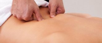
Massage, electrophoresis, exposure to magnets/laser, etc. can be prescribed as physiotherapeutic treatment.
Lumbar spondylosis
Lumbar spondylosis most often affects the sacral vertebrae, which indicates the need to use a more precise formulation of the diagnosis - sacrolumbar spondylosis.
Symptoms of lumbar spondylosis
The following symptoms are usually identified as manifestations of a disease localized in the sacrolumbar region:
- pain of varying intensity
– unpleasant sensations are concentrated in the lumbosacral region, but can also affect the coccygeal region;
- feeling of stiffness in the lower back
– significantly limits the mobility of the spinal column, which negatively affects the patient’s quality of life;
- partial or complete loss of sensation
– a symptom of this kind occurs due to compression (squeezing) of nerve fibers and manifests itself in numbness, tingling or even a local decrease in temperature;
- lameness
– pain while walking, forcing you to stop for short breaks, accompanied by pain in the calf muscles and a feeling of weakness.
Treatment of lumbar spondylosis
It is implemented using an integrated approach, including various types of physiotherapeutic procedures, attending massage sessions, taking medications, as well as interaction with a chiropractor and following the general recommendations of the treating specialist.
Traditional medicine recipes for spondylosis
Now that we have understood all the advantages and disadvantages of traditional medicine, here are some of the most effective recipes that can overcome spondylosis.
Important! Treatment with folk recipes should be supported by therapeutic exercises and proper nutrition. Only in combination with these measures can traditional medicine help overcome spondylosis or at least improve your condition.
- Herbal collection. This recipe includes birch leaves, burdock, sweet clover, knotweed herb, St. John's wort, black elderberry, nettle, peppermint, juniper, parsley, dandelion roots, sage, horsetail, fennel, violets, goldenrod. This herbal mixture has an anti-inflammatory effect,
To treat spondylosis at home, you can use herbal remedies, which act as an auxiliary therapy, help improve metabolic processes in the body, and also have a positive effect on cartilage and joints. This fee should be applied as follows: 1 tbsp. pour a spoonful of dry collection into 0.5 liters. boiling water and leave to infuse in a thermos for 7-9 hours. Then strain and drink hot two hours after meals, four times a day, half a glass. You should drink this decoction for two months, then take a break of 10 days. The course of treatment is 8-10 months; - Red elderberry extract (fruit) . This recipe is suitable for those who have joint deformation, osteochondrosis, spondylosis or arthrosis. It should be used externally, in the form of rubs (or compresses, as you prefer) twice a day. With a large degree of damage, the hood should be used more than twice a day and for a longer time;
- Horsetail juice. This juice is good because it contains a large amount of useful elements (zinc, silicon, iron), so its consumption guarantees improved joint flexibility and reduced pain. There is a contraindication: the juice is prohibited for people with acute kidney inflammation. Take the juice before meals (20-25 minutes before) three times a day, approximately 20-25 drops;
- Extract from comfrey rhizome. Another good anti-inflammatory remedy for those who suffer from diseases of the joints or spinal column. This extract should be taken three times a day, 35-40 drops. If you are worried about pain in a certain area, then the extract can be rubbed into the inflamed area.
Diagnostics
For complaints indicating similar symptoms, a set of diagnostic procedures is used using instrumental research methods. The main method is radiographic examination.
Based on the results of an X-ray examination, the presence of bone tissue growths can be effectively detected and the degree of progression of the pathology can be determined.
Differential diagnosis is carried out taking into account the clinical and radiological picture, taking into account the likelihood of the presence of third-party ailments and the most accurate identification of negatively influencing factors.
General principles of treatment of spinal spondylosis
Treatment of spondylosis is carried out on an outpatient basis. The key goal is to prevent the progression of the disease, eliminate inflammation, relieve pain and, of course, strengthen the patient’s muscular frame.
Features of drug therapy
The use of medications provides an opportunity to reduce the intensity of progression of destructive pathology. The drugs prescribed to patients include:
- NSAIDs (nonsteroidal anti-inflammatory drugs) –
- corticosteroids;
- muscle relaxants;
- psychotropic drugs;
- painkillers;
- chondroprotectors.
A particularly important role in the treatment of spondylosis is played by chondroprotectors, the use of which makes it possible to accelerate regenerative processes and promote the restoration of damaged tissues. One of the most frequently prescribed and very effective drugs in this group is considered to be Artracam, which can be used both in the treatment and prevention of musculoskeletal pathologies.
Treatment of spondylosis with folk remedies
Some people try to avoid medication treatment for various reasons. They can be understood: high prices for drugs, a large number of side effects, many drugs can only be obtained with a doctor’s prescription, and this is also not easy. Therefore, traditional medicine remains a priority alternative to medications.
Advantages and disadvantages of treating spondylosis with folk remedies
Did you know that...
Next fact
Any set of therapeutic measures has its pros and cons, which should be thought about in advance (before starting treatment). Traditional medicine is no exception.
| Advantages: | Flaws: | ||
| Low cost of medicinal components | In our age of rising prices and falling wages, this plus will be very significant. The plant components required for the preparation of ointments, infusions, and decoctions are quite cheap, so that almost everyone can afford them. | Long treatment period | For many doubters, this minus is decisive. Of course, we all want to get rid of the disease as soon as possible, but this does not happen. Traditional medicine requires patience and time; you should not expect a quick effect. But believe me, if you are persistent and patient enough, the results will pleasantly surprise you. |
| Low number of side effects, high safety of treatment | Many people are terribly afraid of all the side effects that medications can give (and rightly so). Sometimes, when you start treating one thing with medications, you cripple another, and you cripple it so much that it can no longer be corrected. Traditional recipes are as safe as possible for the patient. Of course, there are cases of allergies to certain components or individual intolerance, but this is not as scary as, for example, the “fatal” side effect of some antibiotic. | Not all herbal ingredients are compatible with each other | Treatment with folk remedies also needs to be approached wisely. As with medications, many herbal ingredients may be incompatible with each other. This incompatibility can lead to unpleasant consequences, so carefully read the description of all herbal ingredients before throwing them together into a boiling pot. |
| Efficiency and gentleness of treatment | Traditional recipes are good because they do not “shock” your body with one or another treatment program (such as hormonal drugs or blockades). Herbal preparations act gently, gradually affecting the disease and fighting it no less effectively. Plus, folk remedies strengthen the immune system, improve blood flow, tone the body, so they also have pleasant bonuses. | Incorrect dosage may cause immune problems | Yes, this fact occurs in the case of traditional medicine. Like any other treatment, it requires compliance with the dosage, so you should not think that if folk recipes are safe, then you can drink liters of decoctions for six months in a row and get nothing but recovery. |
| Time-tested | Unlike many medications, folk recipes have been tested by quality and time (more than once). | — | — |
Video: “Exercises for the prevention of osteochondrosis and spondylosis”
Massage and exercise therapy
Exercise therapy and massage are key therapeutic and preventive means that can help the patient improve their health and cope with existing problems.
Consistently regular, measured exercise therapy under the supervision of a specialist helps relieve pain and improve blood circulation. In addition, working on the condition of the muscle corset allows you to form correct motor stereotypes, which eliminates the possibility of overloading the affected part of the spine.
Do you want to feel healthy and fully enjoy life? Do not ignore the symptoms of pathological disorders of the musculoskeletal system, try not to neglect preventive measures and enjoy the moments of every day, and Artracam will help maintain the health of connective tissue and create optimal conditions for its replenishment!
Traditional treatment for spondylosis at home
Of course, if you have a problem such as spondylosis, then you should first consult a doctor to at least conduct an examination and find out all the details of your clinical picture.
The basis of traditional treatment for spondylosis is medications and physical therapy (physical therapy) . Massage, physiotherapy, and manual therapy are also sometimes used.
For home treatment, medication and therapeutic exercises are suitable.
Drug treatment for spondylosis
Many patients consult a doctor when they begin to experience pain in the back or cervical spine. In this case, non-steroidal anti-inflammatory drugs . It is advisable that a doctor prescribe them to you in order to avoid possible unpleasant consequences and side effects.
For spondylosis, NSAIDs are used, since the proliferation of osteophytes is caused by inflammatory processes in the spine. The following are standardly prescribed:
- Ibuprofen;
- Diclofenac;
- Ketonal;
- Indomethacin;
- Movalis.
The list of drugs may be shortened or increased, depending on the doctor’s recommendations and the patient’s individual clinical picture. These drugs relieve pain and alleviate the patient's condition.
In addition to NSAIDs, you can use analgesics (Ketoral, Plivalgin), glucocorticoids (Hydrocartisone, Kenalog); less often, blockades with novocaine or lidocaine are suitable for home use, since they are most often used in the form of injections, and this is not always convenient.
Doctors also often prescribe chondroprotectors (chondroxide, hyagin), vitamins (B1, B2, B6).
If the patient has a strong muscle spasm of the back against the background of an existing pain syndrome, which limits mobility and brings discomfort, you can also add muscle relaxants (Mydocalm, Zanaflex) to the list of medications.
All medications should be taken with great caution and it should be understood that it is advisable to use all this “assortment” under the supervision of a doctor. Many drugs can have serious side effects, so caution is a good idea.
Therapeutic exercises for spondylosis
Therapeutic exercise is an integral part of the preventive and therapeutic course for diseases of the spine . But you need to keep in mind that exercises are started only when the acute stage of the disease has already been overcome and there is no severe pain or spasms. We advise you to consult with a specialist before starting the complex.
Here are some simple but effective exercises that will help improve blood flow, tissue nutrition, strengthen the muscle corset and spine, and help improve the flexibility of the spinal column.
Gymnastics should be started with a small number of approaches (1-2) and repetitions (2-3) . Once your spine and muscles are a little stronger, you can increase the number of repetitions and approaches. (4-5 sets of 8-10 repetitions).
Important! The exercises must be performed with great care, slowly, without overloading the spine and muscles. If you feel unwell, pain, or spasms, you should immediately stop doing the exercises and consult a doctor.
Exercises for vertebral deformities are designed to improve blood circulation and mobility of the spine
- Place your hands on your forehead. Press your palms onto your forehead, while offering resistance with your hands. The same exercise should be repeated with your hands on the back of your head;
- Stand up straight, hands at your sides. As you inhale, carefully and smoothly lift your arms out to your sides, squeezing your shoulder blades together. Stretch your arms as high as possible, and then, as you exhale, begin to slowly lower your arms to the starting position;
- Stand up straight, straighten your shoulders, lean on the back of the chair with one hand. From this position, swing your legs back and forth. Try not to bend at the lower back;
- Lie on your stomach, stretch your arms forward, keep your legs closed. From this position, begin to simultaneously raise your legs and arms up to your maximum, hold in this position, and then return to the starting position. Do all movements slowly and carefully, inhale as you rise, as you exhale descend;
- Lie on your stomach, place your hands on the back of your head. From this position, begin lifting only the upper body. At the same time, your legs should be pressed tightly to the floor, your eyes should look at the floor.
Find out more about the development of spondylosis in different parts of the spine:
- What is thoracic spondylosis and how is it treated?
- You can learn about the symptoms and treatment methods for spondylosis in the cervical spine here
- More about lumbar spondylosis on the page
- What is spondylosis deformans?