Osteoarthritis deformans or osteoarthritis (DOA) is a chronic joint disease. In this case, a gradual destruction of the articular cartilage occurs, leading to changes in the surfaces and deformation of the joint itself.
ICD 10 code: M15—M19 Arthrosis
- M15 Polyarthrosis.
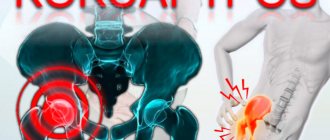
- M16 Coxarthrosis (arthrosis of the hip joint).
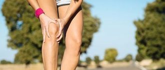
- M17 Gonarthrosis (arthrosis of the knee joint).
- M18 Arthrosis of the first carpometacarpal joint.
- M19 Other arthrosis.
There are also other names for the disease that are synonymous, according to ICD 10 code: arthrosis deformans, osteoarthrosis, arthrosis, osteoarthritis.
The term “deforming osteoarthritis” is more often used in foreign terminology.
Some statistics
Deforming osteoarthritis (osteoarthritis) is the most common joint disease. Its symptoms occur in 20–40% of the world's population, depending on the region. Women are almost twice as likely to get sick. With increasing age, the number of sick men and women becomes approximately the same. Although the disease sometimes occurs in young people, however, it is still the lot of the elderly: among people over 50 years old, almost half are sick, and by the age of 70 – already 80–90%.
The hip joint is most often affected - about 42% of cases, followed by the knee - about 34%. The “top three” is closed by damage to the shoulder joint – in 11% of all DOAs. The share of lesions of other joints accounts for about 13%.
How is coronavirus treated?
There is no specific treatment today: measures are usually symptomatic. The patient takes medications to reduce fever, cough and runny nose, and the body will have to fight the infection on its own. Of course, having a history of arthrosis of the knee joint of the third degree, this will be much more difficult, so it is important to take preventive measures and prevent infection as much as possible.
In severe cases, antiviral drugs, corticosteroids, and specific immunoglobulins are added to the treatment regimen. Sometimes antibiotics are necessary or artificial ventilation is not necessary. There is a positive experience of administering blood plasma to seriously ill patients from people who have recovered from SARS.
There is no vaccine for coronavirus
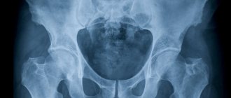
Joint structure
Deforming osteoarthritis (osteoarthritis) causes disruption of normal joint function, which often leads to disability. To understand the pathological processes that occur during the development of this disease, it is worth delving a little into the anatomy of the joint.
Joints are located in those places of the skeleton where distinct movements occur. By the way, there are 360 of them in the human body.
The classification of joints is quite diverse, but we will not dwell on it. It is important to note that joints are always formed by at least two bones - then they are called simple. These include, for example, the shoulder. There are complex joints formed by three or more bones (elbow, knee, etc.).
Articular cavity
The joint is covered by an articular capsule or capsule, which forms its cavity. There are two shells in it: outer and inner. The outer shell has a protective function; ligaments are often attached to it. The inner one has a special layer (synovial membrane), which secretes the so-called synovial fluid. Due to this secretion, the joint is nourished, its surfaces are moisturized and friction is reduced.
Symptoms of coronavirus infection
At the initial stage, coronavirus resembles an acute respiratory infection or the flu. A person may have:
- fever and chills;
- pain when swallowing and sneezing;
- runny nose;
- dry cough;
- headache;
- hypoxia;
- muscle pain.
Some patients notice signs of conjunctivitis or diarrhea. In mild cases, the temperature may remain within normal limits. In severe cases, breathing becomes more difficult, and a persistent non-productive cough intensifies. If a person is undergoing treatment for osteoarthritis or another chronic disease, the condition worsens rapidly, since the body is already exhausted.
With arthrosis, any disease is more severe and more likely to cause complications
How does DOA develop?
Deforming osteoarthritis (osteoarthritis) begins to develop with damage to the articular cartilage. For a long time, the disease goes unnoticed by both the patient and doctors. The appearance of vivid symptoms occurs already with significant tissue damage.
Etiology
The causes of the disease are still not fully understood. Several factors are involved in the development of the disease, of which two dominant ones are identified: excessive mechanical and functional overload of the articular cartilage and a violation of its resistance to normal loads. As a result, pathological degeneration and destruction of articular cartilage occurs.
Risk factors for the formation of DOA:
- There are several risk factors that can contribute to the development of articular cartilage damage and the development of osteoarthritis. These include:
- Hereditary predisposition.
- Overweight, obesity.
- Endocrine and metabolic disorders (for example, lack of estrogen in menopause).
- Professional, sports or household overloads on joints (chronic microtraumatization).
- Various injuries.
- Age from 50 years.
- Concomitant inflammatory and non-inflammatory joint diseases.
Articular cartilage changes
The cause or trigger for cartilage destruction is usually trauma or prolonged microtrauma of the articular surfaces. Also, the onset of the disease can be caused by a change in the congruence (compliance) of the articular surfaces as a result of any pathological processes, for example, dysplasia.
Articular cartilage changes its properties, loses elasticity, becomes rough, and cracks appear on it. This increases the load on the surface of the bones that form it, and their integrity is disrupted.
What happens next?
In order to stabilize the joint, cords formed by connective tissue appear inside it. The amount of synovial fluid, which has a changed composition, increases.
Subsequently, bone growths - osteophytes - form at the edges of the joint. The muscles surrounding the joint become hypotrophied and decrease in size. This leads to further circulatory disorders and aggravation of pathological processes occurring in the joint - the development of contractures (stiffness) and its instability.
Symptoms
The disease does not show itself for a long time. And those few symptoms that may appear in the early stages usually go unnoticed.
Deforming osteoarthritis (osteoarthritis) has several characteristic symptoms that are present in almost all patients.
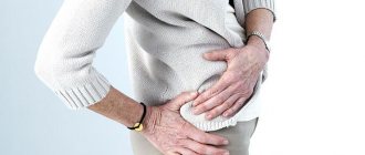
Pain
A complaint of pain in the affected joint is the main reason for visiting a doctor. At the beginning of the disease, it is insignificant and can only occur during walking or physical activity. Pain can also appear when exposed to unfavorable factors, for example, hypothermia or prolonged forced uncomfortable body position.
Gradually, the pain becomes constant and its intensity increases. A characteristic feature of such pain is that at rest its intensity decreases, until it disappears.
Stiffness
In the early stages of DOA, most patients experience a feeling of “stiffness” in the morning. This condition is characterized by a decrease in the range of motion in the joint, decreased sensitivity and pain of varying intensity. With movement, this condition gradually passes.
Crunch
The presence of extraneous sounds - crunching, clicking in the joint, which first appear periodically under unfavorable conditions (long walk, forced position of the body or limb, etc.). Over time, these sounds accompany any movement of the affected joint.
Instability
This symptom is often expressed in the joints of the limbs. Manifests itself as pathological excessive mobility. Mobility in a plane uncharacteristic for joint movement also occurs. There is a decrease in the sensitivity of the limb.
Movement disorder
Deforming osteoarthritis (osteoarthritis) is manifested by a violation of the main function of the joint - movement. Such disorders can be in the form of limited mobility, this is especially pronounced during exacerbation of the disease. Also sometimes there is an increase in mobility - “looseness” of the joint, which is associated with muscle wasting or damage to the ligamentous apparatus.
As the disease progresses, the restriction of movement in the affected joint becomes persistent, contractures occur, and the function of the limb is impaired.
Limb dysfunction
Pathological changes in the joint over time cause dysfunction of the entire limb. There is lameness when walking, limited movement, and a feeling of joint instability. The limb becomes deformed, and as a result of impaired blood supply, sensitivity disorders and other changes occur (a feeling of chilliness or, conversely, burning, coldness of the limb, etc.).
All this ultimately leads to disability.
Other symptoms
In addition to the above, there are less common complaints, mainly of a “cosmetic” nature. These include:
- Decrease or increase in limb circumference.
- Swelling of the joint.
- Presence of fluid in the joint.
- Deformation of a joint or limb.
- Changes in the skin of the limb: increased vascular pattern, pigmentation, etc.
How can you get infected?
You can become infected with the new coronavirus infection from an animal or a person. The danger comes from wild and domestic animals, birds, snakes, and in the case of the new strain, bats. The virus is transmitted through the air, as well as through the fecal-oral route, through contact with nasopharyngeal secretions, vomit, and waste products.
The incubation period is from 1 to 14 days, with an average of 10 days. These days there are no clinical manifestations, however, the person is already a source of infection. This is the main insidiousness of this new strain.
What is the lethality of coronavirus? What is the main danger and why are older people still at risk? An infectious disease doctor says:
Treatment
Treatment of DOA is aimed primarily at improving the patient’s quality of life. This is achieved by reducing pain, restoring impaired joint function and limiting the further development of the disease.
All treatment methods can be divided into three groups:
- Non-medicinal.
- Pharmacological – taking medications.
- Surgical – surgical interventions.
Non-drug treatments
Before treating this disease with medications and other methods, it is necessary to understand that if you follow simple rules in everyday life, a fairly stable remission (weakening or disappearance of symptoms) is possible.
Normalization of body weight
To reduce the load on the joints, the treatment plan must also include measures to reduce excess body weight, which is present in most patients with DOA. This is achieved by a low-calorie diet, special physical exercises, massages, etc.
Security mode
First of all, it is necessary to observe the protective regime. This implies the elimination of factors that influence the development of the disease - trauma (including microtrauma, for example, at home), hypothermia, excessive physical activity.
Load reduction
It is necessary to reduce the load on the affected joint. In the acute period of the disease, when the knee or hip joint is affected, canes or crutches are used. And when the shoulder joint is damaged, immobilization is created using removable plaster, plastic and other splints. Traction can also be used with the help of special devices, which is carried out in a hospital.
In case of DOA of the leg joints, special attention should be paid to shoes, which should be correctly selected and use special (orthopedic) insoles.
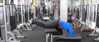
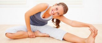
Therapeutic exercise (physical therapy)
The use of special therapeutic physical exercises is invaluable in the treatment of DOA of any location. Exercises must be performed every day. However, performing physical therapy should not bring pain and discomfort.
A set of exercises is usually selected by a doctor taking into account the patient’s physical condition. It includes activities aimed at improving general condition: walking at a moderate pace on a flat surface, swimming in the pool, training on special exercise equipment.
It is also necessary to train and strengthen specific muscle groups. For example, with DOA of the shoulder joint, these are the muscles of the shoulder girdle and upper limb.
Taking medications
The range of medications used in the treatment of DOA is quite extensive. This treatment is used for exacerbations of the disease or in the presence of inflammatory complications - synovitis.
- Medicines that reduce pain. These include various painkillers used in normal dosages.
- Improving blood supply to the affected area and stimulating metabolic processes in it. Drugs that improve microcirculation (drotaverine), improve blood viscosity (chimes), have antioxidant effects (vitamins C, E, B) and other medications are used.
- Non-steroidal anti-inflammatory drugs (NSAIDs) - diclofenac, ibuprofen, indomethacin and others. The use of these drugs has a direct effect on the mechanism of development of the disease, and also has an additional analgesic effect.
- Glucocorticoids are used when other medications are ineffective and their use is limited.
- Chondroprotectors are drugs that stimulate metabolic processes in articular cartilage. Used outside of exacerbation.
A combination of several medications is often used, for example, a combination of taking NSAIDs with chimes.
The routes of drug administration can be different: orally (tablets), injection forms - intramuscular, intravenous. Sometimes - intravenous drip infusions (chimes). If required, the medication is injected into the joint cavity.
Outside of exacerbation, physiotherapeutic and sanatorium treatment is used.
Osteoarthrosis (Deforming arthrosis)
Osteoarthrosis (arthrosis deformans) is a chronic disease of the joints of a degenerative-inflammatory nature, which begins with damage first to the cartilage covering the articular surfaces of the bones, and then to the underlying bone tissue itself, the development of bone growths in the joint - osteophytes, and in later stages - persistent deformation and stiffness of diseased joints. This disease can be found under the names “deforming arthrosis”, “osteoarthrosis”, “osteoarthritis”, but, according to the International Classification of Diseases, 10th revision (ICD 10), these are synonyms.
Pathological changes in deforming arthrosis
Deforming arthrosis ranks first in frequency among joint diseases and is the main cause of disability and disability in middle-aged and older people. The disease significantly impairs the quality of life, causes constant and intense pain, and significantly limits the mobility of the affected joints.
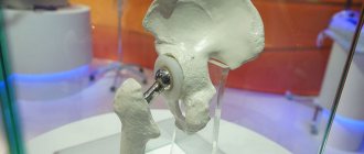
The most severe localization is considered to be damage to the hip, knee, and ankle joints, since the loss of function of these particular joints leads to disability and the need for surgical endoprosthetics - replacing one’s joint with a metal prosthesis.
However, one should not wait for the development of severe irreversible pathological changes in the joint - it is necessary to begin treating deforming arthrosis immediately after the diagnosis is established. And only if complex conservative treatment turns out to be ineffective, the question of surgery is raised.
Causes and risk factors
Deforming arthrosis can be primary or secondary. Primary - diagnosed when there are no obvious reasons for the development of changes in the joint. In this case, the main role is given to genetic predisposition and the presence of risk factors. Secondary osteoarthritis develops as a result of another pathology - for example, post-traumatic arthrosis (after an injury), osteoarthritis against the background of congenital defects of the musculoskeletal system.
Risk factors for developing osteoarthritis:
- Age over 40 years;
- concomitant osteoporosis;
- excess body weight;
- professional sports;
- hard physical labor;
- endocrine diseases and metabolic disorders;
- congenital and acquired diseases of bones and joints;
- previous joint surgeries (for example, for a damaged meniscus);
- hereditary predisposition;
- inflammatory diseases of the joints (arthritis). The disease develops slowly. At the very beginning, those joints that are subject to maximum load suffer, and then others can gradually retract.
Common signs of osteoarthritis:
- Joint pain occurs or intensifies after working or physical labor and goes away at rest. Periodically, acute pain syndrome may occur, which is caused by blockade of the joint by a free intra-articular body (an osteophyte fragment, a piece of destroyed cartilage tissue). As the disease progresses, the pain becomes constant and more intense;
- A crunching sensation when moving the joint (crepitus);
- Joint instability, periodic “wagging” of the legs when the joints of the lower extremities are affected;
- Difficulty walking up stairs;
- Development of various types of deformities;
- Restricted mobility in joints, decreased range of motion;
- From time to time, inflammatory complications—synovitis—occur.
There are 3 degrees of deforming arthrosis:
- Arthrosis of the 1st degree is characterized by a slight limitation of mobility in one direction for the diseased joint; radiographs reveal small osteophytes at the edges of the joint and a moderate narrowing of the joint space.
- Arthrosis of the 2nd degree is characterized by limited mobility in the affected joints, crunching during movements, significant osteophytes and a pronounced narrowing of the gap between the articular surfaces of the bones.
- Arthrosis of the 3rd degree is characterized by significant deformations of the joints, their forced position, a sharp limitation of mobility, the development of ankylosis with the complete disappearance of the joint space.
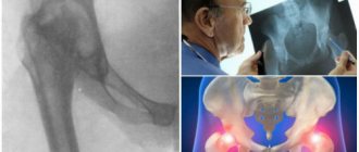
Diagnostics
Diagnosing osteoarthritis is not difficult. A thorough examination by a surgeon or orthopedist, supplemented by X-ray examination, ultrasound of the joint, or the most modern and informative method that allows you to see the condition of both cartilage and bone tissue of the joints - MRI, is sufficient. Typical patient complaints, the presence of risk factors for osteoarthritis, together with an X-ray and/or MRI examination, make it possible to quickly and accurately diagnose the disease and its stage.
Treatment methods
- Treatment for osteoarthritis should begin immediately after diagnosis, and the sooner this happens, the better the result.
- Conservative treatment of osteoarthritis cannot consist of one or two procedures or the intake/administration of one or two medications, but must be comprehensive - using the entire modern arsenal of medications, manual procedures, physiotherapeutic treatment, massage, exercise therapy. Only such a comprehensive program makes it possible to avoid joint replacement surgery!
A comprehensive course of treatment for stage I-II-III osteoarthritis of the knee joints at the OsNova Clinic consists of:
- Initial consultation with a surgeon.
- Determination of body weight and body mass index.
- X-ray + ultrasound (or better yet, MRI of joints).
- If you are overweight or suspect/presence of an endocrine disease, consult an endocrinologist (especially women). If necessary, the endocrinologist prescribes additional examination and treatment.
- Blood test for CRP, rheumatoid factor (to exclude rheumatic diseases).
- If indicated, consult a rheumatologist.
IN THE PRESENCE OF SYNOVITIS:
- If there is effusion in the joint cavity, evacuate the effusion.
- For stages I – II – III of osteoarthritis , depending on the severity of the clinical manifestations of the inflammatory process: prescribing a course of NSAIDs orally or intra-articular administration of anti-inflammatory drugs - relieves pain, restriction of joint mobility for 2-4 hours after administration. The duration of therapeutic action can be 4 or more weeks.
Attention: intra-articular punctures are performed only by a surgeon or orthopedist with the appropriate skills and experience.
- To immobilize the joint - prescribing and selecting an orthosis (a special medical orthopedic device to reduce the load on the diseased joint).
NEXT, OR IN THE ABSENCE OF SPECIFIC SYNOVITIS - IMMEDIATELY:
- Intra-articular administration of chondroprotectors : for example, alflutop intra-articularly into each joint with an interval between injections of 3-4 days. The course of treatment is 5-6 injections into each joint.
- Intra-articular injection of hyaluronic acid preparations . Hyaluronic acid preparations (Otsenil, Femartron, Gilan, Synvisc, Duralan, etc.) are injected into the joint, thereby creating a thin protective film that prevents the cartilage surfaces from rubbing against each other. Injections are carried out only after the acute phase of the disease has passed. As a rule, injections are given several times (but no more than 5) with a break between procedures of 1-2 weeks. After six months, the course can be repeated.
- Manual therapy for arthrosis is joint traction. Gives especially good and lasting results when combined with chondroprotectors and intra-articular injections. Patients with arthrosis should undergo 3-4 courses of manual therapy annually with 3-4 sessions in each cycle.
- Massage for arthrosis is used to alleviate the patient’s condition, improve blood circulation and stop degenerative processes in the joint and surrounding soft tissues. To achieve a lasting result, it is necessary to complete the full course (15-20 procedures) and then repeat it at least 2-3 times a year.
- Physiotherapy aimed at improving nutrition and blood circulation of the joint. The group of physiotherapeutic measures for arthrosis includes a large number of procedures - ultrasound, diadynamic therapy, amplipulse therapy, darsonvalization, interference therapy, therapeutic baths.
- Physical therapy (physical therapy) is one of the main methods of treating osteoarthritis. Regular and proper exercise can strengthen the muscles around damaged joints, which will create excellent support and reduce the load on damaged cartilage. All exercises should be performed only when there is no exacerbation and in the absence of pain. The complex of therapeutic exercises is selected individually - depending on the affected joint/joints, the stage of arthrosis, the patient’s age and his physical fitness.
Diet – no special diet is required for osteoarthritis. It is necessary to ensure a balanced diet, as well as its enrichment with vitamins and microelements. If you are overweight, your diet should be low in calories in order to normalize your weight and reduce the load on sore joints.
Sukhomlin Alexey Konstantinovich Surgeon, oncologist Candidate of Medical Sciences