Joint stability is a necessary condition for its normal functioning. It is capable of making movements of a certain amplitude in certain directions, while all loads are distributed correctly. Stability is ensured by the joint capsule, the ligamentous apparatus of the joint, and the normal state of the articular cavity. Instability of the knee joint leads to redistribution of loads. As a result, not only the joint itself suffers, but also neighboring structures.
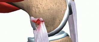
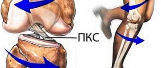
The main cause of knee instability is damage to the cruciate ligaments. They are located in the joint cavity and connect the articular surfaces of the tibia and femur. Often, damage to the cruciate ligaments occurs during an injury and is combined with damage to the menisci and hemarthrosis (accumulation of blood in the joint cavity).
At CELT you can get a consultation with a traumatologist-orthopedic specialist.
- Initial consultation – 3,000
- Repeated consultation – 2,000
Make an appointment
Causes
Often the pathology appears after serious injuries due to a traffic accident or professional participation in the following sports: football, hockey, running, cross-country skiing. The following actions lead to the appearance of the disease:
- Sharp extension and flexion.
- Impacts to the knee joint.
- Dislocations.
People with a genetic predisposition (poor development of the ligamentous apparatus) are most susceptible.
Etiology
Changes in the lateral cartilage of the knee can occur due to several common predisposing factors:
- Injuries affecting the knee and its components, as well as leading to the implementation of one of the pathogenetic mechanisms of injury. They often occur in young people with significant physical activity, as well as in athletes, both amateurs and professionals.
- A decrease in the strength of tissues and cartilage, which mainly develops in older people against the background of degenerative processes in the structures of the musculoskeletal system, the basis of which is the deterioration of their nutrition.
- A congenital disorder of the properties of cartilage tissue, which develops from childhood and is caused by changes in the functional activity of certain genes. In this case, pathology can develop in children against the background of minor loads on the components of the musculoskeletal system.
- Chronic inflammation leading to gradual weakening of the knee cartilage. Its development is primarily initiated by an infectious process or the formation of antibodies by the immune system to its own tissues (autoimmune process).
Determination of the cause must be carried out by a doctor during diagnostic measures. This allows you to prescribe preventive recommendations to ensure the prevention of the pathological condition in the future.
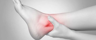
Symptoms of the disease
Immediately after a fall or blow to the joint capsule, severe pain, swelling and stiffness appear, which leads to decreased mobility. After fixing the joint in a trauma center, it is possible to reduce discomfort. This procedure is recommended for preliminary preparation for diagnosis (at an early stage it is impossible due to acute pain).
Typically, a patient comes to the doctor complaining of pain while walking, a feeling as if the knee is “sagging.” With a long course of the disease, the thigh muscles become weaker and decrease in volume.
The doctor examines the patient and conducts special tests. Typically, joint instability is diagnosed during a medical examination without additional testing.
meniscus
meniscus
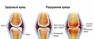
To stabilize and cushion the knee, two cartilaginous components are located in the gap. They are called the lateral (outer) and medial (inner) meniscus. They resemble a crescent moon.
The wide body has small narrowings directed anteriorly and posteriorly and called horns. This structure allows the knee to withstand relatively high loads caused by the pressure of body weight on the lower limbs.
Types and degrees of pathology
The development of the disease often occurs in athletes due to working with heavy weights and due to high loads on the quadriceps femoris muscle. There are 3 degrees of joint instability:
- Mild – there is local deformation of the articular capsule and displacement of the femur and tibia by no more than 5 millimeters. The weakening of the ligaments is minor.
- Moderate – there is a serious deformation of the cruciate ligament and a displacement of a maximum of 10 millimeters.
- Severe – diagnosis reveals a rupture of the posterior or anterior cruciate ligament with severe displacement (more than 10 millimeters).
Note! Displacement is measured not only in millimeters, but also in degrees. For a mild degree it is up to 5o, and for a severe degree it is more than 8o.
The following types of instability are distinguished depending on which ligament is damaged:
- Lateral.
- Medial.
- Combined.
- Rear.
- Front.
Additional subspecies are used:
- Atypical.
- Simple.
- Difficult.
- Total.
- Compensated.
- Subcompensated.
- Decompression.
Knee ligament instability
Home › Services › Knee ligament instability
The ligaments of the knee joint perform a vital function in the human musculoskeletal system. The knee ligaments connect the shin bone to the thigh bone and perform a stabilizing function of the knee joint during various movements (walking, turning, squats, etc.).
The stability of the knee is provided by a large number of ligaments that perform different functions. The most functionally significant ligaments are:
- anterior cruciate ligament (ACL);
- posterior cruciate ligament (PCL);
- fibular collateral ligament (MCL) – external collateral ligament;
- tibial collateral ligament (TCL) – internal collateral ligament;
- patellar ligament.
Knee instability is caused by damaged or torn knee ligaments.
Knee instability is also characterized by impaired muscle trophism, which initially manifests itself as hypotension, and then muscle atrophy. Taking into account the degree of muscle atrophy, the stages of the process are determined. Athletes with joint instability experience the most rapid development of muscle atrophy.
Degrees of knee ligament damage
Ligaments are made up of fibers, and the extent of their damage determines the degree of ligament rupture. In medical practice, gaps of several degrees are distinguished:
- 1st degree – the structure of some of the fibers is damaged, but the overall integrity of the ligament is not compromised;
- 2nd degree - more than half of the fibers are injured, ligament tears occur, and stiffness in movements in the knee joint appears;
- 3rd degree - the ligaments are completely torn, pathological mobility occurs in the knee, due to such changes the structure of the joint is disrupted and its instability occurs. With this degree, damage to other structures of the knee joint - capsule, meniscus, cartilage - is often observed.
In traumatology, cases of simultaneous damage to the cruciate and collateral ligaments are often encountered. With such ligament ruptures, hemorrhage into the knee joint with the formation of hemarthrosis is possible, so the recovery period of the joint functions increases significantly.
Causes of knee ligament rupture
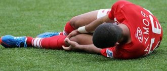
Damage to the knee ligaments occurs most often among young people involved in active sports. Knee ligament ruptures occur due to excessive loads on the joint and active movements in it (for example, twisting the limb along the axis, hyperextension, etc.). Ligament damage can also occur as a result of a direct blow to the knee area or strong pressure on the lower leg.
Certain types of movements can cause a particular ligament to tear. For example, if the shin is in a bent position and directed to the inside, and at this moment the back surface of the knee is subjected to excessive stress, then a rupture of the anterior cruciate ligament occurs. A tear of the lateral collateral ligament is characterized by an inward tilting of the shin and sudden awkward movements of the leg, twisting or stumbling.
Symptoms of knee ligament damage
Signs indicating knee ligament damage are:
- the appearance of sharp pain in the knee (severe pain, as well as hemarthrosis and synovitis are often observed in the acute period of ligament rupture);
- the occurrence of swelling in the knee area (increase in the size of the knee joint);
- limited or, on the contrary, too loose movements in the joint;
- instability of the knee joint, characterized by a feeling of displacement of the bone structures and “weaving” of the legs due to awkward movement or acceleration);
- a feeling of dislocation of the lower leg anteriorly or to the side when injured;
- faint crackling sound when the ligament is damaged;
- moving weight on the injured leg is extremely difficult or impossible when walking, getting out of bed, chair, etc.;
- pathological mobility of the patella, etc.
Diagnosis of knee ligaments
If a knee ligament rupture is suspected, diagnosis begins with an examination of the patient by a traumatologist who palpates the knee joints. Specific stability tests of the knee joint are also used to identify ligamentous disorders. For example, the McIntosh and Houston tests can confirm the diagnosis of a torn anterior cruciate ligament. The "posterior drawer" test is a symptom of a ruptured posterior cruciate ligament, etc.
Traumatologists complement the examination of the patient with instrumental diagnostic methods to make a diagnosis and choose treatment tactics. Very often, specialists resort to the following diagnostic methods:
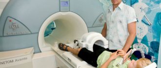
- Magnetic resonance imaging;
- X-ray of the knee;
- Ultrasound of the knee joint;
- diagnostic arthroscopy.
With the help of instrumental studies, it is possible to detect changes in the structures of the knee, confirm or refute the presence of bone fractures and soft tissue damage in cases of knee injury.
Specialists at the New Medicine Clinic select the most optimal treatment for each case, taking into account the results of diagnostic methods and functional tests, the degree of joint instability and the presence of damage.
Treatment of knee ligament instability
When treating ruptures of the ligamentous structures of the knee, conservative and surgical treatment methods are used depending on the severity of the damage. If the patient is diagnosed with grade 1 or 2 damage, then treatment is possible using conservative methods. In cases of grade ligament rupture, surgical treatment is used. For grade 1-2 ruptures, conservative treatment is possible, which consists of immobilizing the joint with a plaster or orthosis, followed by exercise therapy, massage, and physiotherapy. In addition, if hemarthrosis is present, joint puncture is possible.
Conservative treatment of knee ligament instability includes the following methods:
- immobilization of the joint (plaster or orthosis);
- therapeutic exercises (restores muscle strength, has a strengthening effect on the body);
- massage (relieves pain and swelling in the legs);
- physiotherapy (electrophoresis, UHF therapy, paraffin therapy);
- cryotherapy (exposure to cold on blood vessels contributes to their narrowing, which is why;
- drug treatment (use of non-steroidal anti-inflammatory drugs to relieve swelling and inflammation);
- rest (if you receive an injury to the injured leg, it is necessary to reduce physical activity, if possible limit any movements in order to prevent subsequent injuries, reduce pain, relieve swelling and speed up the healing process).
Surgical treatment is indicated in situations where one or more ligaments of the knee joint are completely torn and in cases where conservative treatment has failed. The New Medicine Clinic performs minimally invasive operations using arthroscopy. Arthroscopy restores the integrity of the ligamentous apparatus through 2 micro-incisions using special instruments. All actions inside the knee joint are displayed on the monitor, allowing the doctor to exercise full control over the progress of the operation.
After surgical treatment, the patient must undergo a recovery course, which includes exercise therapy and massage, interstitial electrical stimulation to prevent muscle wasting. The joint begins to function fully after 6-8 months.
You should know that knee ligament instability is a serious pathology that needs to be treated as early as possible. If you receive a knee injury or rupture of the ligaments, an early visit to the doctor will help you avoid complications, choose the most optimal treatment tactics and speed up the process of restoring the functions of the knee joint.
Diagnostics
In order to clarify the diagnosis of instability of the knee joint and exclude other diseases, the following set of studies is carried out:
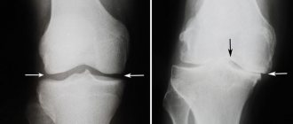
- X-ray of the knee joint.
- Magnetic resonance imaging.
- Arthroscopy - endoscopic examination of the joint cavity - is the most informative diagnostic method. The doctor can examine almost the entire joint cavity and detect pathological changes in the cruciate ligaments and other structures, and also perform therapeutic manipulations during arthroscopy if indicated.
Principles of therapy
Complex treatment includes therapeutic measures with or without surgery, as well as mandatory rehabilitation. The scope and tactics of all measures are determined by the severity and localization of changes determined using modern visualization techniques.
The basic principle of treatment involves the stage-by-stage implementation of all measures with final rehabilitation. This allows you to achieve the main therapeutic goals, namely restoration of the functional state of the knee in full.
Treatment
Treatment of cruciate ligament injuries without surgery can be performed immediately after the injury. Typically, a traumatologist punctures the affected knee joint with a needle, removes blood from it, and applies a fixing bandage to the leg.
In the future, the patient is prescribed physiotherapy, massage, physical therapy (as indicated). If signs of instability persist or appear some time after treatment, surgery is usually required. If the ligaments are completely torn, the traumatologist usually immediately prescribes surgery.
Experienced specialists at the multidisciplinary CELT clinic perform cruciate ligament replacements of any complexity.
Mechanism of change
The cartilaginous components of the meniscus are attached to the bony base of the knee by ligaments. The outer cartilage is fixed less rigidly. It is more mobile in relation to the internal cartilage, therefore, when exposed to significant mechanical force, it moves slightly and is rarely damaged. Changes in the integrity of this component develop as a result of the implementation of several pathogenetic mechanisms, which include:
- A direct blow to the outside of the knee with a blunt object resulting in a severe bruise or a fall on it. Damage is often caused by a fall onto a surface with sharp edges (curb, steps).
- Sudden excessive bending of the knee, leading to displacement of its structures relative to each other.
- Tucked leg, which occurs when the tibia is rotated against a fixed hip, the lateral cartilage is more often damaged in the case of rotation (rotation) inwards.
Also, any changes can develop when the properties are disrupted and the strength of the components decreases. However, they can develop against the background of low loads on the leg.
Prevention
To prevent knee instability, you need to follow 5 rules:
- Choose the right shoes, strictly in size, made from natural materials and with soft soles. Experts often prescribe orthopedic shoes, which are used for flat feet. It is important to firmly fix the foot in a comfortable position.
- It is recommended to use a special bandage during running, football, and hockey. When practicing powerlifting, you need to reduce the load and always use elastic bandages. Training should moderately strain the quadriceps femoris muscle and not cause pain.
- You need to reconsider your diet and add more foods high in magnesium, calcium, zinc, and fluoride. It is advisable to additionally consume a vitamin-mineral complex.
- If weakening of the ligaments and displacement of the joint is detected, you should abandon professional sports and avoid excessive stress.
- The presence of weakness, clicking and pain in the knees is a reason to contact a specialist and undergo an MRI or. Never delay consultation and diagnosis, as the disease can be successfully treated at the initial stage.
In our clinic, this type of surgical intervention is performed endoscopically, without a large incision and without opening the joint. This is the same arthroscopy, during which the surgeon reconstructs the damaged ligament. Modern equipment and endoscopic instruments help to carry out the intervention quickly and effectively, without complications.
Manifestations
Possible changes in the lateral cartilage that occurred as a result of an injury in the recent past are indicated by fairly pronounced symptoms:
- The pain, which is predominantly localized along the outer edge of the knee, is of significant intensity and intensifies with the slightest attempts to perform movements.
- Decreased range of motion, which is accompanied by increased sensations of discomfort. When a part of the cartilage is completely torn off, displaced and wedged, a block of movements occurs.
- Inflammatory signs - any damage is accompanied by the development of an inflammatory reaction, which is characterized by hyperemia (redness), swelling with an increase in the knee in volume, as well as a local increase in temperature (tissues become hot to the touch).
Changes that develop as a result of pathological processes are accompanied by a long and gradual development of clinical signs. The pain becomes more intense after exercise on the leg and usually intensifies in the evening. The movements are accompanied by a characteristic noise in the form of clicks or crunches.
Orthopedics and traumatology services at CELT
The administration of CELT JSC regularly updates the price list posted on the clinic’s website. However, in order to avoid possible misunderstandings, we ask you to clarify the cost of services by phone: +7
| Service name | Price in rubles |
| Appointment with a surgical doctor (primary, for complex programs) | 3 000 |
| Ultrasound of two symmetrical joints (except hip) | 4 000 |
| MRI of the knee joint (1 joint) | 7 000 |
All services
Make an appointment through the application or by calling +7 +7 We work every day:
- Monday—Friday: 8.00—20.00
- Saturday: 8.00–18.00
- Sunday is a day off
The nearest metro and MCC stations to the clinic:
- Highway of Enthusiasts or Perovo
- Partisan
- Enthusiast Highway
Driving directions
Non-surgical therapy
After a small change affecting the lateral meniscus of the knee joint has been diagnosed using instrumental research methods, treatment without surgery is selected by the traumatologist individually for each patient. It involves several directions:
- General recommendations - an important component of treatment, include a diet with a sufficient supply of organic compounds (especially proteins) and vitamins, limiting knee mobility, and giving up bad habits.
- Drug therapy involves the use of drugs, the most popular of which are anti-inflammatory drugs (glucocorticoids or non-steroidal drugs), chondroprotectors (necessary to improve the characteristics of cartilage tissue and prevent its further destruction), vitamins that improve metabolic processes and the patient’s condition as a whole.
- Physiotherapeutic procedures are an important area of therapy, which is advisable for pathological degenerative disorders. It makes it possible to reduce the severity of inflammation and the level of mediators that provoke its development, as well as accelerate tissue regeneration. The procedures usually include magnetic therapy, electrophoresis, and mud therapy.
An alternative modern method of therapy without surgery is the use of platelet mass (a significant number of platelets in saline solution). It is entered directly into the area of modified components. This biological preparation contains bioactive organic compounds (“growth factors”) that accelerate regenerative processes in the altered components of the musculoskeletal system.