What is juvenile idiopathic arthritis?
Considered the most common form of arthritis, juvenile idiopathic arthritis begins before age 16 and involves swelling in one or more joints lasting at least six weeks. JIA includes several types of arthritis formerly known as JRA. However, in recent years, researchers have developed a more sophisticated understanding of the differences between specific types of arthritis, and the terminology and definition of the disease have shifted.
One reason for this shift is that JRA is not, as the term suggests, simply a pint-sized replica that affects adults. In fact, only about 10 percent of children are thought to get the disease, which closely mirrors rheumatoid arthritis in adults. The researchers also concluded that the JRA category was too narrow and should include some related diagnoses, such as ankylosing spondylitis.
Juvenile idiopathic arthritis can include a variety of symptoms, such as tightening of muscles and soft tissues, bone erosion, joint misalignment, and changes in growth patterns. Not all symptoms appear in children with this disease. Moreover, the symptoms of JIA can vary from day to day.
Arthropathy of the left and right knee joint
Arthropathy of the right knee joint is more often diagnosed, since it bears greater physical load. Pathology can develop in adolescents who lead an active lifestyle and engage in outdoor sports.
Acute arthropathy of the left knee joint, on the contrary, occurs in children who tend to lead a sedentary lifestyle. They often have endocrine pathologies and problems with excess body weight. In them, the disease has a long-term progressive nature with gradual destruction of the synovial cartilaginous layer of all the heads of the bones that make up the knee joint.
In the reactive form of the pathology, simultaneous damage to both knee joints is observed. This clinical sign allows you to make an accurate preliminary diagnosis. The main difference from the rheumatoid process or the articular form of ankylosing spondylitis is that two knee joints are affected separately, while the ankle and hip joints retain full mobility.
Diagnostics: research methods
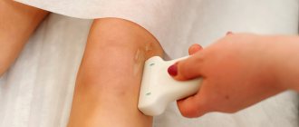
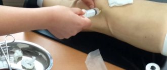
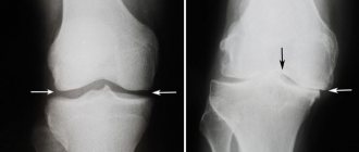
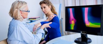
Diagnosis begins with a visual examination by a rheumatologist. Collecting information about the patient's medical history allows the doctor to compile a list of factors that provoke the development of the disease. In addition to mandatory laboratory blood tests, a specialist will prescribe an X-ray examination. Its results make it possible to identify bone corrosion and determine the size of joint spaces. The treating doctor may recommend an ultrasound examination of the affected joint and internal organs.
Polyarthritis:
This type of juvenile idiopathic arthritis occurs when five or more joints are involved within the first six months. About 25 percent of children with juvenile idiopathic arthritis have polyarthritis. Like oligoarthritis, it is more common in girls. But its onset can occur at any time in childhood. Both large and small joints such as fingers and toes may be involved. Your baby may also have arthritis in the neck or jaw, making it difficult for her to chew and open her mouth. Unlike oligoarthritis, polyarthritis more often affects joints on both sides of the body, such as the right and left knees. Children with polyarthritis may face a lower risk of eye inflammation, but will still need to see an ophthalmologist regularly.
The criteria for juvenile idiopathic arthritis also divide children with polyarthritis into two categories, those who test positive for rheumatoid factor (RF), an antibody found in the blood, and those who do not. The RF-positive form of the disease usually occurs in elementary school or later. This is the type most similar to adult rheumatoid arthritis. Children with HPV-positive polyarthritis tend to be more vulnerable to severe disease and associated joint erosion than those who experience rheumatoid factor-negative effects.
Continuation of the article: follow the Articles link.
Causes of knee arthropathy
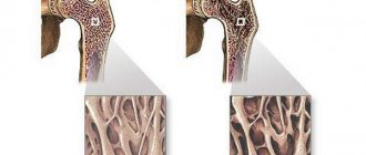
Common causes of the development of arthropathy of the knee joint include a variety of inflammatory and degenerative processes associated with impaired innervation and blood supply to the cartilage tissue of the bone articulation. The knee joint has a complex anatomical structure: it consists of 3 bones (femoral and tibial heads, patella). All bones are covered with synovial cartilage tissue. It ensures ease of sliding, distribution of shock-absorbing load, protection of the heads of bones from destruction and injury. Stability of the knee joint is provided by the posterior and anterior cruciate, lateral and medial collateral ligaments. Nutrition of cartilage tissue is carried out due to the blood supply to the end articular plates and diffuse exchange with the surrounding muscle tissue.
Several negative factors play a role in the development of knee arthropathy:
- disruption of the blood supply to muscle and cartilage tissue, provoked by a sedentary lifestyle or vascular diseases);
- disruption of innervation due to pinched nerve fiber or ischemia due to impaired blood microcirculation (often occurs with compression pressure from post-traumatic hematomas);
- an autoimmune inflammatory reaction in response to the development of tissue decay in any other organ (a reactive form of the disease can occur with tonsillitis, adenoiditis, frequent colds, pyelonephritis, pancreatitis, etc.);
- diathesis and other types of allergic reactions manifested by damage to the skin and connective tissue;
- infectious inflammation in the joint cavity (the spread of bacteria and viruses can occur through the flow of blood and lymphatic fluid);
- aseptic inflammation of joint tissues against the background of ischemia, compression, trauma;
- all types of traumatic effects, including extreme physical activity.
Potential causes of arthropathy may be hidden behind the following pathologies:
- rheumatoid polyarthritis and other types of autoimmune inflammation;
- different types of vasculitis;
- inflammation and narrowing of the blood vessels of the lower extremities;
- infectious diseases (chickenpox, mononucleosis, mumps and rubella are highly likely to provoke the development of knee arthropathy);
- tick-borne encephalitis with predominant damage to the structures of the spinal cord;
- polio and other dangerous infections;
- pathologies of the urinary system with disturbances in the biochemical balance of the blood;
- acute intestinal infections (salmonellosis, shigella, dysentery, food poisoning);
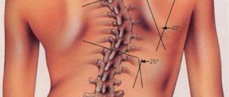
- back and spine injuries;
- displaced fractures of the femur and pelvis;
- ruptures and sprains of the knee joint ligaments;
- surgical interventions in the joint cavity.
Clinical guidelines
At each visit to the doctor, the specialist assesses the degree of inflammation of the joints, the presence of extra-articular manifestations with a diagnosis of reactive arthritis. Recommendations include monthly monitoring of blood parameters (ALT, AST, bilirubin) during long-term use of medications. General recommendations include avoiding factors that can lead to an exacerbation of the disease (hypothermia, physical and mental overload), as well as constant monitoring by a pediatrician, rheumatologist, or ophthalmologist.
Post-traumatic arthropathy of the knee joint
Children may develop post-traumatic arthropathy of the knee joint due to aseptic inflammation. This can be facilitated by:
- sprains of the cruciate and collateral ligaments;
- microscopic ruptures of tendon tissue, resulting in the formation of extensive hematomas that compress small blood vessels and nerve endings;
- fractures of the tibia, fibula, femur and patella;
- damage to the medial and lateral meniscus;
- injuries of joint capsules (bursae);
- ruptures, sprains and bruises of the muscles in the knee joint.
Any traumatic arthropathy of the knee joint requires step-by-step treatment. First, the necessary amount of medical care is provided by a surgeon or traumatologist. Providing complete physical rest is usually recommended. For fractures and cracks of bone tissue, a plaster cast or splint is applied. After the integrity of the damaged tissues is restored, the second stage of treatment must begin - rehabilitation in order to restore the normal structure of the tissues, prevent the formation of scar deformity and contracture.
Treatment of knee arthropathy
It is necessary to begin treatment of knee arthropathy by eliminating the destructive effects of the traumatic factor. If this is an autoimmune process, then preliminary pharmacological therapy is necessary. If you receive an injury, you need the help of a traumatologist. The reactive process requires some effort in treating the underlying disease.
After the acute period has passed, you need to begin restorative rehabilitation. If this is not done, then there is a high probability that the process of formation of rough scar tissue will begin. It will interfere with normal microcirculation of blood and lymphatic fluid. This will lead to further degeneration and deformation of the knee joint.
Without failed rehabilitation, a relapse of arthropathy develops over the next 6 months, and after 12–18 months, MRI examination already shows signs of complete destruction of the cartilaginous synovial layer. This is the second stage of deforming osteoarthritis.
At our manual therapy clinic, treatment for knee arthropathy begins with a correct diagnosis. An experienced orthopedist then develops an individual course of treatment. It may include osteopathy and massage, kinesitherapy and physical therapy, reflexology and physiotherapy, and laser therapy.
If you need treatment for arthropathy, you can sign up for a free consultation with an orthopedist at our manual therapy center in Moscow right now. Call the administrator and agree on a time convenient for your visit.
Diagnosis of Juvenile Idiopathic Arthritis.
Diagnosis of juvenile idiopathic arthritis is based on a physical examination as well as laboratory tests and medical history. In addition to monitoring symptoms for at least six weeks, your child's doctor will wait to see how symptoms develop during the first six months of onset. The number of joints affected during the first six months determines the diagnosis. In addition, juvenile idiopathic arthritis criteria also rely on other results, such as rheumatoid factor blood test results, to help further diagnose children in juvenile idiopathic arthritis subgroups.