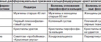
Gout
- one of the oldest diseases described in medicine. In the 5th century BC, Hippocrates called “gout” (from the Greek words “under” - leg, “agra” - trap) acute pain in the foot. It was called “the disease of kings and the king of diseases,” a disease of aristocrats, and was even considered a sign of genius. Such famous people as Leonardo da Vinci, Alexander the Great, many members of the Medici family from Florence, Isaac Newton, Charles Darwin suffered from it. And this list is far from complete.
Gout is a disease associated with a disorder of purine metabolism, which leads to an increase in uric acid in the blood (hyperurecemia) and its deposition in the tissues of the musculoskeletal system and internal organs.
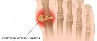
In the human body, purine bases are broken down into uric acid and excreted by the kidneys. When the concentration limit of uric acid in tissues is exceeded, the deposition of its crystals in the form of monosodium urate begins in the joints, kidneys, and soft tissues. Clinical manifestations of arthritis (inflammation of the joint), soft tissue formations (tophi), urate nephropathy (kidney damage) and nephrolithiasis (formation of kidney stones) appear.
Changes in dietary habits and lifestyles have resulted in gout affecting 1-2% of the population in industrialized countries.
Causes of gout development
- Taking medications: diuretics (thiazide diuretics), low doses of aspirin (1g per day), cyclosporines.
- Diseases and conditions that lead to the appearance of gout: diabetes, obesity, coronary heart disease, arterial hypertension, metabolic syndrome, chronic renal failure, lead poisoning, organ transplantation, psoriasis, blood diseases. Exacerbation of gout is facilitated by intravenous administration of contrast (during X-ray examinations), trauma, and surgical interventions.
- The risk of gout is extremely high in people who have a habit of consuming foods containing large amounts of purine bases (fatty meats, seafood, alcoholic beverages, carbonated drinks).
- Gout develops more often in men (age 30-60 years); in women, gout develops much less frequently and mainly during the menopausal period.
Gout symptoms
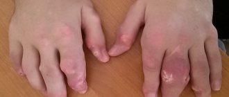
Acute gouty arthritis begins with damage to one joint, most often the big toe. The onset of acute arthritis may be preceded by heavy food consumption, alcohol intake, trauma, or surgery. The disease develops suddenly, with sharp pain, swelling and redness occurring early in the morning, symptoms increase rapidly and reach a peak within 24 to 48 hours. The pain becomes severe, patients often cannot put on socks or touch the sore joint. Acute gout can be accompanied by an increase in body temperature up to 38 degrees. Severe weakness, general malaise. Even without treatment, exacerbation of gouty arthritis disappears gradually within 5 to 7 days.
Acute gouty arthritis
As the disease progresses and in the absence of adequate treatment, attacks of arthritis become more frequent and prolonged, and new joints of the legs are involved. Joint deformation appears due to nodular deposits and bone growths, tophi.
Tophi are uric acid crystals that are deposited in the periarticular tissues and are painless, dense yellowish nodules. Most often they are located in the area of the ears, elbow, ankle joints, joints of the hands and feet.
Joint stiffness gradually appears, making it difficult for patients to move. There is a high risk of fractures. Each new exacerbation of gouty arthritis significantly worsens the course of the disease and contributes to the rapid development of complications.
Uric acid is mainly excreted by the kidneys; the deposition of uric acid salts in the renal pelvis leads to the development of urolithiasis. The passage of concrements (stones) is manifested by intense pain in the lumbar region - renal colic.
This concludes the publication of a series of articles by Evgeniy Mikhailovich Beletsky, a leading specialist in poultry reproduction at the Institute of Animal Husbandry of Ukraine. The review describes in detail a widespread non-communicable disease - uric acid diathesis or gout. The causes of the disease are indicated, special attention is paid to pathogenesis. All currently known medications used to treat gout and preventive measures are presented. Due to the large volume, I took the liberty of removing from the article some biochemical details that make it difficult for non-specialists to understand, and placing them in two posts.
style=”display:inline-block;width:336px;height:280px” data-ad-client=”ca-pub-4037835599918832″ data-ad-slot=”7553000704″>
Uric acid diathesis ( gout , visceral gout, urolithiasis) is a disease associated with metabolic disorders, mainly protein metabolism, accompanied by increased formation of uric acid (hyperuricemia), its accumulation in the body, damage to the urinary tract and deposition of uric acid salts on the visceral surfaces and joints.
Birds of all species, both adults and young, are susceptible to the disease. Although gout occurs in ostriches, these cases are quite rare. More often than others, chickens are affected, especially egg-type chickens aged 100-180 days and chickens of highly productive crosses [25]. The peak of the disease occurs in the cold season. The visceral form of gout also occurs in bird embryos or chicks in the first days of life [16, 13].
Among wild birds, birds of prey are more susceptible to the disease.
In some farms, gout is the main cause of death of birds and can cause the death of 30-40% of the total number of dead birds, although in each herd of turkeys up to 5%, and sometimes up to 15-20% of sick birds are found [7].
In recent years, gout has become widespread among poultry of different age groups of egg and meat crosses in industrial poultry farming. The disease causes significant economic losses, metabolic disorders develop, as a result of which the bird lags behind in growth and development [5, 10, 11, 39].
Although mammals get sick much less often than birds, dogs . Cases of the disease have been reported in horses, cattle and pigs. Cases of gout in reptiles (alligators and snakes) have been described [14, 21].
Gout occurs quite often in humans , especially in men after 40 years of age, and is mainly a hereditary constitutional disease. In case of gout, uric acid salts fall out and are deposited on the articular surfaces of mainly small joints of the arms and legs [32]. The disease also manifests itself as paroxysmal arthritis and hyperuricemia [21].
There are primary and secondary gout. Primary gout is an independent disease, secondary gout is a manifestation of other diseases (myeloid leukemia, psoriasis, hemoglobinopathies, chronic renal failure, congenital heart defects with erythrocytosis) or a consequence of the use of drugs (riboxin, cytostatics, saluretics, etc.) [21].
ETIOLOGY. The main causes of uric acid diathesis are:
- oversaturation of the body with proteins of animal (meat, meat and bone, blood, fish meal, especially with a high acid and/or peroxide value) or plant (high-protein concentrated feed) origin or their low quality due to a lack of vitamins, primarily vitamin A, to the epithelium of the renal tubules occurs . The cause of impaired renal function in chickens can often be a lack of vitamins B6 and/or B12 [25, 23];
— lack of green feed and grass meal ;
— hypothermia (especially in the first days of cultivation), leading to the development of inflammatory processes in the kidneys and intestines, age, insufficient ventilation, dampness, poor living conditions, lack of exercise and sunlight, etc. [6, 13, 26].
- chronic kidney damage under the influence of chemical compounds harmful to the body (grain mordants, endotoxins of microorganisms, fungi, salts of heavy metals, nitrates, sodium chloride, methane, mercuric chloride, aloin, acetone, some organic acids, etc.), leading to reduced urinary excretion acids and urates, liver damage [2, 5, 21];
— violation of the calcium-phosphorus ratio due to excess phosphorus against the background of deficiency of vitamins B2, B4, B6 [27];
— incorrect balance in the diet of acidic and alkaline feeds (acid diet);
- lack or absence of water , especially in the first days after hatching, leading to the formation and deposition of uric acid salts as a result of the use of residual yolk.
— some viral diseases ( infectious bronchitis, Gumboro disease ) and as complications during their specific prevention [7, 8, 9, 20, 45, 62].
Excess glycine can cause necrosis of the renal epithelium and swelling of the renal glomeruli, which predisposes to the deposition of urate salts in the tubules.
In chickens hatched from eggs in conditions of high humidity in the incubator (70%), metabolism may be disrupted and gout may occur.
Feeding chickens a diet unbalanced in protein, arginine, tryptophan, glycine and some other amino acids can be one of the main causes of this disease [29].
PATHOGENESIS . The normal amount of uric acid salts in the blood varies depending on the type, quantity and time of feeding, egg production (in chickens from 20 to 80 mcg/ml, in turkeys - up to 20 mcg/ml ). Uric acid salts are contained in blood serum in the form of monosodium urates (up to 184 μg/ml) [4].
Uric acid salts are excreted by the kidneys only in concentrated form. In case of poisoning, the amount of uric acid salts increases significantly. In case of nephrosinephritis syndrome, an average of 189 mcg/ml of uric acid salts is found in the blood, in case of poisoning in turkeys - up to 160 mcg/ml and up to 200 mcg/ml in the blood serum. The excretion of uric acid salts in turkeys during poisoning ranges from 0.6 to 2.8 g per day [7].
The question of the mechanisms of increasing the concentration of uric acid salts during poisoning in birds has not been fully clarified. Obviously, if the concentration of sparingly soluble uric acid salts crosses the solubility limit, then deposition of salts in tissues and joint cavities begins. With excess protein nutrition, the kidneys and visceral membranes reduce their function in nitrogen metabolism.
Fungal toxins also cause kidney damage. Fatal cases of visceral gout have been described in geese after ingesting moldy corn. Mold toxins caused nephrosis [7].
Day-old chicks may have visceral gout as a result of moisture disturbances during incubation (70% relative humidity instead of 55-60%). Insufficient and late intake of water leads to the fact that uric acid salts, which are formed when using the yolk, are not immediately excreted.
This disease can occur in local, visceral and mixed forms [6].
Under normal conditions, the breakdown products of nucleoproteins (uric acid and its salts) in a dissolved state are excreted from the body mainly by the kidneys. When the metabolism of these complex proteins is disrupted, excessive formation of uric acid occurs and its salts are deposited on the articular surfaces of the fingers of the extremities, in the tendons, cartilage of the auricles, kidneys and serous membranes. At the site of deposition of crystals of uric acid salts, tissues undergo necrosis. An inflammatory focus develops around the dead areas with the proliferation of connective tissue [32].
The disease develops gradually. Nephrosonephritis syndrome occurs due to impaired excretory function of the kidneys due to inflammation of the nephron , the main structural and functional part of the kidney. At the same time, the reabsorption of water and substances soluble in it worsens. Uric acid salts are deposited in organs and tissues. The serous membranes of the peritoneum, internal organs and joints are especially often affected. At the last stage, urates fall into the lumen of the ureters - a sign of urolithiasis [5].
The development of the disease requires a certain time, which depends on the degree of exposure to certain factors. If there is a lack of vitamin A in the diet of chickens, mortality can begin after 26 days. In turkeys, the disease is detected after 56-70 days.
One of the main reasons is the oversaturation of food with proteins and nucleic acids (especially meat) [60], primary kidney damage, insufficient protein and poor quality food, with which acids such as lactic, butyric, acetoacetic enter the body, as well as physical inactivity and stress [24]. A characteristic feature of gout is the accumulation of large amounts of decay products in the blood and tissues. Ammonia occupies a special place here in the sense that it is a very toxic substance [28]. It has been shown that in chickens with gout, the level of ammonia in the blood, liver and kidney tissues increases by 1.5-2.0 times [35]. Ammonia is released in the gastrointestinal tract (the main part), in the kidneys, liver and muscles. This occurs due to the rupture of amide bonds in the protein molecule. The cleaved ammonia binds with glutamic acid to form glutamine [37].
A special place at this stage is given to the enzyme glutamine synthetase. The activity of this enzyme is 10-15 times higher in the liver of chickens compared to other animals. The level of glutamine synthetase activity can change with different protein levels in the diet and is important in the regulation of uric acid formation. The amidation reaction of glutamic acid is extremely important. Firstly, this reaction neutralizes ammonia, and secondly, the resulting glutamine is used further for the synthesis of more complex nitrogen compounds, including the synthesis of purine bases.
It is known that gout is accompanied by impaired ability of the kidneys to excrete uric acid as an end product, and metabolic acidosis is a characteristic feature of chronic renal failure. In this regard, it is important to know how acid-base homeostasis is regulated in the body.
In birds, glomerular filtration is lower than in other animals, and tubular secretion is directed towards the greatest release of waste: for example, tubular cells secrete ammonium, phosphate, uric acid, urates and H ions, i.e. all substances that support renal acid secretion.
In situations where the bird finds itself in stressful situations - high-protein, starvation diets, excess nucleic acids, hyperuricemia becomes acute and the production of uric acid increases. The biochemical mechanism of this phenomenon has not been established. It is possible that amino acids regulate the rate of RNA decay, which, in turn, regulates purine biosynthesis [57].
It is possible that calcium antagonists may have a beneficial effect on uric acid levels.
In birds, whose tissues lack the urease enzyme that breaks down uric acid to allantoin, uric acid is the end product of nitrogen metabolism, and its production is a measure of purity, the balance between purine synthesis, purine breakdown, and purine reutilization.
Hyperuricemia and accumulation of urates in the body of herbivorous and carnivorous farm animals and humans develop due to increased biosynthesis and decreased excretion in urine. This leads to the deposition of urates in tissues. Acute gouty inflammation develops due to the deposition of urate microcrystals in the joint cavity, which can activate the Hageman factor, complement components, and kinins, which leads to an increase in vascular permeability and an influx of neutrophils. Phagocytosis of crystals is accompanied by the release of lysosomal enzymes, resulting in inflammation. Urate crystals are also deposited in the interstitium of the kidneys and tubules, which leads to the development of gouty nephropathy - the second most important clinical sign of gout .
Thus, a significant accumulation of ammonia . Since ammonia is very toxic to the body, it is detoxified by binding to glutamic acid to form glutamine, which can subsequently be used for the synthesis of purine bases.
Because glutamine plays a major role in both ammoniagenesis and purine biosynthesis, there is an association between elevated glutamine levels and purine overproduction in gout. Glutamate is the main precursor of glutamine; Perhaps in gout we have a primary defect in glutamine metabolism [31], which leads to the accumulation of glutamate and, as a consequence, to an increase in the amount of cellular glutamine and subsequent overproduction of purines.
In birds, amine nitrogen is almost completely converted to uric acid. This transformation goes through the adenine nucleotide cycle of processes. On the other hand, the concentration of HCO3—, regardless of changes in pH, may be a major factor in the regulation of glutamine metabolism. It has been established that glucose synthesis decreases with increasing concentration of HCO3— [55].
In addition to disruption of purine metabolism, disturbances of an endogenous nature may occur and so-called secondary gout may occur - in febrile conditions, pneumonia, leukemia, etc. due to the disintegration of leukocyte nuclei rich in nucleoproteins [32].
CLINICAL SIGNS. There are three types of uric acid diathesis: visceral gout - urates are deposited on the serous covers of internal organs, articular gout - urates accumulate mainly in the joints, and a mixed form is also found [7, 20].
During the development of the disease, a certain time passes, which depends on the degree of exposure to factors. The first signs of the disease in geese are observed after 3-14 days, in turkeys - after 56-70 days.
In the acute stage, intestinal dysfunction is observed, droppings are white, the general condition is disturbed, the bird is inactive, egg production decreases, appetite decreases, and comb cyanosis appears. Death occurs within a few days, sometimes without obvious signs. The hatchability and chick hatchability of the resulting eggs is reduced [15]. In hatched chickens, already in the first days, high mortality, refusal of feed, profuse diarrhea, sticking of the cloaca with excreted feces are recorded, the skin around the cloaca is inflamed [5, 7, 20].
The articular form of visceral gout is less common, although it predominates in adult turkeys. Is chronic and disrupted
movement, joints swell, local temperature rises. Later, the skin of the affected joints becomes gray-white. When abscesses are cut, a whitish plaster-like mass is released [6, 7].
Kidney damage and the occurrence of uric acid diathesis in birds cannot be detected by clinical signs. It is installed only after autopsy of apparently healthy, but suddenly fallen individuals. Sometimes, during the chronic course of the disease, uric acid salts accumulate in the cloaca area in the form of pasty lumps.
The clinical symptom of the disease in chickens is short-term diarrhea, and between 7 and 14 days of age they experience growth retardation. In dead individuals, 4-21 days after the onset of the disease, discoloration and swelling of the kidneys and urate deposition in other organs are observed [25].
Gout is especially severe due to a deficiency of vitamin A, which ensures the normal function of the renal epithelium. In the body of a bird experiencing retinol deficiency, the concentration of uric acid salts increases sharply. Thus, in blood serum it exceeds the norm by 8-9 times.
In adult turkeys, the articular form of gout mainly predominates. The first signs: difficulty moving, the bird sits on its feet or lies for a long time. If you pick her up, then after taking a few steps she sits down again. Subsequently, inflammation and swelling of the leg joints occurs. A direct connection has been established between the content of uric acid salts in the blood serum and the severity of clinical signs of the disease. Just like in chickens, egg production and egg hatchability , and unused protein remains in them. In geese, gout is accompanied by diarrhea, difficulty breathing, and often sudden death. The joints swell, become deformed, and dense nodes form - gouty bumps.
Sometimes, long before the appearance of other signs of the disease, gout causes skin damage due to the deposition of uric acid salts in the dermis. Histologically, it is a fine-grained mass of cholesterol,
surrounded by a layer of sodium urate crystals, multinucleated giant cells and a thick fibrous capsule with histiocytes and lymphocytes. Up to 70% of their composition is urate [32].
Most people who suffer from gout are men after 40 years of age. In most patients, the clinically detectable onset of the disease coincides with the sudden development of an acute attack of arthritis. A gout attack most often occurs at night. The patient wakes up from severe, burning, pressing, throbbing or tearing pain in one or more joints. In approximately three-quarters of patients, the first manifestations of gout occur in the metatarsophalangeal joint of the big toe. Less commonly, the onset of the disease occurs as polyarthritis. With decreasing frequency, the joints of the feet, ankles, fingers, knees, elbows, wrists, etc. are involved in the process.
After a few hours, the affected joint swells, the skin over it turns red, becomes hot, tense, and shiny. There is a slight chill and possible fever. The intensity of the pain syndrome is so great that sometimes it cannot be relieved even with drugs. The pain intensifies at the slightest touch to the inflamed surface. Complete painful restriction of mobility of the affected joint occurs. The area of swelling may extend to the surrounding periarticular tissue. Externally, the picture of acute gouty arthritis resembles phlegmon or purulent inflammation of the joint. The intensity of the pain noticeably weakens in the morning, and intensifies again at night. The first gouty attacks usually last no more than 3-4 days, then the symptoms of arthritis and pain gradually disappear. After a few weeks, movement in the affected joint is fully restored. An attack of acute arthritis may recur after a few months. Over the years, the frequency of gout attacks increases. With each repeated attack, more and more joints may be involved in the process.
After 5-10 years from the onset of the disease, the second characteristic sign of gout appears - tophi (tissue accumulations of urates). The favorite localization of tophi (gouty nodules) is the inner surface of the ears, the area of the elbow joint, the joints of the feet and hands. Other localization of tophi is also possible. Their sizes are different and range from 1-2 to 10-15 mm in diameter. The incidence of tophi is directly proportional to the duration of the disease [21].
Repeated gouty attacks in the same joint, accumulation of urate in the periarticular tissues lead to joint deformation, contribute to the development of secondary arthrosis, and sometimes complete destruction of the articular surfaces and epiphyseal parts of bone tissue
Of the extra-articular manifestations of gout, kidney damage is most often observed (50-75% of patients) in the form of urolithiasis, interstitial nephritis, secondary pyelonephritis, glomerulo- and nephrosclerosis. Gouty kidney damage can lead to renal failure, which is the cause of death in almost every fifth patient. Extra-articular manifestations of gout sometimes include tendon lesions, acute forms of lumbago and radiculitis, pharyngitis, iritis and iridocyclitis, allergic reactions, phlebitis, etc. Gout is often accompanied by hypertension, coronary heart disease, and obesity.
In women, gouty arthritis occurs very rarely, mainly during menopause. Their gout, both primary and secondary, is more benign.
The course of gout is long-lasting. Acute joint attacks are replaced by remissions. Gradually, the interictal intervals shorten, and the frequency of joint attacks increases, although their intensity weakens. The severity of the course is determined by the frequency of joint attacks, the speed of joint deformation, the time of occurrence of tophi, bone destruction, and kidney damage. The cause of death in patients with gout is usually cardiac or cerebral complications, renal failure [21].
PATHAL-ANATOMICAL CHANGES. Histological examination of gout in birds is recorded in approximately 6% of cases. In the visceral form of the disease, the kidneys are regularly affected. Upon careful examination, crystals of uric acid salts are found in them in the form of chalky whitish pockets or banded deposits. The serous membranes of the liver, spleen, lungs, air sacs and intestines are also covered with uric acid salts. In the parenchyma of organs they occur in the form of foci. Urates can also be found in subcutaneous tissue [5].
Microscopy reveals radiant urate crystals: the epithelium of the renal tubules is in a state of granular degeneration and necrosis, the stroma is infiltrated with lymphoid and giant cells.
In chickens during embryonic development, a gouty form of dystrophy is also possible, associated with vitamin A deficiency in chickens with excess protein feeding. At the same time, pale yolks of eggs are noted, which subsequently have low fertility and increased mortality. When opening the asphyxiations, hypertrophy of the kidneys and deposition of uric acid salts in them, as well as on the mesentery, heart, amnion, allantoic and yolk sacs are noted. The conclusion is late, the pigmentation of the skin and down is poor [1, 6, 7,]. With visceral gout, crystals of uric acid salts are found in the kidneys. There are two types of kidney lesions: white lobular deposits in the parenchyma and confluent superficial nodular lesions.
Sometimes in embryos and chickens that died from gout, the kidneys, as a result of the accumulation of uric acid salts, have a raspberry color. The ureter contains mucous-white masses and is greatly dilated. Sometimes urates appear as dense, chalky formations; they can fill the small drainage tubules of the kidneys. The serous membranes of the liver, spleen, lungs, air sacs and intestines are also covered with uric acid salts [7].
With uric acid diathesis, damage to the heart is noted. On the pericardium and epicardium there is a coating in the form of powder, in severe cases - plaster-like continuous deposits. Sometimes adhesions of the epicardium and the pericardium are noticeable. Urates are also found in the form of foci in the liver, spleen, cardiac muscles, on the endocardium, endothelium of large vessels, in the intestines, and subcutaneous tissue. In turkeys they reach a value of 0.2-0.4 mm [7].
In newborn mammals who have lived for a week, uric acid renal infarction occurs as a physiological phenomenon, which occurs as a result of a sharp change in metabolic processes during the transition to independent feeding with colostrum. The concentration of uric acid in the blood temporarily increases, which does not have time to be completely excreted from the body in urine. Subsequently, the metabolism is normalized and the animals recover. In case of uric acid infarction of the kidneys, radially located red-yellow stripes are visible macroscopically on the surface from a section in the medulla, representing an accumulation of uric acid salts in the lumen of the straight tubules and in the stroma of the organ [5].
(Visited 4,012 times, 1 visits today)
Diagnosis of gout
- Blood chemistry. Detection of hyperurecemia: increased uric acid levels in men over 0.42 mmol/l, in women over 0.36 mmol/l. Determination of blood creatinine level - to identify renal failure.
- Examination of the synovial fluid of the affected joint. Chemical or microscopic examination reveals uric acid crystals; there is no bacterial flora in the culture.
- X-rays of joints determine changes in chronic gouty arthritis.
- Ultrasound of the kidneys. Identification of X-ray negative calculi (stones).
Recommendations for antihyperuricemic therapy
The goal of antihyperuricemic therapy is to prevent the formation and dissolution of existing monosodium urate crystals by maintaining uric acid (UA) levels below 360 µmol/L.
- Allopurinol
– promotes adequate long-term antihyperuricemic therapy. The drug is recommended at a dose of 100 mg daily, if necessary, the dose is increased by 100 mg every two to four weeks. Patients with renal failure require dose adjustment of this drug.
- Uricosuric agents
(probenecid, sulfinpyrazone) are used as an alternative to allopurinol in patients with normal renal function. These drugs are relatively contraindicated in patients with urolithiasis.
- Benzbromarone
- powerful uricozourik; the drug is more effective than allopurinol. It is used for moderately reduced renal function, but requires monitoring due to hepatotoxicity.
- Colchicine
can be used as a prophylaxis for joint attacks during the first month of antihyperuricemic therapy (0.5-1.0 grams per day) and/or NSAIDs.
It is worth noting that in patients with gout, diuretics should be discontinued if possible (except for cases where diuretics are prescribed for health reasons).
- Losartan and fenofibrate
have a moderate uricosuric effect. These drugs are recommended for use in patients who are resistant to or intolerant of allopurinol or other uricosurics, in the presence of hypertension or metabolic syndrome. However, the clinical significance of such therapy and its cost-effectiveness are still unknown.
At the Clinic of High Medical Technologies named after. N.I. Pirogov patients will be able to determine the serum level of uric acid and other important biochemical blood parameters, as well as undergo clinical blood and urine tests, and receive qualified advice from a rheumatologist on treatment both during the interictal period of the disease and during the attack of an acute gouty arthritis.
Treatment of gout
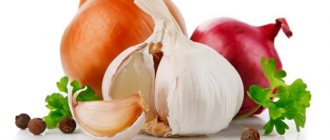
Compliance with dietary recommendations is important to prevent gout exacerbation. This is a restriction on the consumption of: meat and fish products, legumes, sorrel, spinach, cauliflower, raspberries, figs, chocolate, strong tea, coffee. The consumption of alcohol, especially wine and beer, is prohibited.
It is recommended to increase the volume of fluid you drink to 2 liters per day, unless there are contraindications. These are cranberry juice, juices, alkaline mineral waters. Gradual normalization of body weight is mandatory for the patient, as it helps reduce the level of uric acid in the blood.
The goal of drug treatment is to reduce pain during an attack and treat disorders of purine metabolism.
During an acute attack of gout, the patient is recommended to rest completely, especially on the affected limb. You should put your foot in an elevated position, apply an ice pack to the sore joint, and after the pain subsides, apply a warm compress. Nonsteroidal anti-inflammatory drugs (NSAIDs) are used to treat an attack. The drug, its dose, frequency of administration, and duration are recommended by the attending physician.
To achieve a stable decrease in uric acid, which prevents the progression of gout, anti-gout drugs (alopurinol, sulfinperazone, uralit, etc.) are used. These drugs are used for a long time (years). The required drug is selected by the attending physician individually for each patient, depending on age, concomitant diseases, and the level of uric acid in the blood.
When large tophi are formed, tissue ulceration, or the presence of fistulas, surgical treatment is recommended - removal of tophi, since they can no longer resolve when taking anti-gout drugs, and can significantly limit the function of the joints.
How to treat gout of the thumb - methods, medications. What is this.
Acupuncture for pain.
Our specialists work with biologically active points to remove inflammation and pain, harmonize the functioning of the kidneys, liver and pancreas.
Homeopathy for inflammation and swelling.
To relieve an attack and normalize uric acid metabolism, we make microinjections of a complex homeopathic remedy into biologically active points. This method is called pharmacopuncture. An attack of gout, the treatment of which includes homeopathy, goes away faster.
Warming up for gout. Symptoms and treatment. Facilities.
Warming up distant points helps eliminate swelling and swelling of the joint, restore range of motion, improve blood supply to the affected area, and metabolic processes in the joint.
Food as medicine for gout. Nutrition. What you can and cannot eat.
If you follow a diet, your chances of recovering from gout increase. Pay attention to your diet.
An attack of gout, which we treat using this regimen, no longer recurs in the vast majority of patients.
Physiotherapy for gout
In the acute period, applications of dimexide solution are used on the affected joint.
It has analgesic and anti-inflammatory effects. Before starting the procedure, tests are carried out for sensitivity to dimexide (the presence of an allergic reaction), and procedures begin 24 hours after the test. For applications, use a 30-50% solution of dimexide (pure dimexide is diluted with boiled water). A napkin soaked in the solution is placed on the sore joint. Initially, the duration of the procedure is 10 minutes, later it is increased to 2 hours. During the period of remission, applications with mud, paraffin, and ozokerite are used, which helps improve joint function and reduces the urate content in them. During the period of remission, patients are recommended to undergo sanatorium-resort treatment. Health measures have a beneficial effect on the health status of patients and the course of the disease:
- dietary food,
- taking alkaline mineral waters,
- balneotherapy,
- mud therapy