30.01.2021
Spondylolysis of the spine is a defect of the vertebral arch when the arch does not fuse in the area between the joints or in the pedicle due to delayed development of the posterior part of the spine. In 99% of cases, spondylolysis occurs in the lumbar region and in 85% of cases affects the 5th vertebra, in 10% of cases - the 4th vertebra. Simultaneous defeat also occurs.
This is a reversible disease that, if diagnosed at an early stage, can be treated without surgery. Spondylolysis of the spine can be asymptomatic, and, as a rule, its mild forms are accidentally discovered during a comprehensive examination.
Symptoms of spondylolysis[edit | edit code]
Spondylolysis
- Complaints of pain in the lower back or buttocks (not always the case).
- Extensor load on the lumbar spine.
- Spondylolysis gap in the interarticular part of the vertebral arch.
- Possible spondylolisthesis.
Spondylolysis
- vertebral arch defect - occurs in both athletes and non-athletes. Most often, spondylolysis is localized in the L5 vertebra, somewhat less often - in the L4 vertebra. The exact cause of spondylolysis is unknown, but it is generally accepted that persistent hyperextension of the lumbar spine leads to a stress fracture of the interarticular part of the vertebral arch. According to epidemiological studies, the prevalence of spondylolysis in the general population is 4-7%. In athletes, spondylolysis occurs at the same frequency, with the exception of certain sports: diving, gymnastics, weightlifting, wrestling, American football, rowing, where the frequency of spondylolysis is slightly higher. Spondylolysis occurs twice as often in boys as in girls, and prospective studies have identified a hereditary predisposition.
Classification and causes of spondylolysis
Spondylolysis is classified according to its cause and location. Based on their occurrence, spondylolysis is divided into congenital, acquired and mixed.
Congenital spondylolysis occurs during fetal development. The two ossification nuclei, each of which forms half of the arch, do not connect, so the arch does not fuse.
Acquired spondylolysis occurs due to excessive physical exertion, if the bone does not receive or does not receive enough necessary nutrients, or if vertebral shift or dysplasia occurs.
Mixed spondylolysis is a combination of a birth defect and excessive physical activity.
Based on location, spondylolysis is divided into typical, atypical and retrosomatic. In typical spondylolysis, the arch does not fuse in the area of the gap between the joints. In atypical spondylolysis, the arch does not fuse between the base and the joint space. With retrosomatic spondylolysis, the arch does not fuse in the root area (immediately behind the vertebral body).
Acquired and mixed spondylolysis occurs due to high pressure on the spine during flexion and extension of the body. In this case, the presence of cargo is not necessary. The interarticular arch of the vertebra breaks, unable to withstand the load. Such fractures heal well with properly prescribed and timely treatment. The absence of any therapy in 90% of cases leads to progression of the disease and its transformation into spondylolisthesis - displacement or failure of one vertebra relative to another.
Clinical picture of spondylolysis[edit | edit code]
History and complaints[edit | edit code]
Manifestations of spondylolysis vary: from persistent but mild pain in the lower back to severe pain that changes gait or limits the ability to move. The pain intensifies when the lower back is extended and decreases when bending forward. Athletes may complain that the pain prevents them from sleeping or lying on their back. The pain is usually limited to the lower back, but may radiate to the buttock or the back of the thigh. There may also be complaints of tension (spasticity) in the posterior thigh muscles and limited mobility of the lumbar spine. Radicular symptoms are possible but rare with spondylolysis.
Physical examination[edit | edit code]
Physical examination usually reveals few abnormalities. Characteristic limitation of movements in the lumbar spine, mainly extensor. Gentle passive extension usually increases pain. There is a provocative test that helps in diagnosing spondylolysis: the patient must bend back, standing on one leg. With spondylolysis, pain on the affected side increases. On the side where the pain is noted, you can often find tenderness on palpation. If there is no spondylolisthesis, there should be no indentation above the spinous processes. A provocative test for radiculopathy—extended leg elevation (Lacega sign)—is negative, and neurological examination results are usually normal.
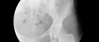
Radiation diagnostics[edit | edit code]
If spondylolysis is suspected, radiography is performed in direct, lateral, right and left oblique projections, and a picture of the lumbosacral joint is taken with the caudocranial direction of the radiation beam. These images will reveal a spondylolysis gap in the pars interarticularis in 85% of cases. With the caudocranial direction of the beam, its central rays pass through the gap between the L5 and S1 vertebrae, and not through the middle part of the lumbar region. In addition, it is necessary to examine radiographs for the presence of spondylolisthesis: it can be visible in 50-75% of initial images. The older the athlete, the higher the likelihood of having spondylolisthesis.
Special methods[edit | edit code]
Athletes with back pain and a pars interarticularis defect on radiographs should undergo bone scintigraphy. If the accumulation of the isotope is not detected, spondylolysis is unlikely to be the cause of the complaints. Single-photon emission tomography is more sensitive and specific than scintigraphy, and it showed accumulation of the isotope in the interarticular part of the arch when scintigraphy gave a negative result. It has also been established that single-photon emission tomography is negative in asymptomatic spondylolysis. If radiography is negative and the history and physical examination suggest spondylolysis, additional testing is indicated. Single-photon emission tomography is considered the method of choice in such cases: due to its high sensitivity and specificity, it allows diagnosing spondylolysis at the stage of a stress fracture.
CT is more sensitive than radiography, and the defect of the interarticular part of the arch can be clearly visible on horizontal sections. The defect can be unilateral or bilateral. The main disadvantages of CT are the difficulty in determining the duration of the defect and its clinical significance. Sclerosis and hypertrophy of the bone ends may indicate a chronic nonunion defect. Because spondylolysis is common, scintigraphy may be required to assess the clinical significance of the defect. If no isotope accumulation is detected, it is unlikely that the cause of the complaint is a defect in the interarticular part of the vertebral arch.
MRI, which is not as sensitive as scintigraphy, is not a routine examination for clinically obvious spondylolysis. In addition, the temporal relationship between clinical cure and resolution of MRI changes is unclear.
General symptoms
Spondylolysis l5 progresses quite quickly and gives the following symptoms:
- The appearance of pain in the sacral region;
- Increased discomfort when standing still for a long time;
- Tension of muscle structures and the development of kyphosis due to load redistribution;
- Reduced height due to curvature of the spinal column and its reduction;
- The appearance of skin folds on the sides and the presence of difficulties when bending forward;
- Noticeable clubfoot and change in gait, which occurs due to improper load distribution.
Similar symptoms are typical not only for spondylolysis l5, but also for many diseases associated with pathologies of the musculoskeletal system. This makes making a diagnosis much more difficult.
Treatment of spondylolysis[edit | edit code]
Conservative treatment[edit | edit code]
Several factors influence the choice of treatment: the athlete's age, severity and duration of symptoms, and radiographic changes. Athletes with a defect in the interarticular part of the arch according to radiography, but without stings, do not require treatment; they can continue to exercise. If the manifestations of the disease are reduced to minor pain in the lower back, it is necessary to strengthen the muscle corset and maintain physical performance. If neurological disorders or other severe manifestations are present, sports activities should be limited and additional examination should be performed. In athletes with back pain and radiological signs of spondylolysis, activity limitation and spinal immobilization with a brace are successful in more than 90% of cases.
The need to wear a corset and the best type of corset are debatable issues. According to observations of seven athletes, rest and limitation of activity without immobilization of the spine led to clinical recovery in all observed. The results of this study were considered equivocal and could not be reproduced. It is usually accepted to recommend short-term immobilization of the spine. Flexible and rigid, lordotic and anti-lordotic corsets are equally suitable for this. It is believed that the anti-lordotic corset (0° curvature) relieves the posterior elements of the vertebrae, and we prefer it. The length of time the brace was worn varied between studies and does not appear to have a direct impact on clinical effectiveness. We currently recommend permanent spinal immobilization during the day (while awake) and allow removal of the brace during sleep. The duration of immobilization is generally 6-8 weeks in combination with restriction of sports activities. For adolescents, repeated X-rays are recommended every 6 months until skeletal maturation (growth stops). For adult athletes, repeat X-rays are indicated only if symptoms return.
The goal of treating athletes with stress fractures of the interarticular part of the vertebral arch (isotope accumulation on single-photon emission tomography in the absence of radiological changes) is fracture healing. A decrease in isotope accumulation can be observed after immobilization (unloading) of the spine. The probability of fusion is maximum with unilateral changes and minimum with bilateral ones. It is important that return to sports activity was not made dependent on achieving bone fusion. It is believed that a strong fibrous (connective tissue) fusion is formed at the site of the defect, ensuring the disappearance of symptoms. Most doctors recommend stopping spinal immobilization and allowing sports activities once symptoms resolve.
After immobilization, the athlete moves on to a rehabilitation program that emphasizes flexion and flexibility exercises. Resuming normal activities is possible over the next 6-8 weeks.
Surgical treatment[edit | edit code]
Due to the effectiveness of conservative treatment, the need for surgery rarely occurs. Candidates for it are athletes whose complaints persist for longer than 6 months despite activity limitation and immobilization (unloading) of the spine.
The reference treatment for spondylolysis at the L5 vertebral level in adolescent athletes is L5-SI in situ posterolateral fusion. The operation is effective, but the mobility of the segment is sharply reduced. Over the past 20 years, there has been increased interest in methods to preserve vertebral mobility. For this purpose, the following methods of osteosynthesis of the interarticular part of the arch have been proposed - Scott wire fixation; wire hooks; fixation with translaminar interfragmentary screws according to Buck and fixation with a screw, hook and pin (considered the most rigid), supplemented by the use of bone graft. The plastic surgery of defects of the interarticular part of the arch from L3 to L5 is described. According to several observations, both the interfragmental method and wire methods gave either good or excellent results and ensured return to sports activity and participation in competitions at the same level as the previous one in more than 90% of cases. It is likely that the success of complex methods is associated with the use of defect closure with a bone graft.
In athletes who have undergone surgery, it is necessary to confirm fusion of the defect before proceeding with rehabilitation. Once fusion is achieved, a rehabilitation program begins, including flexion exercises, development of flexibility and strengthening of the muscular corset.
Possible complications
Spondylolysis is a dangerous pathology for humans, which gradually progresses and can cause irreversible changes in the body. The most severe complication is considered to be the exit of the affected bone segment beyond the conventional line. But besides this, other changes may be observed:
- Serious curvature of the spinal column.
- Excessive pressure of the affected segment on the cartilaginous structure (intervertebral disc).
- Reduction of the lumen in the spinal canal.
- Deformation of nerve endings and compression of the spinal cord.
- Permanent loss of ability to work and disability.
There is a high risk of complications if treatment measures are not taken in a timely manner. Therefore, the key to preventing the development of pathological changes is strict adherence to all doctor’s instructions.
Spondylolysis is a congenital or acquired defect that develops in the vertebral arch. Pathology is detected in both young and elderly patients. Most often it is diagnosed in the fifth lumbar vertebra and is the cause of pain. Properly selected treatment and strict adherence to the chosen tactics can alleviate the condition, stabilize the patient and provide him with a high quality of life.
Return to sports[edit | edit code]
With conservative treatment, sports activities will be allowed to resume after clinical improvement: athletes can return to activities after the disappearance of pain, regardless of the presence of radiological signs of fusion of the interarticular part of the arch. At the same time, participation in high-risk sports - football (including American football) or gymnastics - reduces the likelihood of a favorable outcome by five times compared to low-risk sports - baseball, track and field or swimming.
The timing of return to sports after surgical treatment is controversial. We believe that if fusion is confirmed, there is no pain, range of motion is restored and the muscle corset is strengthened, the athlete can return to training. This may take from 5 to 12 months.
Neurological manifestations
With spondylolysis l5, compression of nerve endings and roots occurs. As a result of such changes in the body, neurological symptoms develop:
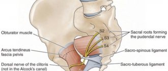
- Decreased sensitivity of the skin in the perineum, genitals and lower extremities;
- There is a tingling sensation and numbness in the legs;
- Characteristic muscle weakness appears.
If no therapeutic measures are taken in time, relative or complete urinary incontinence develops or problems with defecation occur. In severe cases, it is possible that men will develop impotence.
Read also[edit | edit code]
- Abs workout for back pain
- Sports spinal injury - treatment
- Lumbar sprain
- Backache
- Lower back pain
- Herniated disc
- Spondylolisthesis (treatment)
- Fractures of the spine in the lumbar region
- Treatment for back pain
- Rehabilitation for back pain
- Massage for osteochondrosis
- Massage for radiculitis
- Massage for intercostal neuralgia
Establishing diagnosis
What is spinal spondylolysis is clear. How is this disease diagnosed? To begin with, the doctor assesses the patient’s medical history and lifestyle:
- The presence of a depression or protrusion l5;
- Definition of retraction syndrome, indicating movement of the segment;
- Tension of the muscle structures responsible for straightening the spine;
- Presence of Turner's symptom (formation of kyphosis and pathological lordosis);
- Formation of skin folds, protrusion of the chest and shortening of the lumbar region;
- Change in gait - the hip and knee joints are bent, the feet become slightly crossed.