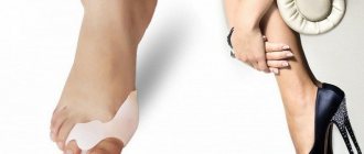
Hammer (claw) fingers
- persistent flexion-extension contracture of the fingers, developing against the background of transverse-longitudinal flatfoot, as well as with spastic and flaccid paralysis. Most often, the second and third toes are deformed, but other or all toes may also change. The disease can be combined with transverse flatness of the toes and deviation of the first toe outward by 30° or more.
Etiology of hammertoe deformity:
- Cerebral palsy,
- Poliomyelitis (with planovalgus foot),
- Myelodysplastic cavus foot,
- hallux valgus
- Muscle imbalance;
- Incorrect gait;
- Genetic predisposition to foot deformities;
- Joint diseases;
- Arthritis;
- Tension of the tendon, which prevents the finger from straightening;
- The second toe is longer than the others;
- Incorrectly selected shoes;
- High arch of the foot;
- Flat feet.
Symptoms
The first alarming symptom is an external change in the shape of the finger: it is bent in an unnatural position. It is either impossible to straighten it at all, or only manually. This condition is accompanied by the following symptoms:
- pain and discomfort while walking;
- the formation of painful calluses in the toe area due to friction with shoes;
- inability to choose normal shoes in size.
If measures to correct the defect are not taken for a long time, the symptoms will worsen.
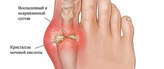
Pathogenesis of hammertoe deformity:
The pathogenesis of toe deformity is based on a decrease in the longitudinal arch of the foot, which leads to excessive tension of the toe flexors, as well as to transverse spreading of the foot with valgus deviation of the first toe, which aggravates this condition. As the foot deformity worsens due to lack of proper treatment, subluxation of the main phalanges of the fingers in the metatarsophalangeal joints may develop.
The appearance of pain in hammertoes is associated with deforming arthrosis in the metatarsophalangeal joints, which are in a subluxated position. Calluses appear on the back surface of the skin in the area of the interphalangeal joints (usually on the second finger), which is associated with constant trauma to the skin from shoes.
Prevention
Since the disease is one of those that is inherited, anyone whose close relatives have experienced hallux valgus should take certain preventive measures, especially girls, to avoid its development. They are:
- wearing comfortable shoes that do not squeeze the foot and have a heel no higher than 4 cm;
- constant use of orthopedic insoles selected individually by an orthopedist;
- compliance with the work and rest regime, especially if the lifestyle and work are associated with increased stress on the feet;
- regular preventive examinations by an orthopedist to correct insoles and timely detection of indications for surgical intervention;
- regularly performing special gymnastics for the feet in order to strengthen the muscular-ligamentous system, which consists of lifting small objects from the floor with the toes, alternately walking on the toes and heels, rolling special massage balls with the foot.
Thus, almost anyone can experience hallux valgus. Therefore, you should think about preventing its development from a very early age (treatment of Hallux Valgus in children), and if signs of deformation occur, do not be afraid to undergo surgery. Timely surgical intervention will allow you to avoid a lot of health problems caused by Hallux Valgus, as well as excruciating pain, swelling and debilitating leg fatigue. But after it is carried out, it is important to strictly follow the doctor’s recommendations. Otherwise, the possibility of a relapse cannot be ruled out.
Clinical picture of hammertoe deformity:
The deformation develops gradually. The fingers bend, and support is redistributed from the fingers to the heads of the metatarsal bones, under which corns form over time. With severe deformation, ulcers are possible. Calluses appear on the back of the fingers due to constant injury from shoes.
At the same time, Hallux Valgus (deformation of the first metatarsophalangeal joint, which is usually called a “bone” or “bump” on the foot) often progresses. As a result of combined deformation, the second toe sometimes falls on the outwardly curved first toe.
The occurrence of hammertoes is accompanied by subluxation of the proximal (located closer to the center of the body) phalanges of the toes and the development of osteoarthritis of the metatarsophalangeal joints. The range of movements in the joints decreases. Patients experience pain when walking or trying to stand on their toes.
Diagnostics
If signs of hallux valgus appear, you should consult a podiatrist. First of all, the doctor examines both legs and asks the patient about the presence of pain in the area of exostosis formation and the characteristics of their appearance. During the examination, the orthopedist pays attention to the flatness of the forefoot, deformation of the big toe, metatarsophalangeal joint, the presence of redness, swelling of the soft tissues and hammertoe deformity of the 2nd and 3rd toes. In this case, the doctor must compare the feet of both legs.
To clarify the diagnosis and degree of Hallux Valgus, instrumental research methods are necessarily prescribed:
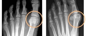
- radiography – pictures are taken in two projections, after which the required angles are calculated;
- plantography – a photograph of the imprints of the plantar parts of both feet, performed on a special device;
- CT and MRI are indicated if necessary to detail changes in the foot and soft tissues.
Diagnosis of hammertoe deformity:
The specialist performs a visual inspection and palpation.
To clarify the clinical picture, X-rays from three projections are used. If necessary, a CT or MRI may be prescribed. If a person’s disease has developed against the background of other acute or chronic pathologies, then first of all they do a full comprehensive examination, apply all the necessary diagnostic standards and carry out a differential diagnosis with diseases such as arthritis; gout; deforming osteoarthritis. Depending on the root cause of the disease, consultation with a rheumatologist, endocrinologist, therapist, or dermatologist may be required.
POLYTRAUMA / POLYTRAUMA
Leonova S.N., Usoltsev I.V.
Federal State Budgetary Institution "Irkutsk Scientific Center for Surgery and Traumatology", Irkutsk, Russia
A NEW METHOD FOR SURGICAL CORRECTION OF DEFORMITY OF THE SMALL TOES
The most common type of toe deformity is hallux valgus [1, 2]. Among the small toes, the second toe is more often subject to deformation, and the remaining toes are less likely to be deformed [3]. With the combination of hallux valgus and varus hammertoe deformity of the second toe (crossed toes), patients experience even greater suffering than with an isolated toe deformity. This is due to a pronounced cosmetic defect, which does not allow the selection of both regular and special shoes and sharply limits movement and communication. In addition, pain associated with deformation, metatarsalgia, formation of calluses, and wounds is of concern. To date, many methods of surgical correction of hallux valgus have been developed, and significantly fewer methods are used to treat deformities of the lesser toes [4-6]. Despite the successes achieved in the treatment of deformities of the 2nd, 3rd, and 4th toes, it is still not possible to avoid such postoperative failures as recurrence of deformity, recurrence of metatarsalgia, transitional metatarsalgia, stiffness and contracture of the metatarsophalangeal joint, and others [7-9]. The lack of effectiveness of the known methods of surgical correction of deformity of the small toes, in our opinion, is associated with the lack of a substantiated approach to the treatment of this pathology. Purpose of the study -
to propose a new method of surgical treatment of varus hammertoe deformity and evaluate its effectiveness.
MATERIALS AND METHODS
The results of examination and treatment of 27 patients with hallux valgus in combination with non-rigid varus-hammer deformity of the 2nd toe were analyzed at the Irkutsk Scientific Center for Surgery and Traumatology (INSCHT) (orthopedic department) for the period from 2016 to 2021. The study did not include patients: with deformation of the 3rd, 4th, 5th toes, under the age of 20 and over 65 years, with planovalgus foot, with elasticity of the foot of the 3rd degree, with systemic connective tissue diseases, systemic osteoporosis, with previous foot surgeries for this pathology. All patients were female and aged from 20 to 65 years, mean age 48.2 ± 12.38 years. The duration of the disease was 20.5 ± 12.14 years (from 2 to 40 years). In 77.8% there was an Egyptian type of foot. The study was carried out in accordance with the “Ethical principles of scientific medical research involving humans” as amended in 2000 and the “Rules of clinical practice in the Russian Federation”, approved by Order of the Ministry of Health of the Russian Federation dated June 19, 2003 No. 266. X-ray examinations were performed in all patients in preoperative and postoperative period in dynamics on the Agfa DX-D 300 apparatus in three projections (dorso-plantar and lateral), standing with a static load, focal length 1 meter. Surgical treatment of patients included correction of deformity of the 1st and 2nd toes. In all patients, the elimination of valgus deviation of the 1st finger was performed according to the author’s method [10]. Two surgical methods were used to eliminate mallet varus deformity of the 2nd finger, and therefore the patients were divided into two groups. The first group included 17 patients who were treated using the known method proposed by Weil [4]. In this case, a longitudinal skin incision is made directly above the 2nd metatarsal bone. After isolating the head and neck of the metatarsal bone, the joint capsule and collateral ligaments are dissected. The toe is then retracted plantar for optimal visualization of the metatarsal head. An osteotomy of the 2nd metatarsal bone is performed from the dorsal surface of its head proximally at an angle to the plantar surface of the metatarsal bone. The plantar fragment of the metatarsal bone is shifted proximally to the required shortening. The shortening depended on the length of the metatarsal bone, as well as the degree of subluxation. The osteotomy was fixed with one screw. The second group included 10 patients in whom the surgical method we proposed was used to correct the deformity of the 2nd finger, which consisted of the following. Before the operation, an X-ray examination of the patient's foot is performed. Using a full-size radiograph taken in a direct projection, the length of the required shortening of the metatarsal bone and the amount of the required lateral displacement of the distal fragment of the metatarsal bone relative to its proximal fragment are determined. On the radiograph of the foot in the deformation zone, draw a line of the longitudinal axis (3) of the metatarsal bone (1), perpendiculars (6, 7) are drawn to this line: one - through the extreme proximal point (4) of the epiphysis of the main phalanx of the finger (2) and the second - through the extreme distal point (5) of the distal epiphysis of the metatarsal bone. The distance between the first and second perpendiculars (6, 7) on the line of the longitudinal axis (3) of the metatarsal bone is the length of the required shortening (8) of the metatarsal bone (Fig. 1).
Figure 1. Scheme for determining the length of the required shortening of the metatarsal bone: 1 – metatarsal bone; 2 – main phalanx of the finger; 3 – longitudinal axis of the metatarsal bone; 4 – extreme proximal point of the epiphysis of the main phalanx of the finger; 5 – extreme distal point of the distal epiphysis of the metatarsal bone; 6 – perpendicular to the longitudinal axis of the metatarsal bone, drawn through the extreme distal point of the distal epiphysis of the metatarsal bone; 7 – perpendicular to the longitudinal axis of the metatarsal bone, drawn through the extreme proximal point of the epiphysis of the main phalanx of the finger; 8 – the length of the required shortening of the metatarsal bone, determined between perpendiculars 6 and 7.
Then two lines are drawn parallel to the longitudinal axis (3) of the metatarsal bone: the first through the midpoint of the articular surface of the head of the metatarsal bone (10), the second through the midpoint of the articular surface of the base of the main phalanx of the finger (9). The amount of required lateral displacement of the distal fragment of the metatarsal bone in relation to the proximal fragment of the metatarsal bone is established along the length of the perpendicular (13) drawn from the point of the middle of the articular surface of the head of the metatarsal bone to the opposite parallel line (Fig. 2).
Figure 2. Scheme for determining the distance of the required lateral displacement of the distal fragment of the metatarsal bone in relation to the proximal fragment: 9 – the middle of the articular surface of the base of the main phalanx of the finger; 10 – middle of the articular surface of the head of the metatarsal bone; 11 – a line drawn through point 9, parallel to the line of the longitudinal axis of the metatarsal bone; 12 – a line drawn through point 10, parallel to the line of the longitudinal axis of the metatarsal bone; 13 – the distance of the required lateral displacement of the distal fragment of the metatarsal bone in relation to the proximal fragment.
After determining the necessary parameters to correct the deformation of the toes, surgical treatment is performed. Under aseptic conditions, under spinal anesthesia with the patient in the supine position, a hemostatic tourniquet is applied to the lower third of the thigh and a dorsal direct projection incision is made over the distal third of the 2nd metatarsal bone, 3 cm long. The edges of the wound are separated and the neck (14) is exposed and the distal part of the diaphysis of the metatarsal bone, the periosteum is dissected and retracted to the sides. After this, using an oscillating saw, an osteotomy (15) of the metatarsal bone (1) is performed in the distal part of the diaphysis in the frontal plane at an angle of 60-70 degrees to its longitudinal axis (3), departing from the neck of the metatarsal bone (14) proximally 10 mm (16), from the dorsum to the plantar surface of the metatarsal bone in the distal direction (Fig. 3).
Figure 3. Scheme for determining the line of osteotomy of the metatarsal bone: 14 – neck of the metatarsal bone; 15 – osteotomy line of the metatarsal bone, at an angle of 60-70 degrees relative to its longitudinal axis; 16 – distance from the neck of the metatarsal bone to the osteotomy line of the 15th metatarsal bone
Using a rasp, the osteotomized fragments of the metatarsal bone are separated, and the proximal fragment is brought out into the wound. In the distal part of the proximal fragment of the metatarsal bone, two intersecting lines are drawn: the first (17) is drawn parallel to the osteotomy plane (15), departing from the distal edge of the proximal fragment at a distance equal to the length of the required shortening (8). The second line (18) is drawn perpendicular to the first line, starting from the distal edge of the proximal fragment, departing from the lateral surface of the metatarsal bone at a distance equal to the required lateral displacement (13) (Fig. 4a).
Figure 4.
Scheme of formation of a step-like recess on the proximal fragment of the metatarsal bone, comparison and fixation of osteotomized fragments: a) osteotomy plane and intersecting lines drawn in the distal part of the proximal fragment of the metatarsal bone: 17 – line drawn parallel to the osteotomy plane (15) of the metatarsal bone; 18 – a line drawn perpendicular to line 17, starting from the distal edge of the proximal fragment of the metatarsal bone; b) step-like recess of the distal part of the proximal fragment of the metatarsal bone (19); c) final position of the proximal and distal metatarsal fragments after compression (20); d) osteosynthesis of the compared fragments of the metatarsal bone and fixation with a screw (21).
Then, from the medial side of the distal part of the proximal fragment of the metatarsal bone, the step-shaped notch (19) obtained by marking between the two lines (17, 18) is cut out, forming a lateral protrusion on the lateral side of the distal part of the proximal fragment of the metatarsal bone (Fig. 4b). The proximal lateral corner of the distal fragment of the metatarsal bone is placed behind the cut of the formed lateral protrusion and compression is performed along the axis of the metatarsal bone until its fragments are in complete contact (20) (Fig. 4c). The fragments of the metatarsal bone are fixed with a cannulated screw (21) in an oblique direction from the head to the wall of the diaphysis of the metatarsal bone (Fig. 4d). In the postoperative period, all patients walked in orthopedic boots from the first day. The results of surgical treatment in all patients were assessed immediately after surgery and 6 months after surgery. Statistical processing of the results was carried out using the Statistica 10.0 program. In the compared groups, the arithmetic mean (M) and standard deviation (± SD) were determined. When performing a comparative analysis of the obtained quantitative data between groups, the t-Student criterion was used. Differences were considered statistically significant at p < 0.05.
RESULTS
In all 27 patients, intervention on the 1st ray made it possible to completely eliminate the valgus deviation of the 1st toe without loss of correction and relapse of the disease. The Weil technique was used to correct varus hammertoe deformity of the 2nd toe in 17 patients of the 1st group. In this case, Weil osteotomy of the 2nd metatarsal bone was used in combination with lengthening of the tendon of the long extensor of the 2nd finger, which made it possible to straighten the 2nd finger in all 17 patients, that is, to achieve a certain correction of the hammertoe deformity. However, during a clinical assessment of the result immediately after surgery, in 5 cases the elevated position of the main phalanx of the finger (dorsal deviation of the finger) was visually determined. According to the radiograph, 10 patients had dorsal subluxation of the metatarsophalangeal joint. In 12 patients (70.6%), deviation of the 2nd finger to the medial side (varus) was noted, while the radiograph revealed subluxation of the main phalanx of the finger in the horizontal plane. Taping was used to correct these deviations of the 2nd finger in the postoperative period. In the remaining 5 patients (29.4%) after surgery, a straight position of the 2nd finger was visually noted, which indicated that correction of varus deformity had been achieved, but the medial subluxation of the main phalanx of the finger remained on the radiograph. 6 months after surgery, during clinical and radiological examination, all 17 patients had medial subluxation in the metatarsophalangeal joint, 12 patients had varus deviation of the 2nd finger, 10 patients had dorsal subluxation, and 5 had dorsal deviation of the finger. Pain syndrome in the forefoot persisted in 14 patients, 10 of them had rare moderate pain, 4 patients suffered from intense daily pain in the 2nd metatarsophalangeal joint. In 7 patients, hyperkeratosis was detected on the plantar surface in the projection of the head of the 2nd metatarsal bone. In 5 cases, transitional metatarsalgia was observed in the subcapitate space of the 3rd metatarsal bone. 11 patients complained of limited physical activity at various loads. 12 (70.6%) of 17 patients were satisfied with the result of the treatment. 10 patients were operated on using the proposed method (group 2). No additional intervention on the tendon apparatus was used in this group. Clinical and radiological assessment of the treatment results immediately after the operation showed that in all patients it was possible to extend and straighten the 2nd toe, that is, to achieve correction of hammertoe and varus deformity of the 2nd toe. Subluxation in the 2nd metatarsophalangeal joint was eliminated, and the congruence of the articular surfaces of the head of the metatarsal bone and the base of the main phalanx of the finger was restored. During an observation period of 6 months, 3 patients experienced rare, moderate pain in the 2nd metatarsophalangeal joint; the remaining patients were not bothered by pain. Hyperkeratosis was eliminated. According to clinical and radiological data, all 10 patients did not reveal subluxations in the metatarsophalangeal joint, varus or dorsal deviation of the 2nd toe. In 1 case, transitional metatarsalgia was noted in the subcapitate space of the 3rd metatarsal bone. Two patients were concerned about the limitation of physical activity due to excessive exercise. All patients were satisfied with the results of the surgical treatment, a significant improvement in the appearance of the foot, its function and the ability to wear normal shoes. In patients of both groups, postoperative wounds healed in all cases without complications, and satisfactory fusion of bone tissue was achieved after osteotomies. A comparative analysis of the results of surgical treatment of varus hammertoe deformity of the 2nd toe in two groups of patients revealed the advantages of the proposed method (Table).
Table.
Comparative assessment of the incidence of indicators characterizing unfavorable results of surgical treatment in patients of two groups with varus hammertoe deformity of the 2nd toe 6 months after surgery (n = 27)
| Index | Frequency of occurrence | |||
| Group 1 (n = 17) | Group 2 (n = 10) | |||
| Abs. h. | % | Abs. h. | % | |
| Persistence of pain | 14 | 82.5 | 3 | 30 |
| Hyperkeratosis on the plantar surface | 7 | 41.2 | 0 | 0 |
| Limiting physical activity | 11 | 64.7 | 2 | 20 |
| Dorsal subluxation of the 2nd metatarsophalangeal joint | 10 | 58.8 | 0 | 0 |
| Subluxation in the 2nd metatarsophalangeal joint in the horizontal plane | 17 | 100 | 0 | 0 |
| Dorsal deviation of the 2nd finger | 5 | 29.4 | 0 | 0 |
| Varus deviation of the 2nd finger | 12 | 70.6 | 0 | 0 |
| Transitional metatarsalgia | 5 | 29.4 | 1 | 10 |
| M±SD | 10 ± 4.56* | 0.5 ± 0.84 | ||
Note:
* – significant differences between groups according to Student’s t-test at p < 0.01.
The significantly lower frequency of occurrence of the analyzed indicators by a factor of 20 and the minimal number of their manifestations in patients of group 2 serves as evidence of the greater effectiveness of using the proposed method of treatment compared to the known method. Thus, when using the known method of treatment, the majority of patients retained pain in the forefoot, there was varus deviation of the 2nd toe, subluxations in the metatarsophalangeal joint, and limitation of physical activity, which indicated insufficient correction of hammertoe and, to a greater extent, varus deformity 2nd finger. The use of the proposed treatment method made it possible to completely eliminate hammertoe and varus deformity of the 2nd toe, subluxations in the metatarsophalangeal joint, hyperkeratosis on the plantar surface, restore physical activity in 80% of patients, relieve pain in 70% of patients, that is, improve treatment results. Despite the advantages of the proposed method, there were no significant differences in the number of patients who were satisfied with the treatment result, which may be due to the initial severity of the deformity. We present a clinical example of a patient who underwent surgical treatment using the proposed method. The patient gave voluntary informed consent for the publication of the clinical case.
Clinical example
Patient D., 69 years old, applied to the clinic of the Institute of Chemistry and Traumatology and Traumatology. She complained of severe deformation of the 1st and 2nd toes of the left foot, the presence of a corn and sharp pain on the plantar surface in the projection of the head of the 2nd metatarsal bone, the inability to pick up and wear shoes, and fully use the left foot. foot. The patient was examined and radiography was performed (Fig. 5a, b, c).
Figure 5. Patient D., 69 years old. Photo of the left foot before surgery: a – top view; b – front view; c – radiograph in direct projection.
Main diagnosis:
“Transverse-longitudinal flatfoot of II-III degree on the left. Valgus deviation of the first toe of the left foot, varus-hammer deformity of the second toe of the left foot. Metatarsalgia." Based on the radiograph, the required shortening and lateral displacement of the 2nd metatarsal bone were calculated, which were equal to 4 mm and 3 mm, respectively. Surgical treatment began with correction of the deformity of the 1st finger, then correction of the deformity of the 2nd finger was performed using the proposed method (Fig. 6a, b, c).
Figure 6. Patient D., 69 years old. Photo of the left foot after surgery: a – top view; b – front view; c – radiograph in direct projection.
After the operation, the patient walked in an orthopedic boot on her left foot for 4 weeks and returned for follow-up examinations. A follow-up examination 6 months after surgery revealed: no complaints, satisfied with the result of treatment, no metatarsalgia, deformity of the 1st and 2nd toes of the left foot was completely eliminated (Fig. 7 a, b, c).
Figure 7. Patient D., 69 years old. Photo of the left foot 6 months after surgery: a – top view; b – front view; c – radiograph in direct projection.
The use of the proposed method of surgical treatment made it possible to achieve correction of the valgus deviation of the 1st toe and the varus hammertoe deformity of the 2nd toe of the left foot.
THE DISCUSSION OF THE RESULTS
The results obtained in the study indicate that the use of the Weil technique for the correction of varus hammertoe deformity is low. This technique involves performing a radical release of the metatarsophalangeal joint with dissection of the collateral ligaments, which leads to its destabilization. This contributes to the fact that the dorsal subluxation of the main phalanx of the finger persists; the finger tends to take an elevated position above the support or return to the hammer-shaped position. In addition, traumatic intra-articular intervention on the metatarsophalangeal joint can lead to contractures and/or joint stiffness in 10-20% of patients [8] and provoke pain. The classic Weil technique involves shortening the metatarsal by moving the metatarsal head proximally to correct hammertoe deformity. To correct varus deformity of the finger, it is necessary to perform a lateral displacement of the metatarsal bone fragment, which is not provided for by the Weil technique. That is, the use of the known technique does not eliminate varus deformity of the finger, but can only be effective in cases of hammertoe deformity. The proposed method of surgical treatment used new well-founded approaches, preliminary calculations, and techniques that made it possible to eliminate hammertoe and varus deformities of the finger. Preliminary determination from a radiograph of the foot of such parameters as the length of the required shortening and the amount of required lateral displacement of the distal fragment of the metatarsal bone relative to its proximal fragment, allows us to take into account the individual characteristics of the deformation of the fingers of each patient, and during the operation to accurately mark the lines in the distal part of the proximal fragment of the metatarsal bone and make cuts along them to properly eliminate the deformation of the toes, restore its anatomical state and static-dynamic function without relapses in the postoperative period. Osteotomy of the metatarsal bone is performed in the distal part of the diaphysis at an angle of 60-70 degrees to its horizontal axis, departing proximally 10 mm from the neck of the metatarsal bone, so that the osteotomy plane passes only in the diaphysis zone, without injuring the head of the metatarsal bone and the subcapitate space. As a result, when the diaphysis of the proximal fragment is compared with the diaphysis of the distal fragment of the metatarsal bone, the regenerative potential of the bone fragments and the nutrition of the head of the metatarsal bone are maximally preserved. In this case, the contact area of the fragments during osteotomy at an angle of 60-70 degrees is sufficient for stable fixation and successful fusion of osteotomized fragments of the metatarsal bone. Drawing two intersecting lines in the distal part of the proximal fragment of the metatarsal bone before performing an osteotomy allows you to accurately determine the distance by which it is necessary to shorten the metatarsal bone in order to straighten the bent toe, and the distance by which it is necessary to move the distal fragment of the metatarsal bone medially to restore the anatomical relationship the articular surface of the head of the metatarsal bone and the articular surface of the base of the main phalanx of the finger, achieving congruence at the metatarsophalangeal joint. Achieving congruence in the metatarsophalangeal joint and gentle intervention in the joint ensure restoration of movements in the joint, prevention of subsequent contractures, stiffness, and arthrosis. The recess, formed in the form of a rectangular step, makes it possible to more easily and clearly immerse the proximal lateral corner of the distal fragment of the metatarsal bone into it for complete contact of the proximal and distal fragments of the metatarsal bone, increase the rigidity of the fixation of the proximal and distal fragments of the metatarsal bone, and reliably stabilize the distal fragment relative to the proximal one. Displacement of the distal fragment of the metatarsal bone in a given direction allows you to unload the subcapitate space, remove the head of the metatarsal bone from the load of body weight and eliminate pain due to metatarsalgia. The use of the proposed method contributes to the successful correction of two types of toe deformities at once: varus and hammertoe deformities. Surgical treatment using the proposed method allows you to eliminate a cosmetic defect, pain, restore normal foot function, and provide the ability to wear regular shoes.
CONCLUSION
The proposed method of surgical treatment of deformity of the small toes provides a more accurate correction of varus-malleus deformity due to the pre-calculated necessary shortening and displacement of the metatarsal bone, taking into account the individual characteristics of the patient's foot condition, restoration of congruence of the articular surfaces of the metatarsophalangeal joint, reduces the risk of postoperative relapses of toe deformity and metatarsalgia , that is, it is clinically effective.
Funding and conflict of interest information
The study had no sponsorship. The authors declare that there are no obvious or potential conflicts of interest related to the publication of this article.
LITERATURE REFERENCES:
1. Kardanov A. The schemes and the pictures of surgery of the anterior part of the foot. M.: Medpractica, 2012; 20-25. Russian (Kardanov A. Surgery of the forefoot in diagrams and drawings. Medpraktika. M, 2012. P. 20-25) 2. Kondrashova IA, Kondrashov AN. Clinical and radiological aspects of diagnosis of hallux valgus and transverse platypodia. Injury
.
2003; 14(4): 81-86. Russian (Kondrashova I.A., Kondrashov A.N. Clinical and radiological aspects of the diagnosis of hallux valgus and transverse flatfoot // Trauma. 2013. T. 14, No. 4. P. 81-86) 3. Privalov AM. Modern possibilities of surgical correction of hammer toe. Kazan Medical Journal.
2017;
98(2): 296-299. Russian (Privalov A.M. Modern possibilities of surgical correction of hammertoe deformity // Kazan Medical Journal. 2021. T. 98, No. 2. P. 296-299) 4. Trnka HJ, Mühlbauer M, Zettl R, Myerson MS, Ritschl P. Comparison of the results of the Weil and Helal osteotomies for the treatment of metatarsalgia secondary to dislocation of the lesser metatarsophalangeal joints. Foot Ankle Int.
1999;
20(2): 72-79: 74-75 5. Espinosa N, Maceira E, Myerson MS. Current concept review: metatarsalgia. Foot Ankle Int
. 2008; 29(8): 871-879. doi: 10.3113/fai.2008.0000x 6. De Prado M. Minimally invasive foot surgery: a paradigm shift. In: Minimally invasive surgery of the foot and ankle. Maffulli N, Easley M, editors. London: Springer London. 2010. P. 3-11. doi: 10.1007/978-1-84996-417-3_1 7. Petrosyan AS. Surgical management of postsurgical complications and secondary deformations of anterior foot: abstracts of candidate of medical science. M., 2013; 24 p.m. Russian (Petrosyan A.S. Surgical treatment of postoperative complications and secondary deformities of the forefoot: abstract of thesis ... candidate of medical sciences. M., 2013. 24 p.) 8. Cherevtsov VN, Tadzh AA, Protsko VG, Tamoev S.K. Treatment of central metatarsalgia syndrome. Modern science: actual problems of theory and practice. Series: natural and technical sciences. 2017; (7-8): 139-144. Russian (Cherevtsov V.N., Taj A.A., Protsko V.G., Tamoev S.K. Treatment of central metatarsalgia syndrome //Modern science: current problems of theory and practice. Series: natural and technical sciences. 2021. No. 7-8. pp. 139-144) 9. A way of removing of non-rigid hammer deformations of the fingers 2, 3, 4 in transverse platypodia: patent 2610335, Russian Federation; MPC 7 A61 V 17/00. Ketov MS, Protsko VG, Zagorodniy NV, Ketova DV, Tadzh AA; applicant and patent owner Ketov MS, Protsko VG, Zagorodniy NV. No.2016102039; application January 22, 2016; published on February 9, 2021. Bulletin No.4. Russian (Method for eliminating non-rigid hammertoe deformities of the 2nd, 3rd, 4th toes with transverse flatfoot: Pat. 2610335, Russian Federation; MPC7 A61 B 17/00 / M.S. Ketov, V.G. Protsko, N.V. Zagorodniy, D.V. Ketova, A.A. Taj; applicant and patent holder M.S. Ketov, V.G. Protsko, N.V. Zagorodniy. – No. 2016102039; application 01/22/2016; publ. 02/09/2017. – Bulletin No. 4) 10. A way of surgical treatment of valgus deformation of the toe: patent 2592604, Russian Federation; MPK7 A61 V5/055 /Usoltsev IV, Leonova SN. Applicant and patent owner INtsKhT. – No.2015119292/15; application May 21, 2015; published on July 1, 2016. – Bulletin No.21. Russian (Method of surgical treatment of hallux valgus of the first toe: Pat. 2592604, Russian Federation; MPK7 A61 B5/055 / I.V. Usoltsev, S.N. Leonova; applicant and patent holder INCHT. - No. 2015119292/15; application. 05/21/2015; published 07/01/2016. – Bulletin No. 21)
View statistics
Loading metrics...
Links
- There are currently no links.
Treatment of hammertoe deformity:
Conservative treatment. In the initial stage, it is recommended to wear loose shoes and apply an adhesive bandage to the back of the foot.
In addition, the means of conservative treatment of hammertoe deformity are special orthopedic devices, such as a comb or splint for hammertoe. The use of such orthopedic devices helps to gradually straighten deformed fingers and relieve pain. It is also possible to use protective caps or bandages with gel, which prevents friction of fingers on shoes.
Surgical treatment consists of eliminating pronounced flexion contracture of the interphalangeal joints.
Percutaneous treatment method
Through small skin punctures 2mm. A special cutter with side sharpening is introduced and an osteotomy of a certain area of the bone occurs to eliminate the deformity. As a rule, this technique does not use metal structures or fixators, be it knitting needles or screws. The fingers are fixed with an elastic bandage in a given position. This is very convenient for the patient and does not require repeated surgery to remove the fixator. The following video provides an example of such an operation.
In most cases, after surgery, the patient goes home the same day, but sometimes stays for a day. The next day, the first dressing is changed with a doctor, then the patient can change the dressing independently. After 12-14 days, the sutures are removed.
Laser removal of finger callus. Laser surgery is a popular method, however, the laser is used primarily on soft tissue (not bone). Therefore, this method is used in addition to surgical instruments.
Surgical treatment of hammertoe deformity:
- Symptomatic. This treatment aims to remove all bone growths and protrusions. This surgical intervention achieves excellent treatment results in a short period of time. But there are cases when finger deformation after this type of treatment appears again after a few years.
- Radical. This treatment is aimed at deep plastic surgery of bones and ligaments. During surgical intervention, the transverse arch of the foot is formed, and flat feet (one of the main causes of hammertoe deformity) are also eliminated. This treatment has a longer recovery period than symptomatic treatment, but it is much more effective.
It should be noted that if surgical intervention on the feet is contraindicated for the patient, then individual production of orthopedic shoes is necessary, and in case of hammertoes deformities, a soft upper is additionally created on the shoes.
Care for hammertoes:
As a rule, the patient does not require additional care and is able to care for himself independently.
Prevention of hammertoe deformity:
The main prevention strategy is the timely detection and treatment of transverse flatfoot and hallux valgus (foot bunions). You should also give preference to comfortable shoes with loose toes instead of tight and uncomfortable shoes.
What is a hammertoe?
A condition in which the finger becomes deformed, resembling the shape of a hammer.
Any finger except the thumb can become deformed into a classic hammertoe. There is a true hammertoe deformity, and there is a deformity caused by constant pressure in the shoe. Let's consider true deformation using the example of the 2nd toe.
Rice. 1
- When transverse flatfoot progresses, the 2nd metatarsal bone moves down, and the proximal phalanx is forced to rise due to the traction of the ligaments and tendons (Fig. 1). In this case, the area of the proximal interphalangeal joint rubs in the shoes and a characteristic callus is formed.
Rice. 2
- If the second toe is longer than the first, then in the shoe it begins to rest against the toe and it is forced to bend (Fig. 2). At first we don’t feel this until a painful core callus forms, which can only be removed with a scalpel or surgical laser. This deformation is elastic and decreases when changing shoes over a period of 6 months. But there are patients who resort to shortening the 2nd toe in order to be able to wear dress shoes.