Home > Articles > Pathology
Qualified hand surgeons perform a full range of reconstructive surgeries on children and adults with congenital and acquired pathologies of the hand, as well as the entire upper limb.
The hand is one of the most important parts of our body. For a young patient, it is also a tool for actively exploring the world and developing intellectual abilities. Therefore, the sooner the full function of the hand is restored, the more actively your baby will develop.
Hand surgery is an independent branch of surgery that deals directly with the reconstruction and restoration of hand function.
The hand has a complex anatomical structure and provides highly coordinated movements, which are important for a full life.
Congenital pathology of the hand in children is a separate narrow area in hand surgery, requiring deep knowledge of the structural features of the child’s body, the variety of forms of congenital pathology, and the peculiarities of surgical interventions in childhood.
The specialists of our clinic have many years of experience in reconstructive operations in children with congenital and acquired hand pathologies. Anesthesiological support makes it possible to perform complex hand surgeries in children without the use of “harmful” medications through the use of regional blockades. In the shortest possible time after the operation, the child will be able to return to the usual rhythm of life without any health risks.
The hospital is provided with comfortable rooms and equipped with the most modern equipment from the best European companies.
Clinodactyly of the fingers
Clinodactyly is a disease characterized by deviation of the phalanges of the finger at an angle relative to the axis of the finger.
It can be congenital (in the presence of an additional phalanx) or acquired (due to injury). The most common type is congenital clinodactyly of the first and fifth fingers.
A small angle of deviation of the phalanx of the finger (up to 10 degrees) occurs quite often and does not require mandatory surgical intervention. Of course, if you want to improve the overall aesthetic perception, you can also correct minor angular deformations of the finger.
Congenital clinodactyly with a deformity angle greater than 20 degrees should be corrected promptly to improve hand function and eliminate the cosmetic defect
Clinodactyly of the little finger is a fairly common deformity, but in most cases it is only a minor cosmetic defect that requires correction only if there is a desire to improve the aesthetic appearance of the hand. The treatment methods we use allow us to preserve the function of nearby joints and reduce rehabilitation time. Clinodactyly requires correction to improve both the functional and cosmetic condition of the hand.
This pathology was assigned an ICD code M20. It refers to congenital diseases that develop due to developmental abnormalities during the prenatal period.
This anomaly is quite common in newborns and most often appears on either the arms or legs. In rare cases, articular joints on both types of limbs may be affected.
Most often, patients have only aesthetic problems with this condition, since physiologically the condition usually does not limit life activity. Only in rare cases can the disease significantly affect the quality of life and limit performance.
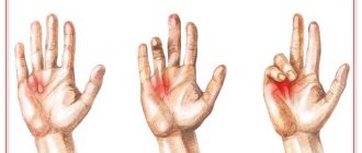
The disease most often manifests itself in the area of the little fingers, but other joints of the hands or feet in the area of the fingers can also be affected. Typically, clinical manifestations are symmetrical, affecting both limbs. When pathology manifests itself, the axis of the finger shifts sideways approximately at the level of the middle phalanx, if the condition is correlated with the axis of the hand.
In childhood, this disease is difficult to identify. The curvature process takes a fairly long period. Doctors are able to examine the real manifestations when the patient is about 18-20 years old. Accordingly, therapy begins around this age.
In the photo there is clinodactyly of the little fingers
Stenosing ligamentitis
Stenosing ligamentitis (or trigger finger) is a common disorder of the digital flexor tendon in which the child is unable to straighten the finger independently. This pathology significantly limits the function of the hand and causes severe discomfort to the baby.
Most often, stenosing ligamentitis of the first finger develops in the first year of life, in some cases on both sides. Damage to the remaining fingers is much less common. The cause of stenosing ligamentitis (snapping finger) is a fusiform thickening of the flexor tendon and its “closure” in the annular ligament.
When examining a child, parents can independently identify a club-shaped thickening at the base of 1 finger or on the palm with ligamentitis of 2-5 fingers)
If flexion contracture occurs in the interphalangeal joint and it is impossible to independently straighten the finger, it is necessary to consult a specialist involved in hand surgery to determine the optimal treatment tactics.
Description and classification of pathology
Syndactyly (photo) is a congenital anomaly (developmental defect) of the musculoskeletal tissues of the fingers and toes, characterized by their complete or partial fusion. In some cases, they talk about non-separation of the fingers at the stage of embryogenesis. The disease can affect one limb or several. It is distinguished by a variety of clinical manifestations.
Common types of syndactyly according to the International Classification of Diseases, 10th revision (ICD-10):
- fusion of fingers;
- webbed fingers;
- fusion of toes;
- webbed toes;
- polysyndactyly;
- syndactyly, unspecified (synphalangia NOS).
According to the type of fusion, syndactyly occurs:
- soft tissue or skin - represents the fusion of the fingers without deformation of the phalanges and synostosis (continuous pathological connection of the bone tissue of different fingers), with almost complete preservation of movement;
- bone - fusion of fingers with synostosis.
Syndactyly is diagnosed based on its length (depending on the number of affected phalanges):
- total (complete) - all phalanges of the fingers are fused;
- basal (incomplete) - proximal and/or middle phalanges are fused;
- terminal (end or distal) - nail phalanges are fused.
According to the degree of involvement of limb tissues in the pathological process, syndactyly can be:
- simple (isolated or separate) - represents the fusion of correctly developed fingers without accompanying deformities, detected in 40% of all recorded cases;
- complex - is part of a variety of syndromes of congenital anomalies of the musculoskeletal system, accompanied by fusion of the phalanges with the bones of the metatarsus and metacarpus, flexion contractures, torsion deformities and other defects.
Diseases with which syndactyly can be combined:
- ectrodactyly;
- polyphalange;
- polydactyly;
- aplasia of the fingers and/or toes;
- ectodermal dysplasia;
- cleft lip and/or palate;
- abnormalities in the development of the lacrimal glands and lacrimal ducts;
- urogenital defects (hypospadias);
- conductive hearing loss.
Disease statistics: frequency - 1 case per 2-4 thousand births, sex ratio - 50:50, unilateral syndactyly is detected 2 times more often than bilateral.
The condition is classified according to the reasons for its manifestation. The primary type indicates that the disease is an independent anomaly that developed due to a hereditary factor or random mutation. But the secondary type usually indicates the presence of more serious pathologies. Accordingly, their clinodactyly is only part of the clinical picture.
Congenital polydactyly of the hand
Polydactyly (from the Greek "poly" - many, "dactylos" - finger) is a congenital pathology of the hand, characterized by the presence of additional fingers. Polydactyly can occur as an isolated developmental defect and as part of genetic syndromes.
According to the degree of severity, the pathology can be represented either by a simple, additional skin rudiment or by a fully formed additional finger.
Thus, according to the type of duplication, polyphalanx, polydactyly and ray duplication are distinguished. In this case, polyphalange is understood as doubling of the nail or nail and middle phalanges, polydactyly is the doubling of the finger, and doubling the ray is an increase in the number of all segments of the finger and the corresponding metacarpal bone.
Polydactyly occurs in both the first (thumb) fingers and the fifth (little) fingers. Duplication of one of the central fingers is much less common and, as a rule, is combined with syndactyly (fusion) of the additional finger with neighboring ones (synpolydactyly). Another developmental anomaly, which is manifested by a significant increase in the number of fingers (up to 10 on one hand), is called “mirror hand.”
Surgical treatment of polydactyly in most cases is a complex reconstructive operation, during which the removal of the accessory segment, elimination of the underlying bone deformity, and reconstruction of the ligamentous-capsular apparatus are performed. Even when the extra toe dangles on a thin stem, simply cutting it off or tying it with thread can result in a painful scar in the area where the extra toe was located, since the nerve leading to it can be sealed into the skin scar.
The optimal time for treatment of polydactyly and polyphalanx (except for skin rudiment) is from 1 year to 1.5 years.
A preliminary consultation with a specialist will allow you to choose the best surgical treatment option for your child.
Syndactyly of the fingers and toes is a congenital developmental anomaly consisting in the fusion of one or more fingers with impaired function and cosmetics of the hand. This developmental defect can occur in isolated form: in these cases, the fingers are fully developed, but there is soft tissue or bone fusion between them. In most cases, syndactyly is a symptom accompanying the main diagnosis.
This pathology, alone or in combination with other deformities, including hyperplasia, clefting, clubhand, etc., according to a number of authors, accounts for more than 50% of all congenital hand anomalies.
The absence or limitation of differentiated movements of the fingers with congenital syndactyly is a great obstacle to the normal harmonious development of the child due to impaired grasp and, accordingly, psychomotor, and in some cases, intellectual development.
Congenital syndactyly of the hand is characterized by a variety of clinical manifestations.
By length, depending on the number of captured phalanges, the following are distinguished:
- incomplete form of syndactyly;
- full form of syndactyly.
Depending on the type of fusion, the following are distinguished:
- soft tissue form of syndactyly;
- bony form of syndactyly.
According to the condition of the affected fingers:
- simple form of syndactyly;
- complex form of syndactyly.
If this developmental anomaly is detected in a child, it is necessary to consult an experienced surgeon. A diagnostic test will require radiography.
To eliminate this congenital developmental anomaly, reconstructive plastic surgery is indicated, aimed at eliminating the fusion of the fingers of the hand, eliminating the cosmetic defect and restoring its function.
If there are concomitant bone deformities during surgery, additional simultaneous correction of the bone component with metal fixation is possible.
After surgery, a small light-colored scar remains, which subsequently becomes almost invisible with proper rehabilitation treatment.
Surgical treatment of complex forms of congenital syndactyly of the hand in children should begin at the age of 6 months - 1.5 years, before the development of a stereotype of using the affected hand and the appearance of secondary deformities
Clinical picture
The condition is manifested by the following symptoms:
- The first stage is manifested by a deviation of the finger by 10°;
- Next, shortening of the affected finger occurs, but, as a rule, it is insignificant;
- The finger is severely deformed;
- The tilt angle reaches 20°.
There is no pain with clinodactyly. The mobility of the affected part is limited. The patient's finger activity may decrease and writing becomes difficult. It is harder to hold some objects in your hands.
Disease prevention
The condition of clinodactyly is congenital. This anomaly is usually inherited. The main cause of the defect is a change in the epiphysis in the middle part of the finger. Its growth begins to resemble a triangle or trapezoid.
It is worth noting that this condition can be independent or be part of the clinical picture of other pathologies. These include the following pathologies:
- Klinefelter's syndrome;
- Turner syndrome;
- Down syndrome;
- Trisomy on the 18th pair of chromosomes.
These conditions are quite severe, and therefore clinodactyly itself and its treatment, as a rule, fade into the background. But more often than not, the pathology is hereditary. Moreover, if in one parent the disease manifests itself at the first stage, then in the child it can grow to the second.
Important! The disease is transmitted in an autosomal dominant manner. But a gene mutation can also occur spontaneously.
In most cases, syndactyly is of an endogenous nature and is inherited in an autosomal dominant manner, when one of the parents is a carrier of the corresponding disease “defective” gene in a heterozygous state on one of the paired chromosomes. However, sporadic cases are also known. The deformity originates in the initial period of intrauterine development, between 4 and 8 weeks of pregnancy, when the embryo develops fingers and toes.
Changes in the human genome responsible for the development of syndactyly:
- duplication of chromosome 2q35 - causes skin fusion of the 3rd and 4th fingers, 2nd and 3rd toes, synostosis of the nail phalanges;
- heterozygous mutations in the HOXD13 gene - are the cause of two types of complex synpolydactyly involving the phalanges and soft tissues of the fingers, metacarpus and metatarsus in the pathological process;
- mutations of the GJA1 gene - cause complete bilateral syndactyly of the 4th and 5th fingers, hypoplasia/aplasia of the middle phalanx of the little finger;
- mutations in the ZRS regulatory element of the SHH gene, located in the region of intron 5 of the LMBR1 gene, provoke the development of complete bilateral syndactyly of the hands in combination with polydactyly (cup-shaped hand syndrome);
- mutations in the LRP4 gene cause deformation of the phalanges of the hand with complete syndactyly (spoon-shaped hand).
In case of inheritance of a mutation, the possibility of transmitting the disease to children from a sick parent is 50%. In the case of a primary (sporadic) occurrence of a defect in a child, the risk of relapse in the second and subsequent children is minimal and amounts to no more than 1%.
Causes of new mutations in genes leading to syndactyly:
- injuries of the uterus and fetus;
- oligohydramnios, increased pressure of the uterine walls;
- inflammatory processes in the mother's body;
- taking certain medications during pregnancy;
- exposure to chemicals or radiation.
Slowing down the development of the embryo under the influence of negative external and internal factors increases the risk of congenital defects several times.
There are no methods for preventing hereditary syndactyly as such. Modern genetic studies of the genome of future parents and close relatives make it possible to identify carriers of a defective gene even at the stage of planning a child. Cases of simple fusion are not an indication for termination of pregnancy, as they are completely cured in 80-90% of cases.
You can prevent the development of sporadic mutations:
- undergoing the necessary examinations during pregnancy in a timely manner;
- following the advice of your doctor;
- eating well and properly;
- avoiding external and internal negative influences on the body.
When a fetus is diagnosed with a severe syndrome of congenital pathologies, including syndactyly, parents should carefully consider the decision to continue pregnancy or terminate the pregnancy. In this case, one should take into account the possibilities of modern medicine, the psychological and financial state of the family.
Reasons for the development of the anomaly
Clinodactyly can be an independent disease, or it can act as a symptomatic sign of various pathologies.
You should know what underlies the occurrence of the disease. Treatment of clinodactyly depends on the severity of the disease and the factors that provoked its development.
In most cases, the disease is hereditary. If the mother was diagnosed with type 1 clinodactyly, then the daughter may have a clinical picture of the 2nd stage of the development of the disease. Numerous studies have shown that the disease is transmitted in an autosomal dominant manner.
If we consider clinodactyly as a component of more serious genetic pathologies, then the disease can occur in the following cases:
- Down syndrome;
- Klinefelter's syndrome;
- trisomy in the 18th pair of chromosomes;
- Turner syndrome.
Doctors say that the disease can develop due to a spontaneous gene mutation.
In this case, the most difficult thing is to identify the disease in a timely manner and take the necessary measures. Doctors claim that clinodactyly can be diagnosed during routine ultrasound screening during pregnancy. The woman may be referred to geneticists, who will carry out a procedure that involves determining the child’s karyotype to subsequently exclude chromosomal abnormalities.
Indications for surgery
In addition to aesthetic discomfort, syndactyly of the hands and fused toes in a newborn cause limitation of the normal functioning of the fingers and the entire limb as a whole. Impaired hand function and difficulty in grasping activity negatively affect the psychomotor, speech, and in some cases intellectual abilities of the child, his full and harmonious development. This greatly complicates the learning process and limits the possibilities for self-realization and choice of a future profession.
Syndactyly of the toes in children is rarely complicated by functional disorders and requires surgical treatment only if the supporting functions of the foot are reduced.
In adulthood, such an aesthetic “defect” as fused fingers can bring psychological suffering to a person. Such patients often complain of an inferiority complex, isolation, lack of self-confidence, psychological stress, and difficulties in communicating with the opposite sex. Those with syndactyly of the toes are embarrassed to go to the gym or pool, do not go to baths and saunas, never expose their feet in hot weather, and experience discomfort on the beach. All these factors negatively affect the quality of life of a person with syndactyly, which results in a visit to the surgeon.
Types of surgical treatment
Elimination of syndactyly is carried out surgically. The type, duration and number of surgical interventions depend on the subtype of the disease, the degree of damage to soft tissues and bones, and the presence of associated deformities.
Recommendations for timing of operations for newborns:
- simple skin thermal (partial) fusion - at 3–4 months;
- basal or complete membranous fusion - at 5–7 months;
- complex syndactyly with synostosis - no earlier than 12 months;
- complex bone syndactyly with the presence of accompanying deformities - from 1.5–2 years.
The essence of the surgical intervention is to separate the fused phalanges and restore maximum functionality of the limb. Each operation requires careful preparation, which consists of preoperative diagnosis. To clarify all the nuances of deformation, the following is carried out:
- a thorough physical examination;
- ultrasonography of the arm and/or leg;
- Dopplerography of limb vessels;
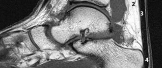
- X-ray examination in at least two projections;
- magnetic resonance, computed tomography.
Modern medical equipment can correct syndactyly using gentle, minimally invasive endoscopic or microsurgical methods, which can significantly shorten the rehabilitation period and significantly reduce the percentage of possible complications.
Basic and additional types of surgical reconstruction measures for limb syndactyly:
- dissection of the skin membranous fusion without plastic surgery;
- dissection of fused fingers, covering defects with donor skin flaps from the patient with the formation of an interdigital fold;
- multi-stage operations with restoration of bones, tendons, muscles and skin;
- excision of scar tissue after unsuccessful operations;
- filling defects with skin grafts;
- excision of tumors (if detected) followed by sending the material for histological examination.
All types of operations to eliminate the disease in children are performed under general anesthesia; dissection of simple membranous syndactyly in adults is allowed to be performed under local anesthesia.
Diagnostics
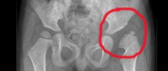
After birth, the condition is determined by visual examination, as well as by x-rays.
In the process, it is also important to differentiate the primary type from more serious diseases.
During pregnancy, according to some doctors, a similar anomaly in the fetus can be determined by screening using the CA marker. This is usually done at a routine ultrasound around 19 weeks.
If a woman is suspected of such a condition, she is referred to a geneticist, who carries out manipulations to determine the karyotype. In this way, it is possible to exclude or confirm chromosomal abnormalities.
X-ray of a child's hand in clinodactyly
What should I do if I have a similar but different question?
If you did not find the information you need among the answers to this question, or your problem is slightly different from the one presented, try asking an additional question to the doctor on the same page, if it is on the topic of the main question. You can also ask a new question, and after a while our doctors will answer it. It's free. You can also search for the information you need in similar questions on this page or through the site search page. We will be very grateful if you recommend us to your friends on social networks.
We answer 96.45% of questions.
Clinodactyly is a congenital condition in which an abnormal development of the musculotendinous system leads to limited mobility of the fingers or toes. At the same time, the brush itself takes on a unique appearance.
It is impossible to regard this condition as a defect from an aesthetic point of view, since the curvature of the fingers is not immediately noticeable.
Postoperative rehabilitation
The recovery period after surgery can last from 2 to 6 months. A set of rehabilitation measures, individually designed for each patient, can significantly reduce this period and completely restore the function of the limb.
Immediately after surgery, the two fingers are immobilized (fixed) in the correct position using a removable cast. To prevent re-fusion, special spacers are inserted into the interdigital space.
After the sutures are removed, rehabilitation measures begin, the goal of which is to restore full finger movements.
Physiotherapeutic treatment includes:
- massage of the operated limb;
- electrophoresis;
- ultraphonophoresis;
- thermal applications with ozokerite, paraffin;
- acupuncture;
- mud applications.
Patients are recommended to attend physical therapy classes. Following the recommendations of specialists, it is advisable to continue to perform exercises and develop the limb at home.
Types of hand exercises that can be performed after eliminating syndactyly:
- careful flexion and extension of each phalanx;
- rotation of the operated fingers;
- grip training (sequential connection of the thumb with other fingers);
- folding small objects (puzzles, beads, coins, buttons);
- rolling a rolling pin on the table;
- modeling from plasticine, salt dough;
- rolling the ball in the palms.
Performing exercises is permissible until pain appears. Loads should be increased gradually.
How to treat
Considering that in most cases the disease is hereditary, it can be understood that medications are extremely ineffective in treating the condition. If we talk about exercise therapy and physiotherapy, then such measures of influence can give minimal results.
Clinodactyly is largely an aesthetic disorder, and therefore the issue of eliminating such an anomaly is resolved by the appropriate method - plastic surgery.
Important! This type of operation has no special contraindications, and therefore can be used even in childhood. But still, doctors recommend waiting until the formation of the skeleton and musculo-ligamentous apparatus is complete.
Surgical treatment photos before and after surgery
Forecast
In the absence of therapy, most often the prognosis is relatively positive. That is, the defect will persist, but will not greatly affect life activity. In rare cases, such an anomaly can somewhat impair the functionality of the hand and the ability to hold specific and small objects.
When performing plastic surgery, the aesthetic defect can be corrected. The disease progresses and leads to disability in isolated cases.
Features of hand plastic surgery, recommendations and reviews in our video: