Wrist sprain
happens quite often, since hands are the main human tool. Provoking factors are:
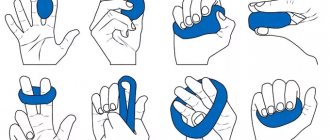
- physical exercises performed incorrectly;
- Careless movements, including in children due to the fault of adults;
- unsuccessful falls, blows;
- costs of the profession if it is necessary to repeatedly repeat movements and constant physical activity.
Factors predisposing to complications:
- old age, when the elasticity of connective tissues is lost;
- congenital tendon pathologies;
- inflammation of tissue in the joint area;
- prolonged sitting;
- insufficient physical development.
Causes
The main cause of inflammation is prolonged tension in the wrist joint, which appears due to microtrauma and increased physical activity. Constant load on this place contributes to the deposition of salts in the tissues of the tendon and cartilage.
This occurs due to monotonous, frequently repeated movements. The disease can be called occupational; people of certain professions are susceptible to it:
- musicians;
- seamstresses;
- builders;
- athletes (rowing, tennis, golf);
- people employed in agriculture;
- mining workers;
- if the profession involves working on a computer.
Styloiditis can develop after bruises, fractures, dislocations and any other injuries. Sometimes the cause is a local metabolic disorder, heredity or a complication after infectious diseases or other inflammations.
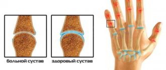
Symptoms and location of pain
For each type of injury listed above, there are characteristic symptoms, based on which a qualified specialist will establish an accurate diagnosis and prescribe the necessary treatment. The main signs of damage to the wrist joint include:
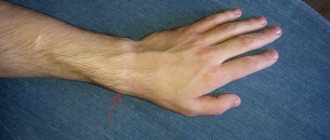
- Joint pain of varying intensity and location
- Edema and swelling of the injured area
- Changes in the appearance of the hand and wrist
- Redness of the skin in the damaged area
- Hemorrhage at the site of injury
- Joint contracture (severe limitation of joint mobility)
- Increase in overall temperature
- Weakness, malaise, chills
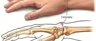
- The appearance of cones (with hygromas)
Symptoms of the disease
What is the characteristic of styloiditis of the wrist joint?
- aching pain in the wrist area, which can extend to the elbow or hand;
- increased discomfort in the joint when moving the thumb or hand, at night or when weather conditions change;
- if the cause was injury, pain is felt even if you just touch your hand;
- the appearance of all signs of inflammation - redness, swelling, increased temperature;
- the joint crunches when moving;
- the tendon tightens and becomes hard;
- decreased sensitivity, including pain;
- problems with fine motor skills and finger movements, difficulty grasping and holding objects;
- fingers become numb, tingling or burning may be felt;
- muscle tone and strength deteriorate.
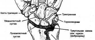
Midcarpal joint, art. mediocarpea
Located between the bones of the proximal and distal rows of the wrist. The structure of the joint is complex; its shape is a combination of two spherical joints.
Movements: increases the range of movements in the wrist joint - mainly around the frontal axis: flexion and extension; to a lesser extent abduction and adduction around the sagittal axis.
Ligaments that strengthen the joint: a) palmar intercarpal ligaments, ligg. intercarpalia palmaria; b) dorsal intercarpal ligaments, ligg. intercarpalia dorsalia; c) interosseous intercarpal ligaments, ligg. intercarpalia interossea.
The hand joint is a combination joint that combines the radiocarpal joint and the midcarpal joint. The movements in them occur simultaneously and complement each other.
Carpometacarpal joints of the hand, art. carpometacarpales
1. Carpometacarpal joint of the thumb, art.carpometacarpalis pollicis. Formed by the articular surfaces of the first metacarpal bone and the trapezium bone. The joint is simple in structure, saddle-shaped in shape, and biaxial in function.
Movements: around the frontal axis - flexion and extension; around the sagittal axis – abduction and adduction. When these movements are combined, complex movements occur - opposition and reposition.
2. Carpometacarpal joints of the II-V fingers, art.carpometacarpales II-V. Formed by the bases of the II-V metacarpal bones and the bones of the distal row of the wrist (trapezoid, capitate and hamate). The joints are complex in structure, flat in shape, and inactive in function.
Ligaments that strengthen the joint: a) dorsal carpometacarpal ligaments, ligg. carpometacarpalia dorsalia; b) palmar carpometacarpal ligaments, ligg. carpometacarpalia palmaria; c) interosseous carpometacarpal ligaments, ligg. carpometacarpalia interossea.
The hard base of the hand is the bones of the distal row of the wrist and the II-V metacarpal bones, connected by inactive joints and strengthened by ligaments.
Metacarpophalangeal joints, art.metacarpophalangeae
Formed by the articular surfaces of the heads of the metacarpal bones and the base of the proximal phalanges of the fingers. The joints are simple in structure, spherical in shape, but biaxial due to the presence of collateral ligaments and the absence of rotator muscles.
Movements: around the frontal axis - flexion and extension; around the sagittal axis – abduction and adduction; circumduction.
Ligaments that strengthen the joints: a) deep transverse ligaments of the metacarpus, ligg. metacarpalia transversa profunda; b) collateral ligaments, ligg. collateralia; c) palmar ligaments, ligg. palmaria
Diagnostics
It is difficult to identify styloiditis, since it is practically no different from other diseases of the joint and its tissues. During the examination, sometimes only some clinical signs are revealed. To accurately determine styloiditis, a comprehensive examination is necessary:
- visual inspection;
- collecting anamnesis for occupational, sports or domestic injuries;
- ultrasonography;
- radiography;
- laboratory research;
- CT scan;
- Magnetic resonance imaging.
Prevention
Preventive measures are necessary, especially if there is a possibility of styloiditis occurring in connection with professional activities:
- do not make sudden movements, use the entire hand to grasp the object;
- warm up muscles before physical education or sports;
- wear special gloves when working with vibrating tools;
- when working with a computer, rest every hour for 5 minutes, stretch your fingers and hands;
- the position of the body and hands at the computer should be as natural as possible;
- do special exercises if work involves a risk of developing a disease;
- try not to do monotonous work for a long time without a break.
If prevention is not carried out, the likelihood of developing styloiditis of the wrist joint increases.
There is no need to turn a blind eye to pain: identifying the problem in the initial stages will help you get rid of it quickly and without problems. Depending on the form and severity of the disease, the doctor will select suitable treatment methods. At the initial stage, you can easily and quickly get rid of styloiditis using shock wave therapy without surgery.
Types of damage
Common types of injuries to the wrist joint include:
- Fractures. The radius bone is especially often injured. There are two types of fracture of the radius at the wrist joint:
- Smith's fracture (flexion fracture). The cause of injury is a fall on an outstretched arm, on its back side. The bone breaks and at the same time there is a displacement of bone fragments towards the palm.
Colles' fracture (extension fracture). Damage occurs when a person falls on the palm, and there is a displacement of bone fragments towards the thumb and the back of the hand.
. They can occur against the background of previous injuries, hormonal disorders, excessive joint tension, infections, etc.
- Tenosynovitis is an inflammatory lesion of the tendons and surrounding membranes in the wrist area.
Treatment
Features of treatment of the initial stage of the disease:
- The hand is immobilized for 14 days. To do this, apply a splint or splint made of plaster.
- To relieve pain, use external ointments: diclofenac, voltaren, fastum-gel.
- For severe pain, nonsteroidal anti-inflammatory medications are needed: movalis, ibuprofen, ketorol, diclofenac.
- Glucocorticoid injections: hydrocartisone, diprospan, kenalog.
- It is necessary to apply a cold compress of ice 3-4 times a day. This is usually done in the first few days.
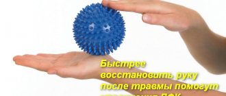
- Physical therapy will help improve blood flow and metabolism in the problem area. It can be used when there is an acute inflammatory process.
Of the conservative methods of treatment, many physiotherapeutic methods will also be useful:
- electrophoresis using lidase;
- magnetic therapy;
- ultrasound therapy;
- ozokerite;
- laser treatment;
- mud and paraffin compresses;
- massage with special ointments, manual therapy.
Sometimes the disease cannot be detected in the early stages. In severe and advanced cases, when conservative methods do not work, surgery is performed by excision of the problem part.
Full recovery after surgery occurs within 3 months. Athletes can engage in their activities after 3-4 months. To speed up tissue renewal and restoration, you need to actively move the brush. After the operation, you can immediately develop the thumb, and after a few days - the entire hand.