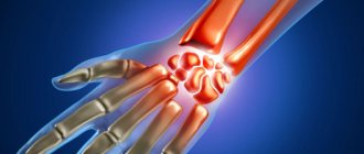
In the earliest stages, the disease may manifest itself as minor aching pain in the wrist joint. As the disease progresses, the pain becomes regular, especially intensifying with physical activity.
In advanced stages, it is impossible to easily move the hand, not only because of acute pain, but also because of cartilage degeneration.
Arthrosis is a disease caused by degeneration of cartilage and adjacent bone tissue, and if appropriate treatment is not started in time, this can lead to joint deformation and complete immobility of the hand. Since it is almost impossible to restore a destroyed joint to its original healthy state, it is necessary to stop its destruction in a timely manner using medication and physiotherapy.
In the most advanced stages, surgical intervention will be required to partially restore the functions of the wrist, but is it worth neglecting your health so much that you don’t find time to undergo diagnostics in our clinic and begin treatment.
Causes and mechanism of occurrence
This pathology is always a consequence of some underlying disease. The conditions that most often result in the development of deforming arthrosis of the wrist joint include:
- osteoarthritis;
- rheumatoid arthritis;
- psoriatic arthritis;
- traumatization.
Osteoarthritis
Osteoarthritis develops due to physiological wear and tear of the constituent components of the wrist joint. This is especially true for people who have a family history of arthritis. The disease occurs in middle-aged patients, although it occasionally occurs in young people.
Osteoarthritis involves the gradual wearing away of the smooth, slippery articular cartilage that covers the ends of bones. In patients with DOA after wrist osteoarthritis, the articular surfaces of the radial, scaphoid, lunate and triquetral bones become thinner, and the joint space narrows. Since the surface of the cartilage has virtually no blood supply, its regenerative ability in case of damage and wear is somewhat reduced. When cartilage wears down, it becomes rough and the protective space between bones decreases. This leads to deterioration in joint function and proliferation of connective tissue, which further limits its mobility and deformation.
Rheumatoid arthritis
Rheumatoid arthritis is a chronic autoimmune disease characterized by multiple joint lesions. As a rule, the disease affects small joints, and often such changes are symmetrical. With this disease, damage to the small joints of the hands is often observed.
In rheumatoid arthritis, immune system cells infiltrate the synovium that covers the joint surfaces. This creates aggressive connective tissue called pannus. It releases substances that promote the destruction of bone, cartilage and surrounding ligaments. Due to damage to the fixing articular apparatus, minor subluxations are observed. As a result, the joints lose their configuration, which leads to their deformation and subsequent loss of function.
Drug therapy
Non-steroidal anti-inflammatory drugs are used to treat arthrosis of the wrist joint. This group of products is aimed at eliminating inflammation in the area of injury and providing mild pain relief. It includes Aspirin, Diclofenac, Indomethacin, Ibuprofen, Ketoprofen and others.
NSAIDs are considered relatively safe drugs for the body, but taking them for a long time is not recommended due to the possibility of adverse reactions. Medicines have contraindications: diseases of the stomach, heart, kidneys, liver. Long-term use of drugs from this group has a corrosive effect on the mucous membranes.
Corticosteroid drugs are powerful hormonal medications. In the treatment of dystrophic disorders of the osteochondral apparatus, this group of medications is very effective. Medicines are prescribed for severe pain when non-steroidal drugs do not give the desired result. According to the intensity of action, all corticosteroids are of several types:
- weak - Prednisolone, Hydrocortisone;
- moderate action - Fluorocort, Lorinden;
- strong - Ekolom, Advantan, Triderm;
- very strong - Klovate.
The stronger the remedy, the more caution it should be taken.
Treatment with corticosteroid drugs should only be prescribed by a doctor. Their spontaneous use is dangerous to health.
Medicines in this group also have contraindications. They should be used with caution in case of diabetes, hypertension, stomach ulcers, and mental disorders. It is better not to use potent drugs for treatment if there are bleeding disorders, infections, or severe forms of osteoporosis. Corticosteroids are ineffective if the patient has a transarticular fracture, with progressive destruction and deformation of the joint that is incurable with medication.
Adverse reactions include nervous excitability, irritability, muscle spasms, uncontrolled weight gain, growth retardation in children, and problems with puberty.
Chondroprotectors are drugs that improve the condition of cartilage and promote their restoration, nourishing and moisturizing joint structures. The first results are observed after a long time from the start of taking medications of this group, since regeneration processes proceed very slowly. Chondroprotectors are most effective in the treatment of arthrosis of the wrist joint in the early stages. If the process of deformation of the joint has begun, then it no longer makes sense to use these drugs.
The list of means for restoring cartilage structures is extensive. Chondroxide, Teraflex, Arthrodar, Alflutop and others are widely used in the medical practice of osteoligamentous pathologies. There are no absolute contraindications to their use. Relative ones include pregnancy, breastfeeding and allergies to individual components of the drug.
Psoriatic arthritis
Psoriasis is an autoimmune disease that primarily affects the skin and secondarily the joints and kidneys. The development of inflammatory changes in joint tissues during psoriasis is most often observed in patients aged 26–54 years.
Often this process is multicentric, and damage to the interphalangeal joints of the hands is observed.
Psoriatic arthritis, which develops in 30% of psoriasis cases, often entails the development of deforming osteoarthritis.
Injury
Post-traumatic arthritis, which leads to deformation and the development of osteoarthritis, can develop:
- in case of primary traumatic injury if the correct regimen is not followed during healing (continued loads on the joint, secondary traumatization);
- under constant load, often professional (painters, musicians, fencers).
These two factors lead to perverted regeneration of the fixing apparatus of the joint - the lateral radial, lateral ulnar, dorsal radiocarpal, palmar radiocarpal, intercarpal interosseous ligaments. This leads to degenerative changes and deformation of the radiocarpal joint.
Symptoms
As a rule, the following symptoms are noted with DOA of the hands:
- pain;
- edema;
- decreased joint mobility;
- weakness;
- actual deformation.
Don't miss: MRI of the wrist joint
The presence of these symptoms, as well as their severity, will be influenced by the underlying disease.
The most common and first sign is pain. If we talk about arthritis, when degenerative changes are still reversible and not visually noticeable, pain in the wrist joint usually worsens during movement. As the disease progresses, it gradually intensifies.
Swelling is often a manifestation of inflammatory changes and accompanies pain in cases of exacerbation of chronic arthritis.
The mobility of the joint can also gradually deteriorate, until the development of ankylosis - immobility of the joint.
In rare cases where a patient has hyperelasticity, such as in Ehlers-Danlos or Marfan syndromes, the wrist may maintain a wide range of motion despite severe degenerative changes.
The pathognomonic symptom for DOA is deformity. In existing rheumatoid arthritis, the deformity may be associated with subluxation of the radiocarpal joint.
Pain and deformity lead to loss of hand function. It becomes difficult for the patient to perform usual activities related to the load on the wrist. Considering that degenerative changes also affect the anatomical canals formed by the tendons of the flexor and extensor muscles, numbness of the fingers, their weakness and deterioration in sensitivity may be observed. This occurs because as connective tissue grows in the carpal and ulnar canals, the transmission of nerve impulses along the ulnar and median nerves will deteriorate.
FIND OUT MORE:
Deforming arthrosis of the knee joint: etiology, clinical picture, diagnosis and treatment
Treatment of deforming arthrosis of the foot
Deforming arthrosis of the shoulder joint grades 1, 2 and 3 - traditional treatment and folk remedies
Treatment of deforming arthrosis of the hip joint grades 1, 2 and 3
Etiology and pathogenesis
There are a large number of reasons for the development of arthrosis of the wrist joint, but the most common of them are associated with injury. For this reason, the disease is considered occupational among athletes, agricultural workers, seamstresses, builders, and typists. The negative impact on the joint is aggravated by vibration that occurs when working with a jackhammer, drill, and other tools.
The post-traumatic form appears several weeks and sometimes months after the injury. By this time, all the consequences in most cases have healed, and the pain syndrome has passed.
Other causes of the disease:
- aging processes;
- injury;
- birth defects;
- inflammatory processes, autoimmune diseases (rheumatoid form);
- diseases of the endocrine system, hormonal imbalances;
- hypothermia;
- lack of nutrients,
- connective tissue diseases.
Cartilage and joint tissues begin to deteriorate due to mechanical damage. Against the background of the processes of destruction of cartilage tissue, joint deformation occurs, and specific growths in the form of spines (osteophytes) form on the tissues. Inflammation occurs almost immediately - with rheumatoid and other forms of arthritis.
Due to calcification of ligaments, the elasticity of cartilage decreases, and there is a risk of damage to nerves and blood vessels by formed osteophytes.
In the later stages of the disease, damage occurs to the periarticular tissues that are located outside the boundaries of the joint capsule.
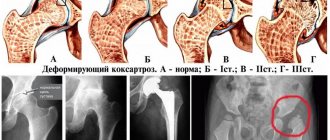
Staging: 1, 2 and 3 degrees
In total, there are three degrees of DOA of the hand joints. These stages reflect the severity of osteoarthritis itself, based on the nature of the pain syndrome and physical examination data.
1st degree
At this degree, the pain is aching in nature. As a rule, it passes at rest and does not hinder movement in the joint. At this stage there is no need to use drug therapy. Both physiotherapeutic measures and folk remedies are appropriate. At this stage, it is possible to permanently stop deforming arthrosis.
2nd degree
At this degree, the pain becomes stronger and more intense. It worsens the patient’s quality of life and begins to disturb him at night. When examining the joint, compactions may be observed. At this stage, dystrophic changes are already irreversible.
3rd degree
With grade 3 DOA, there is obvious deformation of the joints, often nodular growths are observed in them. Patients almost always experience pain and paresthesia.
Establishing diagnosis
To make a diagnosis of DOA of the wrist joint, it is necessary to collect an anamnesis, a qualitative physical examination of the patient, as well as data from instrumental and laboratory research methods.
Anamnesis
When collecting anamnesis, the doctor needs to clarify the nature of the pain syndrome, what the patient associates with the appearance of this pain, how long ago it appeared and as a result of what treatment measures it goes away.
The doctor also clarifies the presence of similar joint changes in someone in the patient’s family, because some inflammatory and degenerative processes are associated with a hereditary predisposition.
A summary is made of existing diseases. Let's say that if a patient has already been diagnosed with rheumatoid arthritis or psoriasis, it will be quite easy to guess the nature of the pathological changes in the joint. This will also help the doctor decide on the treatment method.
Don't miss: Treatment of rheumatoid arthritis in the hand joints
The most important issue for a doctor is the profession of the patient. If he works with tools associated with constant load on the wrist joint (sports, construction work, playing musical instruments) or any other negative physical factor (for example, vibration when using a drill), there is a high probability of traumatic DOA.
Physical examination
During examination, the doctor may find:
- decreased range of motion in the wrist area;
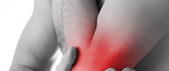
- weakness and pain in certain areas of the hand and the joint itself;
- signs of instability of the wrist joint;
- edema.
Diagnostics
Diagnosis begins with examination and palpation of the affected area, the doctor compares the joints on both hands, paying special attention to anatomical features (protrusions, folds, dimples). The disappearance or formation of defects in a non-standard area indicates the presence of a pathological condition, but this is not always arthrosis.
The presence of an inflammatory process is indicated by redness of the skin and increased temperature.
Using the following diagnostic methods will help you make an accurate diagnosis:
Radiography is one of the methods for studying the disease
- radiography - allows you to examine the distance of the gap between the joints, recognize the presence of bone processes, the articular structure of the joint;
- clinical blood test, rheumatic test;
- Ultrasound - helps to obtain information about the condition of nearby bone tissues;
- MRI is performed if there is doubt about the diagnosis.
Based on the data obtained, the doctor makes a diagnosis.
Additional research methods
Additional methods include laboratory and instrumental studies. If laboratory diagnostics in the case of this disease are rather indicative, then instrumental data help to establish the severity of the process. However, laboratory testing can determine the underlying disease that led to DOA.
Laboratory research
As a standard, all patients are prescribed a complete blood count (CBC). It will show the general condition of the body, the presence of inflammatory changes, their intensity, character (level of leukocytes, leukocyte formula, in particular, the ratio of different forms of mature and immature neutrophils). The presence of inflammation may suggest an autoimmune disease.
A biochemical blood test helps clarify the etiology of the inflammatory process and its severity. In particular, the presence of rheumatoid factor (RF) in the blood serum indicates the presence of an autoimmune disease.
It is noteworthy that RF is not a specific marker of rheumatoid arthritis, as was previously believed. It can be increased in psoriatic arthritis, systemic lupus erythematosus, and ankylosing spondylitis.
Instrumental research
The most justified methods of instrumental diagnostics for DOA include X-rays. X-rays give the doctor detailed images of dense structures such as bones.
CT scans can be useful in identifying changes in bones and joints very precisely, but they are not commonly used.
Magnetic resonance imaging (MRI) is useful in assessing instability caused by ligament pathology.
Physiotherapy
The goal of physiotherapeutic treatment is to stimulate metabolic processes, improve blood circulation, and strengthen cartilage tissue. The list of treatment procedures is quite wide. To eliminate inflammatory processes, mud therapy, electrophoresis, magnetic therapy, cryotherapy, massage using anti-inflammatory agents (gels, ointments, creams), wraps, and compresses are used.
Anton Epifanov about physiotherapy for joints:
- Mud therapy is a safe technique. Its influence on the course of the disease is not effective enough, but it allows you to remove unpleasant painful symptoms. Most often, mud therapy is combined with drug treatment and used as rehabilitation measures after surgical operations. It is better not to use it as an independent treatment. There are no contraindications, except for possible allergic reactions to the composition of the medicinal mixture;
- electrophoresis is the procedure of applying electric current to the affected area. It allows you to relieve a person from severe pain in the wrist. If treatment is carried out in combination with anti-inflammatory gels or ointments, a good therapeutic effect can be achieved;
- Magnetic therapy is aimed at reducing pain and slowing down the degenerative process that accompanies arthrosis of the wrist joint. It is advisable to integrate the technique with active physical exercises that help strengthen the muscles near the damaged area. Electrical stimulation can be used together with magnetic treatment to improve muscle strength without overloading the joint during the patient's rehabilitation period;
- cryotherapy is aimed at eliminating pain. The technique is a treatment with cold temperatures. Localized and continuous application of cold keeps the muscle fibers in contraction. These manipulations help control wrist pain and reduce the effects of injury. Most effective for mechanical damage to the joint, accompanied by swelling of the soft tissues and impaired blood circulation in them;
- massage should begin no earlier than 3-5 days after the end of the acute period of the disease. A good therapeutic effect is achieved by using topical anti-inflammatory drugs during sessions. The purpose of the massage is to increase blood circulation in the area of damage and improve tissue nutrition. This will lead to the restoration of cartilage tissue, increase the regeneration of synovial fluid, reduce the manifestations of the inflammatory process and increase joint mobility.
The specificity of physiotherapeutic techniques is that they are prescribed only in a non-acute period. If this requirement is not taken into account, then the patient’s condition can be aggravated and harm the joint.
Treatment methods
Initial treatment for arthrosis deformans of the hands is nonsurgical and is intended to minimize symptoms.
The following is recommended for therapy:
- Change in activity. Limiting or stopping activities that increase pain.
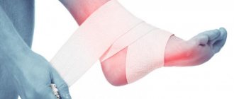
- Immobilization. Wearing an external splint for a short time will help support the ligaments and relieve stress on the joint.
- Medicines. Nonsteroidal anti-inflammatory drugs (NSAIDs), such as Aspirin, Naproxen, and Ibuprofen, can help reduce both pain and swelling. Local NSAIDs can be applied as compresses to the joint area.
- Exercises. Certain exercises can help improve range of motion and wrist function.
- Physiotherapeutic procedures (electrical stimulation, ultrasound therapy).
Nutritional supplements (dietary supplements) and chondroprotectors have no proven effectiveness in the treatment of this disease.
Treatment with folk remedies is permissible only in the early stages and in the absence of concomitant pathology.
Nutrition for arthrosis
Diet is fundamental for effective and deep restoration of cartilage tissue and ligaments. It is very important to change your gastronomic habits so that they contribute to healing in combination with conservative treatment.
The diet should contain the following products:
- raw fresh vegetables (in the form of salads, vegetable juices). They are rich in enzymes, vitamins, and minerals that are beneficial to humans. Eating them triggers detoxification processes in the body, removes waste and toxins, improves tissue nutrition at the intercellular level;
- fruits are very nutritious and help reduce joint inflammation by removing toxins and strengthening muscle fibers. All this has a positive effect on the healing process. You can eat any fruit, the only condition is that they cannot be mixed with other foods, you need to take breaks between taking different foods for half an hour. Different foods mixed in the stomach are poorly digested and lead to inflammation of the joints;
- Grains and whole baked goods are rich in fiber, which helps cleanse the intestines gently and effectively. Abuse of baked goods and sweets leads to slagging. Toxin breakdown products accumulate in joints and cause health problems;
- clean water is part of proper nutrition, especially with arthrosis. You need to drink at least 2 liters of clean liquid per day. This will ensure activation of oxidative reactions in the body and cleansing of toxins.
During treatment and for the prevention of arthrosis of the joints, it is necessary to completely exclude or at least limit the consumption of sugars, baked goods made from white flour, fried, smoked, and salty foods from the diet. Do not overuse red meat, whole cow's milk, cheeses, drinks containing caffeine, or alcohol. All these products disrupt metabolism, contribute to the accumulation of toxins in joint structures, and negatively affect their condition.
Arthrosis of the wrist joint is effectively treated with conservative methods of therapy if the disease is detected at an early stage. In the case when an irreversible process of deformation has begun in the joint, only a surgical scalpel will help. The task of any patient is not to start the disease and avoid surgery.