What does kyphosis mean? This is a curvature in the upper spine. The disease is pathological, but it is also possible that it develops against the background of a physiological abnormality. In the first case, development is observed in the thoracic region. As the nerve roots are compressed, pain occurs, the body suffers, its functionality is impaired, weakness in the legs and pelvic disorders appear. The severe form occurs with complications in the functioning of the lungs and heart. Diagnosis takes place in the form of examination, laboratory tests, and radiography.
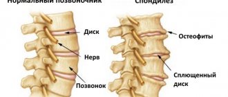
In order for a doctor to accurately diagnose thoracic kyphosis, the angle of curvature must be from 45°. This is already a pathology, a deviation from the norm. The disease develops both independently and against the background of other diseases, for example, scoliosis. Most often, thoracic kyphosis develops as a result of a vertebral fracture.
There are several types of kyphosis: arcuate and angular. If the patient is sick with tuberculosis, then the appearance of angular kyphosis is possible. This type is characterized by protrusion of the chest and the appearance of a hump. If the kyphosis is arched, then the entire thoracic spine is deformed and a C-shaped defect is formed.
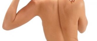
Are you experiencing symptoms of kyphosis?
Only a doctor can accurately diagnose the disease. Don't delay your consultation - call
Causes
If something went wrong with the development of the fetus during pregnancy, then it is possible that in the future the baby will have a deviation in the form of a curvature of the spine. Kyphosis is also caused by heredity, surgeries performed on the spine, and various injuries.
Take into account! In the absence of physical activity, for example, if you do not play sports or do exercises, the muscles lose tone and become weak, and this leads to the development of spinal kyphosis.
Thoracic kyphosis is often diagnosed in older women; it occurs as a result of compression fractures of the thoracic vertebrae. Such a fracture occurs against the background of decreased bone density and the development of osteoporosis.
Spinal kyphosis can develop with the progression of infectious diseases; cases of kyphosis with spinal tumors are isolated. In rare cases, in practice there are diseases that appeared as a result of radiation therapy (especially children are at risk if they underwent radiation therapy at an early age).
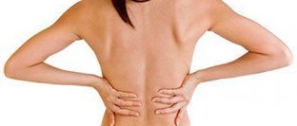
Kyphosis of the cervical or thoracic spine negatively affects the functionality of the entire body and the functioning of many organs. The body reacts especially sharply in the acute form of the disease. In addition to pain, patients complain of disruptions in the functioning of the heart, constipation, as there is pressure on the abdominal cavity, and the pelvic organs suffer.
Prevention
Prevention of the disease is of great importance. It consists of eliminating provoking factors : injuries, infections, muscle weakness.
Prevention in children should begin at an early age. It is necessary to monitor the child’s posture, select the right furniture and school bag, and avoid sitting at the computer for a long time. It is important to strengthen the back muscles; playing sports and outdoor games help with this. The child should also receive the necessary vitamins and minerals.
To prevent kyphosis, it is necessary to monitor posture. In adults, prevention is to prevent physical inactivity and excess weight gain.
If your profession involves staying in one position for a long time, then it is recommended to take breaks every hour and perform simple exercises. You should “make friends” with physical education and walking.
Elderly people are recommended to take calcium supplements to prevent osteoporosis, as well as light exercise and walking.
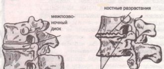
Any diseases of the spine - osteochondrosis, protrusions - must be treated in a timely manner to prevent complications in the form of kyphosis.
People involved in strength sports are recommended to take vitamin complexes and wear special support corsets during training, which reduce the load on the spine.
Following these simple recommendations will help you avoid encountering a dangerous disease.
Classification
Traumatologists and orthopedists give a classification of pathological kyphosis. It can be congenital, functional, paralytic, degenerative, post-traumatic, juvenile.
When contacting JSC “Medicine” (clinic of Academician Roitberg), after examination, a specialist will take into account the angle of curvature and only after that will determine the degree of the disease. Kyphosis may be normal, with an increased or decreased angle. If the patient has been diagnosed with increased kyphosis, then its degree is already determined:
- I – curvature angle 35°;
- II – curvature angle from 31 to 60°;
- III – angle of curvature 60° or more.
In order to correctly make a diagnosis and determine the degree of kyphosis, doctors at JSC “Medicine” (academician Roitberg’s clinic) conduct a number of studies using modern techniques and modern equipment.
Kinds
There are several types of kyphosis in adults.
Functional
This is incorrect posture, which is formed with deviations as a result of insufficient muscle tone. Pathology can also develop against the background of psychological disorders. When the disease is diagnosed, the patient is prescribed a set of simple exercises, and the doctor also recommends sleeping on a hard surface. Patients are required to lie, sit, and walk correctly.
Youthful
It is difficult to say exactly for what reasons this deviation develops. At the same time, experts were able to establish that the development of this type is influenced by heredity. Also the cause is many micro fractures of the spine. The kyphosis angle can reach 75°.
Congenital
Occurs when there are developmental disorders of the fetus at the time of formation of the vertebrae. Against this background, other diseases may develop.
Paralytic
If you damage the back muscles or cut them, this will lead to paralysis. In this case, kyphosis may be accompanied by other diseases, such as scoliosis. It is treated with a conservative method, the patient is prescribed massage and physical therapy.
Post-traumatic
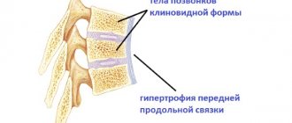
If the patient has suffered a fracture of the lumbar, cervical, or thoracic vertebrae, this can lead to the development of kyphosis. This is the situation most patients encounter, accounting for about 40% of cases. Not in all situations, such injuries lead to kyphosis; it all depends on the level of severity, the nature of the damage, and the injury. Also, post-traumatic kyphosis can be the result of a neurological disorder. It is treated mainly with surgical intervention, but it all depends on the age of the patient and his general condition.
Symptoms
The fundamental factor is the degree of curvature.
- Stage 1 kyphosis is characterized by fatigue and mild muscle pain, mainly at the end of the day. You can also observe poor posture and stooping.
- Kyphosis 2 degrees, when the kyphosis worsens and is complemented by compensatory lordosis, pain and stoop increase, the head and shoulders drop down.
- Kyphosis of the 3rd degree is accompanied by limited mobility of the spine, disturbances in the functioning of internal organs appear as a result of their compression by the deformed chest.
Diagnostics
By contacting JSC “Medicine” (academician Roitberg’s clinic), patients receive a full consultation, the doctor conducts an initial examination, communicates with the patient, which allows him to learn about the symptoms, condition, and well-being. Only after obtaining a complete picture is treatment prescribed.
Previously, the patient undergoes a number of procedures:
- inspection;
- laboratory research;
- ultrasound examinations;
- computed tomography;
- MRI.
An important role in making a diagnosis is played by communication with the patient, from whom the specialist learns about the development of the disease, how and when the problems began, how long the disease lasts, what new symptoms have appeared recently, what treatment was used before, whether the disease is accompanied by pain. The back and neck must be palpated to determine how well the muscles are developed and how sensitive the skin is. Also, the doctor is faced with the task of determining the neurological status; for this, a number of tests are performed.
Another mandatory stage of the study is radiography (this is one of the most important stages). They take pictures both straight and side or aimed. The perspective can be different, even non-standard - this is all so that the doctor can get a complete picture, predict the further development of the disease and prescribe the most effective treatment.
Treatment
Surgery is the most extreme case; in general, treatment of kyphosis is a conservative method. Exercise therapy is often prescribed for kyphosis; it is carried out by qualified doctors at the medical center. The patient is also prescribed massage, wearing a corset, physiotherapy and manual therapy.
In order to reduce pain and alleviate the patient's condition, wearing a corset is prescribed. But this is in extreme cases and for a short period, because the corset is unable to correct posture, while it contributes to weakening of the muscles, and this will only aggravate the development of the disease.
There are cases when conservative treatment does not give the desired result, the disease begins to progress, developing into a new degree of severity. In this case, surgery is prescribed. The main indications for surgical intervention include:
- severe pain, which cannot be relieved with conservative treatment methods;
- rapid development of the disease, when the angle of curvature becomes greater than it was before conservative treatment;
- a cosmetic defect that cannot be eliminated using conventional treatment methods.
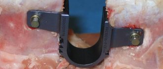
If back kyphosis prevents a person from leading a full life, going to work and performing professional duties, an operation is prescribed, the purpose of which is not only to improve appearance, for example, to reduce the angle of curvature, but also to do everything possible to prevent the disease from progressing in the future.
Before the operation is performed, the patient is fully examined and the tolerance to anesthesia is determined. General anesthesia is given. After the first operation, it will become clear whether additional surgical interventions will be required or not. The postoperative period is also important; doctors monitor the body’s reaction – whether it rejects the metal structures used or not. If the body does not perceive them as a foreign body, then the structure that fixes the spine can stand for several years.
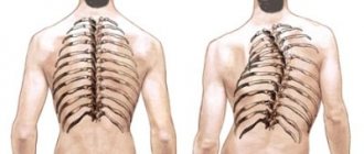
Excessive thoracic kyphosis: much more than poor posture
Signs of PCG include pain, breathing problems, limited function, decreased athletic performance, gastrointestinal distress, and increased mental stress (Rolf 1989; Myers 2001; Hanna 1988; Price 2010). This article explains how to identify PHC, identifies some of the most common causes, discusses potential symptoms, and suggests ways to resolve the problem.
Detection of excessive thoracic kyphosis
Excessive thoracic kyphosis is directly related to the functions of the chest, as each rib is attached to the vertebrae of the thoracic spine. The musculoskeletal assessment described below can determine whether or not your client has PCH.
Grade
Place the index finger of one hand on the notch of the sternum, the depression between your collarbones at the lower front of your neck. Then place the index finger of your other hand directly under the most prominent spinous processes of the vertebrae, which are usually found at the base of the neck (see also Appendix 1). Turn your head to the side and compare the position of your fingers in the mirror. Ideally, your fingers should be at approximately the same height. If the finger that is in front, at the sternum, is lower than the finger at the back at the base of the neck, your thoracic spine is tilted forward and your ribcage is lowered, then you have PCG.
Possible causes of PCG
The most common causes of PCG:
- Musculoskeletal compensation
- Environmental factors
- Exercise/Activity Selection
- Psychological stressors.
Musculoskeletal compensation
Musculoskeletal imbalances in any part of the body can trigger PCI. For example, moving the head forward, a useful position for focusing attention on a small object such as a hand-held electronic device, causes the thoracic spine to lean forward to help support the head (Kendall, McCreary & Provance 2005). It's like a big fish on the end of a flexible fishing rod. The rod bends forward to accommodate the additional load. Over time, a forward head position can lead to PCI.
CHC can also occur from habitual overpronation, where the feet roll inward. With the feet rolled inward, the shins and legs generally turn toward the midline of the body. Internal rotation of the legs causes a posterior displacement of the hip joints, and the pelvis rotates forward. Rotation of the pelvis results in an increased arch in the lower back (excessive lumbar lordosis) and the mid and upper back rounds backward to maintain balance in the body (Price, 2010).
Myofascial adaptations and restrictions can also lead to PCI. For example, changes in the length-tension relationship of the biceps brachii muscle, which originates at the outer edge of the scapula and inserts into the forearm, can directly provoke such an imbalance. When the arms are bent at the elbows for long periods of time during activities such as cooking, driving and typing, the biceps muscle is permanently shortened. If you straighten your arms after this, the muscle cannot lengthen properly. This causes the forearms to pull the shoulder girdle forward (through the biceps muscles) across the chest. A position in which the shoulder girdle is tilted forward ultimately results in a forward tilt of the chest, increasing thoracic kyphosis (Myers 2001).
Environmental factors
Excessive rounding of the upper back is usually associated with a sedentary lifestyle or occupations that involve prolonged periods of sitting, such as playing computer games or working on a computer. However, some less obvious environmental factors can also cause PGC. Respiratory diseases (asthma, COPD) and allergies to air pollution can affect the body's breathing mechanism and the ability of the diaphragm to contract and relax correctly. Restrictions in the chest wall associated with chronic breathing problems can cause immobility of the thoracic spine and ultimately lead to PCI (Rolf 1989).
Food intolerance is another stimulus for the development of excessive thoracic kyphosis. Intolerance to gluten, lactose, fructose and ethanol can lead to inflammation in our digestive tract (Harker 1998). The intestines are separated from other soft tissues and muscles by connective tissue, which is attached to the spine. Thus, chronic intestinal inflammation can cause myofascial restrictions and adhesions in the trunk, leading to PCG.
Exercise/Activity Selection
Exercise and activity choices may also promote CHA. Prolonged periods of spinal flexion required for cycling and/or stationary cycling, freestyle swimming, and hobbies such as knitting and gardening are major contributors to excessive thoracic kyphosis.
Psychological stressors
When the brain perceives a threat, whether it is real or not, the body prepares to counter the threat—the “fight or flight” response. Typical responses include tensing the jaw, tightening the abdominals, holding one's breath, and rounding the shoulders (Hanna 1988). Consequently, current psychological stressors—career dissatisfaction, relationship problems, money or family difficulties—can lead to fascial restrictions, neuromuscular adaptations, and musculoskeletal changes in the body, which in turn will lead to PCI.
Symptoms of PCG
PCH is a postural problem, but can also manifest itself through many other symptoms:
- pain
- function limitation
- poor health and illness
- increase in mental stress.
Pain
PCG is usually accompanied by excessive lumbar lordosis—hyperextension of the lower back, which leads to low back pain, disc degeneration, and nerve compression (McGill 2002). It can also cause excessive cervical lordosis, a neck position in which the head moves forward, leading to tension headaches, jaw pain, and/or neck and shoulder pain. PCG may also lead to pain in the lower extremities.
Previously, compensatory movement patterns that arise as a result of CHA (forward head displacement, lumbar lordosis, excessive pronation, etc.) have been described that can cause problems, for example: sciatica, pain in the sacroiliac joints, hip bursitis, iliac syndrome. tibial tract, knee pain, Achilles tendinitis, plantar fasciitis and ankle pain (Kendall, McCreary & Provance 2005; McGill 2002; Price 2010).
Function limitation
Daily movement and athletic performance may be impaired due to PCI. The excessive flexion of the middle and upper back that characterizes PCH limits shoulder function, which can cause difficulty with movements such as lifting your arms to wash your hair, stacking dishes in wall cabinets, or putting on a sweatshirt. CHA may also affect movements that require extension of the thoracic spine, such as butterfly swimming and upward-facing dog yoga poses.
Poor health and disease
CHG is a protective posture that limits body movements. As discussed earlier, PHA can make breathing difficult, which in turn can limit blood flow to internal organs. The downward movement of the diaphragm, as it contracts, “massages” the liver and facilitates blood supply to other vital organs. Therefore, the tendency of CHC to impair breathing may also lead to intestinal problems and/or diseases (Rolf 1989).
Increased mental stress
When the body adopts a posture with excessive thoracic kyphosis, the nervous system interprets this as a threat and activates the stress areas of the brain. Over time, increased activity in the "stress-sensitive" areas of the brain causes fatigue, and the body's (and brain's) inability to effectively cope with stress (Lawlis 2008).
Means of combating ChGK
There are several ways to reduce BGK:
- corrective exercises
- breathing training
- relaxation
- lifestyle change training.
Corrective exercises
Self-myofascial release techniques are very effective in reducing fascial restrictions and improving posture.
Two tennis balls for the upper back
You will need tennis balls and a pillow for this method of targeting BHC. The self-massage technique shown promotes extension of the thoracic spine.
Lie on the floor on your back and bend your legs. Place tennis balls under each side of your spine, level with the line connecting the bottom edges of your shoulder blades. Place a large pillow under your head so that you don't feel too much pressure from the tennis balls. Cross your arms over your chest, hugging yourself. Find the pain points and maintain pressure until relief appears (10 - 15 seconds). Then move the balls to the other pressure point, moving your pelvis down so that the balls move up along the spine. Adjust the pillow every time you move it. Spend 2 – 3 minutes every day on this region.
Tennis ball under belly
This technique uses one tennis ball under the abdomen. Self-massage reduces tension in the abdominal muscles and hip flexor muscles/fascia, increasing trunk mobility and extension of the thoracic spine. Lie face down on the floor. Place the tennis ball at the lower edge of your ribcage to the right of your navel. Find the painful point and hold pressure until relief (5 - 10 seconds). Then move the ball to a new point by moving your body up so that the ball moves down. Continue along your belly from your navel to your upper thighs. Repeat the manipulations for the left side. Spend 1 to 2 minutes on each side.
Breathing and relaxation techniques
Diaphragmatic breathing techniques will help increase chest mobility, strengthen the diaphragm, supply blood to organs, facilitate relaxation, and reduce stress.
Expansion of the lower chest
Hands should be placed with palms on the lower part of the ribs. Lie on your back with your legs bent (use a small pillow to support your head if necessary).
Wrap your palms around your lower chest and breathe through your nose. As you inhale, focus on expanding your lower ribs and pressing them into your palms. Hold your breath (contraction of the diaphragm) for a few seconds and then exhale slowly. Perform 10 – 15 breathing cycles every day.
Lifestyle Change Training: Know Your Stress Triggers
In addition to the causes of PCG, mental stress can manifest as muscle pain, breathing problems, dizziness, gastrointestinal distress, and skin problems (hives, eczema, and rashes). These are symptoms that precede a situation of negative stress. Record every physical activity during the week that caused stress and then led to inflammation or problems. Also pay attention to your thoughts and actions when the symptom became more noticeable, for example, “the pain in my back became worse when I thought about the recent arguments in the argument” or “the headache occurred after discussing the client’s wishes.” Once you become aware of your stress triggers, focus on limiting your exposure to them in similar situations.
Keep PHC under control
Our musculoskeletal and neuromuscular systems are designed to respond to constantly changing conditions. The key to managing PHA is to pay attention to symptoms such as musculoskeletal pain, mental stress, gastrointestinal distress, and performance fluctuations that indicate overload or imbalance in your physical or mental state. The strategies listed above can mitigate the impact of your lifestyle and activity, helping to avoid potentially harmful exposure to PHC.
Excessive Thoracic Kyphosis: Much More Than Just Bad Posture Justin Price, MAIDEA Author/Presenter
Source: ideafit.com
Translation by FPA expert S. Strukov
Sources:
- Hanna, T. 1988. _Somatics: Reawakening the Mind's Control of Movement, Flexibility and Health._Cambridge, MA: Da Capo.
- Harker, M. 1998. Health and Healing (3rd ed.). Waipu, New Zealand: Wings of Waitaha.
- Kendall, F. P., McCreary, E. K., & Provance, P. G. 2005. Muscles: Testing and Function with Posture and Pain (5th ed.). Baltimore: Lippincott Williams & Wilkins.
- Lawlis, F. 2008. The Stress Answer. London: Penguin.
- McGill, S. 2002. Low Back Disorders: Evidence-Based Prevention and Rehabilitation. Champaign, IL: Human Kinetics.
- Myers, T. 2001. Anatomy Trains: Myofascial Meridians for Manual and Movement Therapists. Edinburgh: Churchill Livingstone.
- Price, J. 2010. The Fundamentals of Structural Assessment: Module 1. The BioMechanics Method.www.thebiomechanicsmethod.com. Rolf, IP 1989. Rolfing: Reestablishing the Natural Alignment and Structural Integration of the Human Body for Vitality and Well-Being (Revised ed.). Rochester, VT: Healing Arts Press.
How to make an appointment with a traumatologist-orthopedist
There are several ways to make an appointment with a specialist at our clinic, for example, by calling. If you have a difficult situation, use the phone number to call an ambulance. Specialists will arrive in the shortest possible time, telephone lines work around the clock. You can also make an appointment using the website. On each page you will see an appointment form in the top corner.
JSC "Medicine" (clinic of academician Roitberg) is located in the center of Moscow near the metro stations Belorusskaya, Mayakovskaya, Novoslobodskaya, Tverskaya, Chekhovskaya. Address: 2nd Tverskoy-Yamskoy lane, 10.