Back pain - most adults don't need an explanation of what it is. Many have encountered this symptom at least once in their lives. By the way, according to statistics from American researchers, back pain is the most common reason why people visit doctors and take sick leave.
Our expert in this field:
Lashch Natalia Yurievna
Neurologist of the highest category, candidate of medical sciences, associate professor. Laureate of the Moscow City Prize in the field of medicine.
Call the doctor Reviews about the doctor
94% of adults have various problems with the spine. Sometimes they do not cause any particular problems, but sometimes they make a person disabled. All these diseases are united by a common term - dorsopathies.
Types of spinal dorsopathy
The human spinal column consists of different sections. Depending on this, dorsopathies of the cervical, thoracic and lumbar spine are distinguished. There are also lesions in multiple parts of the spine, only the back of the head and the first two vertebrae, the sacral, sacrococcygeal sections.
In the International Classification of Diseases - a document that all doctors should rely on when making a diagnosis - dorsopathies are divided into three types:
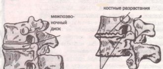
- Deforming dorsopathies. Associated with curvature of the spinal column. This group of pathologies includes lordosis, kyphosis, scoliosis, osteochondrosis, etc.
- Spondylopathies. This group includes: ankylosing spondylitis (ankylosing spondylitis), inflammatory and infectious diseases of the spine, osteoarthritis, arthritis, spondylosis.
- Other dorsopathies. The group includes diseases that are accompanied by damage to the intervertebral discs, as well as back pain of other origins.
Dorsopathy - what is it? Reasons and mechanism of its development
Dorsopathies are a group of diseases of the musculoskeletal system and connective tissue, the leading symptom complex of which is pain in the back and limbs of non-visceral etiology.
The term dorsopathy is translated from Latin as back disease. The spine is a complex structure that can withstand significant loads associated with the vertical position of the human body. Until the age of 30, the processes of anabolism (synthesis and restoration of tissues) prevail in the body; later, the process of involution and degenerative changes in the tissues of the spine, primarily the cartilage of the intervertebral discs, occurs. This leads to deformation of the vertebrae, pinching of the spinal roots, and inflammation of the paravertebral muscles and tissues.
Treatment of spinal dorsopathy
Treatment of dorsopathy of the neck, thoracic and lumbar spine has the following main goals: eliminating pain, restoring normal functioning of the spinal roots and slowing the progression of pathological changes in the spinal column.
Nonsteroidal anti-inflammatory drugs (NSAIDs), such as ibuprofen, are used to relieve pain. Drugs called muscle relaxants help relax tense muscles in the neck, back, and lower back. Chronic pain leads to a person becoming nervous and irritable - in such cases, a neurologist prescribes sedatives.
What factors contribute to the occurrence of dorsopathy?
- Heredity means there is a high probability of developing this pathology in children whose parents suffered from spinal diseases.
- Increased static load on the spinal column - persons in certain professions associated with prolonged standing (surgeons, hairdressers). In these cases, the process develops in the lumbar spine.
- Strong simultaneous load on the spine associated with lifting significant weights
- Congenital curvatures of the spine
- Metabolic disorders in the body
- Alcohol abuse and smoking
- Chronic infections
- Insufficient and unbalanced nutrition
- Sedentary lifestyle
All causative factors lead to changes in the height of the intervertebral discs, changes in the shape of the vertebral bodies and inflammation of the paravertebral tissues with spasm of the striated muscles and pinching of the nerve fibers of the spinal cord. As a result, these processes become irreversible
Traction and physiotherapy for dorsopathy
To unload the vertebrae and intervertebral discs, traction treatment is used. The patient is placed on a special bed with a raised headboard and secured with special loops behind the head or armpits. Sometimes underwater traction is used: a person is in a pool, a float is attached to his neck, and a weight is attached to his legs.
Physiotherapy procedures include electrophoresis, magnetotherapy, phonophoresis, darsonvalization, diadynamic currents, exposure to ultraviolet radiation, and ultrasound. After the pain subsides, massage and physical therapy are prescribed. In some cases, they resort to manual therapy, acupuncture, and osteopathy.
To normalize the functioning of the affected nerve roots, the doctor prescribes vascular medications and B vitamins.
In each individual case, treatment for dorsopathy is selected individually, depending on what disease it is caused by. Sometimes - in rare cases - surgery may be required.
Dorsopathy is usually a chronic disease; if not treated correctly, over time it can make a person disabled. If you are worried about back pain, visit a neurologist as soon as possible, make an appointment by calling +7 (495) 230-00-01.
The main symptom of all types of dorsopathies is pain in the back (or in the neck, lower back, depending on which parts of the spine are affected by the pathological process).
The pain is most often aching, constantly bothering, becoming stronger during physical activity, when a person lifts weights, staying in a monotonous uncomfortable position for a long time, or with sudden movements. Sensitivity disorders (numbness, tingling, “goose bumps”) and movements (paresis, paralysis) in the area of innervation of the affected nerve roots may also occur.
The symptoms of dorsopathy have some characteristics depending on which part of the spine is affected.
Book a consultation with a neurologist today
Message sent!
expect a call, we will contact you shortly
Spinal traction
Clinical studies have shown the high effectiveness of spinal traction treatment. During the procedure, the height of the intervertebral discs increases. Negative pressure is created in the intervertebral space, retracting the protruding part of the disc. The pressure on the nerve roots decreases or disappears completely.
During traction, conditions are created that are favorable for the regeneration of the torn area of the annulus fibrosus, and blood flow improves. The shock-absorbing properties of the vertebrae are restored, the load is removed from the muscles and spine.
Traction is carried out after examining the patient on MRI or CT on a multifunctional traction-massage, inversion tables and Professor Velikanov’s device. The patient is placed on the simulator, the limbs are secured with belts, and the spine is stretched using a sliding table or belt traction. All parameters of the procedure are determined by the doctor, taking into account the characteristics of the patient’s body.
You can stretch the spine yourself - using a horizontal bar. The exercise is useful, but it practically does not affect the cervical and first thoracic vertebrae.
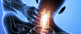
Symptoms of dorsopathy of the cervical spine
Characteristic manifestations of damage to the cervical spine:
- Headaches, pain in the neck, shoulders, arms.
- Dizziness.
- Noise, ringing in the ears. “Floaters” and colored spots flash before your eyes.
- Feeling of numbness, weakness in the arms.
- Tension in the muscles of the neck and shoulder girdle, a feeling as if “someone was riding on your shoulders.”
Clinical manifestations of dorsopathy
Clinically, dorsopathy manifests itself as:
- reflex syndrome (90% of cases)
- compression syndrome (5-10% of cases).
Reflex syndromes in dorsopathy (muscular-tonic, neurovascular and neurodystrophic) arise due to irritation of pain receptors in the back muscles as a result of the action of any pathological factors (pinching, inflammation or irritation) and are accompanied by a reflex muscle spasm. However, muscle spasm itself causes an additional pain impulse, resulting in a vicious circle of “pain - muscle spasm - pain”.
Compression syndromes in dorsopathy are caused by the mechanical effect of a hernial protrusion, bone growths or other pathological structure on the roots, spinal cord or blood vessels. Accordingly, compression syndromes are divided into radicular (radiculopathy - pinching of the spinal nerve root), spinal (myelopathy - compression of the spinal cord) and neurovascular (compression of the vertebral artery).
As for myelopathy, it is more often observed in the cervical, less often in the lumbosacral spine.
Myofascial pain in dorsopathy
In the diagnosis of dorsopathy, the role of myofascial pain syndromes (myositis or myalgia, affecting 35 to 85% of the population) is often underestimated. The essence of myofascial pain syndrome is that the muscle suffers primarily, and not after morphological or functional disorders in the spine. Any muscle or muscle groups can be involved in the pathological process.
One of the most common causes of myofascial pain is acute muscle overstretching. Usually the patient remembers exactly what movement or action caused the pain. Myositis can also develop against the background of constant overstrain of a muscle group, or hypothermia.
To make a diagnosis of myofascial pain syndrome, it is necessary to identify the following clinical signs:
- on palpation the muscle is spasmodic;
- within the spasmed muscle, zones of even greater muscle compaction are clearly defined - trigger points that are particularly painful.
Symptoms of dorsopathy of the thoracic spine
The vertebrae in the thoracic region are connected to the ribs, and through them to the sternum. The thoracic region has less mobility compared to the cervical and lumbar regions, and therefore some pathologies are relatively rare here. Possible symptoms of thoracic dorsopathy:
- Back pain.
- Pain in the heart, “in the internal organs.” This can be misleading: a sick person believes that he has problems not with the spine, but with the heart, stomach, and other organs.
- The pain may worsen during deep breaths. Sometimes it can be very strong, it feels as if “a stake has been driven into the chest.”
Over time, damage to the thoracic spine leads to limited mobility of the chest. Because of this, breathing and heart function are impaired.
What is dorsopathy?
Dorsopathy (from the Latin dorsum - back) is a generalized name for various pathologies of the spine, soft tissues of the back (paravertebral muscles, ligaments, etc.).
A general symptom of various types of dorsopathy is pain syndromes in the body area (cervical spine, back, lower back), not associated with diseases of the internal organs. The cause of pain in dorsopathy is degenerative-dystrophic diseases of the spine, as well as damage to the soft tissues of the back (paravertebral muscles, ligaments, etc.). The main manifestations are back pain and limited mobility of the spine.
Dorsopathies are characterized by a chronic course with exacerbation of pain.
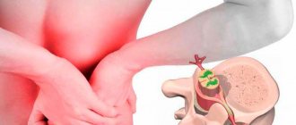
Symptoms of dorsopathy of the lumbar spine
Evolution has made man the most intelligent creature on earth, but it also had some “side effects”. Due to upright walking, our lower back experiences heavy loads. In addition, the lumbar spine has quite a lot of mobility. This increases the risk of certain types of dorsopathies.
Symptoms that occur with dorsopathy of the lumbosacral spine:
- Lower back pain – can radiate to the buttock, thigh, lower leg, foot.
- Decreased sensitivity, movement disorders (paresis and paralysis) in the legs.
- In severe cases, the functions of the pelvic organs are disrupted. A sick person partially or completely loses control of the bladder and rectum.
Treatment options
In general, spinal dorsopathy and its complications can be treated with medication. Surgical intervention is rarely used: in cases of severe pain, severe limitations in functionality and the absence of positive results from conservative treatment.
Drug treatment of dorsopathy involves taking NSAIDs: they suppress inflammatory processes in the area of the affected vertebrae and help reduce pain. To relax the back muscles, the patient is prescribed muscle relaxants. When treating nonspecific dorsopathy the following is used:
- Manual therapy and massage. With their help, blocking of the lumbosacral joints is eliminated.
- Transcutaneous electrical neurostimulation on the paravertebral area of the back.
During the period of remission, physical therapy is permitted. Classes (group or individual) are conducted under the guidance of a doctor or instructor. The task of exercise therapy is to teach the patient a rational motor regimen and return him to his usual professional and everyday motor activities. Patients with chronic pain learn to overcome their fear of movement. Special exercises strengthen the muscles of the lower back.
When dorsopathy is accompanied by psychoemotional disorders, psychological correction is carried out.
If conservative treatment does not have an effect, neurological complications of dorsopathy progress, compression myelopathy occurs, defecation and urination functions are impaired, and surgery is prescribed.
Treatment of dorsopathy symptoms
The main symptom of all types of dorsopathies is pain. It is relieved with the help of non-steroidal anti-inflammatory drugs, novocaine blockades (procedures during which an anesthetic solution is injected into the area of damaged nerve roots), and physiotherapy.
They try to restore movement and sensitivity during rehabilitation treatment. It includes massage, physical therapy, mechanotherapy, and physiotherapy.
The above is only an indicative list of general areas of treatment. Of course, dorsopathy can be effectively treated only after its cause has been clarified and a diagnosis has been established. Therapy should be aimed not only at the symptoms, but also at the cause of the disease.
Correspondence consultation with a neurologist
Message sent!
expect a call, we will contact you shortly
Dorsopathies are a fairly large group of diseases. They are united by two common symptoms: they are all associated with lesions of the spinal column and are accompanied by pain in the neck, back, and lower back. All these pathologies are divided into three groups:
- Deforming dorsopathies.
- Spondylopathies.
- Other dorsopathies.
Prevention
To prevent the development of pathology in the spine and back, it is necessary to lead a correct lifestyle, eliminate sudden loads, and keep the muscles of the torso and back in good shape. Regular sports, gymnastics, and walks help with this. If you sit for a long time, you need to get up periodically to stretch your spine. Make time for active recreation: swimming, exercising on the horizontal bar to unload the spine (a simple hang is enough). A good prevention is morning exercises. It is recommended to perform simple exercises daily: slight bends and turns of the torso. Try to include more dairy products in your diet, which contain calcium, which strengthens bones and joints. If you experience regular pain in the neck, back, or tailbone area, you should consult a doctor. Disease prevention can be divided into primary and secondary. The primary measures include the following:
- playing sports on a regular basis;
- visiting a massage parlour;
- exercise therapy classes with a qualified trainer;
- eliminating factors leading to obesity;
- generally healthy lifestyle;
- elimination of existing pathologies correctly and in a timely manner.
Secondary prevention includes measures to prevent recurrence of the disease:
- exercise therapy classes with a trainer on a regular basis, in a group or individually;
- visiting the pool and massage sessions;
- regular visits to the doctor and careful adherence to all his instructions and recommendations;
- creating the right diet;
- exclusion of strength sports and, in general, limitation of significant physical activity;
- course intake of vitamins on the recommendation of a doctor.
Causes of deforming dorsopathies
Kyphosis (except congenital) is a curvature of the spine in the form of an arch, the apex of which is directed backward. Acquired kyphosis can be a consequence of rickets, tuberculosis, spinal injuries, and poor posture.
Lordosis (except congenital) is a curvature of the spine in the form of an arch, the apex of which is directed forward. The main causes: poor posture, pathological hip dislocation, inflammation, spinal tumors.
Scoliosis is a lateral curvature of the spine, in which twisting and deformation of the vertebrae occurs. In 80% of cases, the exact cause of the disease is unknown - this is the so-called idiopathic scoliosis.
Osteochondrosis is a degenerative-dystrophic disease in which the spine prematurely “wears out” and “ages.” The pathological process begins in the intervertebral disc, then spreads to the vertebrae, intervertebral joints, and ligaments. Mostly older people are affected, but now dorsopathy caused by osteochondrosis has begun to occur at the age of 20–30 years.
How to treat dorsopathy?
Medications and various types of physical exercises can be prescribed as treatment. The complex is compiled by the attending physician or instructor, taking into account the degree of damage and the capabilities of the human body.
To get a good effect, you can use special simulators. They restore muscle tone and affect the causes of the disease.
Let's look at the main types of treatment for dorsopathy.
The course of treatment is usually carried out comprehensively. At the same time, medications, physical therapy exercises, and physiotherapy can be used.
Drug treatment
Drugs that relieve inflammation are used. All products should be used under the supervision of a physician. Ointments and gels help relieve pain.
If the pain is very severe, I prescribe non-steroidal drugs in the form of injections.
The following are used as medicines:
- Ketoprofen;
- Ibuprofen;
- Diclofenac;
- Meloxicam;
- Celecoxibs.
The doctor may prescribe the use of hormonal drugs. Intra-articular injections and blockades are performed. Diprospan is used for this.
It is important to remember that all drugs have significant side effects.
Taking some medications will not completely relieve the disease; for this, other methods should be used.
Physiotherapeutic treatment
Physiotherapeutic procedures are a good addition to taking medications. There are a number of treatment methods.
These include:
- Electrophoresis;
- Laser therapy;
- Massage;
- Electropulse therapy.
All these procedures allow you to restore the spine and its functions. If the cause of dorsopathy is osteochondrosis, it is recommended to use UHF. Massage is most often used as a course.
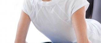
Exercise therapy
Health-improving exercises will help support your spine and improve your condition. Therapeutic gymnastics is indicated for people at any age. It helps relieve pain in the lower back.
Attention!
You should not do gymnastics during an exacerbation or the presence of “lumbago”
You should start exercising after the pain has passed.
Complexes of health-improving exercises help cope with the disease.
A detailed example of treating dorsopathy using exercise can be seen in this video:
Prevention of dorsopathy
For prevention purposes, you can do gymnastics or exercises every day. It should be borne in mind that any load on the spine must be dosed. To avoid lower back pain, you should choose the right posture for work, lead an active lifestyle, and choose hard orthopedic mattresses for sleeping.
Causes of spondylopathies
Ankylosing spondylitis (Bechterew's disease) is a chronic disease in which inflammation develops in the joints of the spine and mobility in the intervertebral joints is impaired. Not only the spinal column, but also other joints and internal organs can suffer. Young men are most often affected.
Inflammatory processes in the spine. They can be caused by various infections, vertebral osteomyelitis, purulent inflammation of the intervertebral discs. Sometimes the cause of the inflammatory process cannot be detected.
Spondylosis is “premature aging” of the spine. Pathological changes in the spinal column can lead to compression of the nerve roots, resulting in pain, movement disorders, and sensitivity.
Forestier's ankylosing hyperostosis is a rare disease in which bone tissue grows in the ligaments and tendons of the spinal column. His mobility is impaired.
Spondylopathies are also caused by vertebral tuberculosis, brucellosis, enterobacterial spondylitis, syringomyelia and other diseases.
Diagnostic features
The doctor carries out a set of measures to determine the causes of the disease and make a diagnosis. The specialist clarifies with the patient at what time the pain sensations appear, what their nature is, and their intensity. The doctor prescribes a number of tests, including:
- Computed tomography
is a high-precision method that allows you to examine tissue in several planes. The equipment creates a three-dimensional image of the inspected area. - MRI
is used to diagnose deformation changes in the cartilage of intervertebral discs. With its help, blood vessels, nerve endings and ligaments are visualized. - X-ray of the spine
in different projections - performed to confirm or exclude spondylopathy, provides good visualization of hard bone tissue.
Other causes of dorsopathy
Pain can be caused by lesions and displacements of intervertebral discs, and degenerative processes in them.
Cervicocranial syndrome is chronic pain in the upper neck and headaches. They can be caused by circulatory disorders as a result of injury or constant overload of the neck.
Cervicobrachial syndrome - pain in the neck, shoulders and arms. Common causes: collarbone injuries, shoulder dislocations, pathologies of the 4th-7th cervical vertebrae, constant arm tension (most often associated with the profession).
Spinal instability is pathological mobility in the spinal column, due to which it cannot bear normal loads. Occurs due to injuries, osteochondrosis, congenital underdevelopment of ligaments.
Get a consultation with a doctor
Message sent!
expect a call, we will contact you shortly
The diagnosis of “dorsopathy of the cervical, thoracic or lumbar spine” is actually not one, but a group of different diseases. Identifying the cause is not always easy. A neurologist who diagnoses and treats these pathologies must understand the causes as accurately as possible and prescribe appropriate treatment.
Poor posture, scoliosis, osteochondrosis, spondylitis, degenerative diseases of the spine - this is just a short list of the most common causes of dorsopathy. In fact, there are many more of them.
What happens to the intervertebral disc during Dorsopathy?
The nucleus pulposus of the disc is under constant pressure. The height of the disc decreases due to the pressure of the body weight, which is in an upright position during the day. When the discs are young and healthy, they contain a lot of water - so much that at night, when they are saturated with moisture and we straighten out, we add up to two centimeters in height.
The “straightening” of the disc occurs due to the properties of the nucleus pulposus and the elasticity of the ligaments. Liquids, as we know, are practically incompressible, so any pressure acting on the core is transmitted in all directions. The annulus fibrosus holds the core, that is, the pressure of the core is balanced by the tension of the annulus fibrosus, the ligaments surrounding the spine and the tone of the back muscles. The opposition of these two forces, according to doctors, is the key to understanding the degenerative-dystrophic osteochondrosis processes occurring in the spine. The muscles also play a huge role in this counteraction, creating a kind of tension field around the spine.
The emergence and development of dorsopathy is promoted by:
– physical inactivity (low motor activity);
– monotonous or meager food;
– constant work in unfavorable weather conditions, when low temperatures are combined with high air humidity;
– vibration;
– an imperceptible uneven load on the spinal column due to a variety of factors (for example, incorrect position of the spine during work and other daily activities);
– prolonged stay of the body in physiologically uncomfortable positions: sitting for many hours, bent over a desk at school, college, home, at work; sitting at the steering wheel of a car, at a computer console, at a drawing board, standing at a machine, at a counter, etc.
As a rule, age-related changes in intervertebral discs appear after 45 - 50 years. However, they can also occur at an earlier age, especially if there have been macro- and microtraumas, infectious lesions, or metabolic disorders. The causes of the disease can be an unfavorable hereditary predisposition, and weakness of the musculoskeletal system, cartilage structure, and muscle corset. The general situation is worsened by excess body weight.
At the same time, the blood supply and provision of nutrients to the tissues adjacent to the spine, vertebral bodies and intervertebral discs is almost 30 times worse than with a rational motor mode. As a result, the content of microelements, enzymes and vitamins in the tissues of the intervertebral discs, ligaments and muscles of the corresponding part of the spine decreases. All this gives rise to the emergence of the entire complex of disorders, which culminates in degenerative-dystrophic changes either in an individual intervertebral disc and vertebra, or in a group of discs and vertebrae, or in the entire spine as a whole.
Over time, the disc loses 70% of its previous properties and can no longer serve as a strong and reliable shock absorber. With age, the soft core of the intervertebral disc loses water and ceases to perform its functions. The mass of the core is reduced, thereby reducing the height of the disk.
The extensibility and elasticity of the disc shell (fibrous ring) also decreases. During this period, small cracks may appear in the fibrous ring around the nucleus and disc, and the substance of the nucleus will begin to press into these cracks. These cracks also appear as a result of malnutrition of the intervertebral discs and stress on the spine.
This is the first stage of the disease - chondrosis, when the pathological process is limited to the disc. It is very difficult to recognize the disease. At the beginning of the disease, a person experiences mild discomfort. X-ray does not show any changes.
Starting from the second stage - dorsopathy, characterized by further spread of the process to the bone base of the spine, adjacent vertebral bodies and intervertebral joints. Further destruction of the fibrous ring occurs, the fixation of the vertebrae among themselves is disrupted, and their pathological mobility appears. Further disc degeneration inevitably leads to a decrease in the height of the intervertebral space, thereby compressing the neurovascular endings, blood and lymphatic vessels, resulting in increased pain.
In the third stage of the disease, if a complete rupture of the fibrous ring occurs (for example, as a result of significant physical activity - heavy lifting), intervertebral hernias are formed (the deformed nucleus pulposus breaks through (falls out) through the fibrous ring outside the disc). Under pressure from the hernia, the irritated nerves send impulses to the central nervous system, and the person experiences severe pain. During this period of the disease, a fixed deformity of the affected parts of the spine may develop in the form of kyphosis (convexity of the spine backward), lordosis (bending of the spine forward) and the initial stage of scoliosis (lateral curvatures) of the spine. The physiological line of the spine is disrupted.
The fourth stage is characterized by painful compaction and displacement of the vertebrae. A deformed intervertebral disc does not provide complete connection of the vertebral bodies, and thus, slight displacements occur relative to each other, which, in turn, causes a reaction of the surrounding paravertebral muscles, which painfully contract and spasm.
The result is a restriction of movement in a specific segment, called a “blockade”. Sometimes such blockades occur suddenly. For example, in the cervical spine when turning the head - in bed, when reversing a car, or when moving the head suddenly (during a car collision). “Rejuvenation” of cervical osteochondrosis is associated with an intense increase in the frequency of motor vehicle injuries of the spine.
As a result of the described changes in the lumbar spine, the vertebrae are displaced posteriorly (pseudospondylolisthesis, or retrolisthesis), and in the cervical spine, subluxations occur. Feelings of pain and discomfort in the back or neck tend to intensify in uncomfortable positions. The connection between the vertebrae is disrupted, and the spine loses its flexibility and mobility.
If this entire process continues, which is inevitable if the load on the spine continues, then the vertebrae react with the formation of pathological bone growths (osteophytes), narrowing the intervertebral foramina. All this leads to irritation, compression and inflammation of the nerve roots (sciatica), vasospasm (impaired lymph circulation, arterial and venous circulation in the spine), compression (compression) of the spinal cord, resulting in damage to the central and peripheral nervous system.
A reduction in the space between the vertebral bodies leads to the development of a deforming disease of the spine, spondyloarthrosis (arthrosis of the intervertebral joints). The disease leads to a real disaster: motor activity decreases, the mobility of the spine is impaired, sudden movements cause acute, sometimes literally unbearable pain. In advanced cases, disability may occur at this stage of the disease.
All four stages of dorsopathy are inherent in any type of dorsopathy - cervical, thoracic, lumbosacral. The largest number of people suffer from dorsopathy of the cervical spine, since 40% of the height of the cervical spine falls on cartilaginous intervertebral joints, while in the thoracic region they account for only 20%, and in the lumbar region - about 33%. If we compare the specific loads on the discs, then in the cervical region it is 11 kg/sq. cm, and in the lumbar region - 9.5 kg/sq. cm. Therefore, the neck is most susceptible to this disease.
By taking on the weight of the head (support function), the cervical spine largely neutralizes shocks and concussions of the brain. The cervical spine is the most mobile, since the neck provides a large range of movement of the head, and therefore its muscles get tired much faster than the muscles of the rest of the back corset. This leads to accelerated wear of the intervertebral discs of the cervical spine. Degenerative changes in the discs are more common in the most mobile lower cervical parts of the spine (at the level of the 5th - 7th cervical vertebrae).
The cervical spine differs from other sections primarily in that, in addition to the canal for the spinal cord, there is a canal for the vertebral artery. At the level of the 6th cervical vertebra, the artery enters the canal of the transverse processes, and at the level of the 2nd vertebra it exits and, penetrating the cranial cavity, participates in the blood supply to the brain.
With pathological changes in the cervical spine, the vertebral artery spasms, thereby reducing the flow of blood to the brain. In addition, the vertebral artery is intertwined with fibers of the sympathetic nerve, and its functions include the transmission of pain signals. With osteochondrosis of the cervical spine, when the nerve fibers are irritated, a stream of pain impulses hits the brain. It is as a result of these features of the cervical spine that at the slightest changes in it we are tormented by headaches, weakness, increased muscle fatigue, and sleep disturbances.
Noise in the ears, numbness of the fingers, weakness of the hands, discomfort and pain in the arms, in the scapula, shoulder, in the heart area, impaired hearing, vision, fluctuations in blood pressure, impaired coordination of movements, dizziness and even fainting (syncope) - these are also neurological manifestations of osteochondrosis of the cervical spine.
Damage to the intervertebral discs with subsequent spondyloarthrosis of the lower parts of the cervical spine can affect the nerve roots, the fibers of which lead to the shoulder joints and upper limbs. Pain from the cervical spine radiates to the shoulder joint and arm, sensory disturbances occur, and the fingers of the affected hand become unruly. If the nerves serving the shoulder joint are damaged, its mobility is limited.
It is known that all the controlling and controlling neural connections of the brain with our organs pass through the spinal cord. As a result of dorsopathy (due to compression, that is, compression of the spinal cord), this connection is disrupted, the functions of vital organs, that is, the work of our entire body, are disrupted. That is why the spine is called the pillar of life.
To diagnose spinal diseases, clinical and radiological studies are carried out, sometimes using contrast agents (discography, myelography) and computed tomography and magnetic resonance imaging methods.
How does a neurologist diagnose dorsopathy? What happens in the doctor's office?
First of all, it is important for your doctor to understand the symptoms that are bothering you. Therefore, your appointment at the neurologist’s office will begin with you being asked some questions:
- How long have you had back pain? How often do they occur? After what do they usually intensify? After what do they subside?
- Have you noticed that your neck and lower back have become less flexible?
- Are you bothered by pain in your arms and legs? Have your legs and arms become weaker or moved worse? Do you experience a feeling of numbness or unpleasant tingling?
- Where and who do you work for? Do you often have to lift heavy loads? Sitting at an office desk for a long time?
- Have you recently had a back injury? Have you had any illnesses?
This will be followed by a neurological examination. The doctor will check the strength and tone of the muscles of your arms and legs, skin sensitivity, feel the back muscles to detect their tension, and press on certain points in the spine to identify soreness. After this, you will be given an examination.
Treatment
Faced with back pain, many are interested in what is dorsopathy and how to treat it? First you need to change your lifestyle: avoid stress, distribute forces correctly, and unload the spine. The back muscles should be kept in good shape by doing morning exercises. Nutrition should include foods high in calcium and microelements.
Treatment using traditional medicine methods involves taking medications to restore blood circulation, physical therapy, physiotherapy and wellness. Therapy is divided into conservative and surgical treatment. Most patients are indicated for conservative treatment, in which pain is relieved, measures are taken to prevent the chronic stage of the disease, the risks of relapse are eliminated, and rehabilitation is carried out. Patients are prescribed medication. Physiotherapeutic procedures show high effectiveness. Hydromassage, ultrasound, as well as electrophoresis, paraffin applications, and UHF help well.
If conservative therapy is ineffective, surgical treatment is indicated. The use of modern equipment allows operations to be performed with minimal injury. After surgical interventions, a rehabilitation complex is carried out. To avoid relapses, you must follow your doctor's recommendations.
Radiation diagnostics of dorsopathy
For dorsopathy, the following radiation diagnostic methods are used:
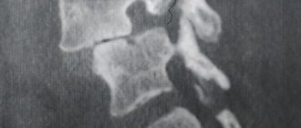
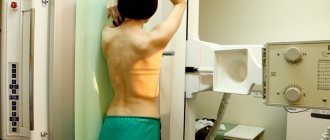
- Radiography. Typically, photographs are taken in two positions - full face and profile; if necessary, they can be taken in other positions. X-rays help assess the condition of the vertebrae, the presence of bone growths on them - osteophytes, and the height of the intervertebral discs.
- CT scan. A more accurate diagnostic method that helps to study this or that part of the spinal column in more detail and take pictures in which layer-by-layer sections will be visible. CT scans, like radiography, use x-rays.
- Magnetic resonance imaging. This method helps to assess the condition of the intervertebral discs, spinal ligaments, and surrounding soft tissues. During the study, a strong magnetic field is used.
Classification of dorsopathies
Dorsopathy can be caused by degenerative and inflammatory processes of various structures of the spine: intervertebral disc, intervertebral joints, spinal ligaments, paravertebral muscles. Involvement of the spinal roots or spinal cord in the process produces focal neurological symptoms.
According to international standards, all types of dorsopathy can be divided into three large groups:
- deforming dorsopathies - pathological deformations of the spinal column caused by dystrophic changes in the intervertebral discs (without violating the integrity of the fibrous ring, without protrusions and hernias of the nucleus pulposus). This group includes lordosis, kyphosis, scoliosis, spondylolisthesis (displacement of one of the vertebrae relative to another), osteochondrosis and subluxations;
- spondylopathies - include all types of inflammatory, degenerative and traumatic spondylopathies;
- other dorsopathies are discogenic dorsopathies with progressive degenerative-dystrophic changes in the intervertebral discs (fibrous ring and nucleus pulposus) with protrusion, intervertebral hernias, as well as various types of dorsalgia, i.e. pain syndromes in the neck, trunk and limbs without displacement of the intervertebral discs, without dysfunction of the spinal roots or spinal cord.
Other methods for diagnosing dorsopathy
Depending on the pathological changes identified during the examination, radiography, CT and MRI, the doctor may prescribe other diagnostic methods for dorsopathy, such as lumbar puncture followed by examination of the cerebrospinal fluid, spinal column scintigraphy, biopsy, biochemical, immunological and other blood tests.
These studies are carried out for special indications, when there is a suspicion that the symptoms of dorsopathy are caused by a particular disease.
The material was prepared by Natalya Yurievna, a neurologist at the international clinic Medica24, Candidate of Medical Sciences Lasch.
Prevention of dorsopathy
To protect the spine from diseases, you need to move more - but without overload, dress for the weather and eat right. If acute pain occurs in the spine, it is necessary to give the body a rest. You can only lie on a hard, flat bed. Duration of rest - from one to several days.
If you already have a spinal disease, you should not lift or carry heavy objects. A healthy person needs to learn how to work with a load correctly: when lifting, you need to bend not your back, but your legs and not allow your torso to turn.
The doctors of our clinic have modern equipment for diagnosing dorsopathy and are proficient in all methods of its treatment. They are ready to give recommendations on how to prevent dorsopathy. You can make an appointment on the website or by phone.
Degenerative
The first place in this group of diseases is occupied by osteochondrosis - a set of disorders that affect the normal structure and size of the intervertebral discs.
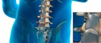
The lumbar region is represented by five vertebrae (L1-L5), separated by cartilage “pads” - intervertebral discs that act as shock absorbers and soften the load on bone tissue when the body moves. From birth, a child's intervertebral discs are very elastic, easily absorb and release fluid, due to which the hydrostatic resistance of the spine to loads is realized. Initially, their nutrition is carried out through their own blood vessels, but by the age of ten the vessels close, and the intervertebral disc becomes a more isolated system.
All fluid movements and exchanges occur by diffusion between the vertebrae and intervertebral discs. Therefore, it is not surprising that as we age, the nutrients in this enclosed space become less and less. Cartilage loses water and elasticity, becomes more rigid or even “shrinks out”. A decrease in the height and elasticity of the cartilage pad increases the load on the vertebral bodies, leads to their damage, and also contributes to pinching between adjacent segments of blood vessels and spinal nerve roots that exit the spinal canal.
In the lumbar region, at the level of the middle of the L2 vertebral body, the spinal cord ends, and the nerve roots are collected in a plexus, which is called the “cauda equina.” Also, the roots of the spinal nerves exit through the intervertebral foramina of the lumbar region:
- iliohypogastric (T12-L1);
- ilioinguinal (T12-L1);
- femoral cutaneous (L2);
- femoral-genital (L2);
- obturator (L2-L3-L4).
Next comes the sacral plexus, in which the greatest interest is the greater sciatic nerve, the largest nerve in the human body.
The development of osteochondrosis of the lumbosacral spine occurs most often. After all, it carries the maximum static and dynamic load, being a support for the entire human body. Lumbar dorsopathy accounts for 75% of cases of this pathology, while degenerative changes in the cervical spine approach 20%, and thoracic changes approach 5%.
Pain in osteochondrosis of the back is defined as acute if it lasts from 4 to 12 weeks and chronic if it persists for 12 weeks or longer. In approximately 20 percent of patients with acute pain, the process becomes chronic with severe pain symptoms within one year.
The pain syndrome that occurs with degenerative dorsopathy of the lumbar and sacral regions is associated with pinching of the spinal nerve roots (radiculopathy)
Compression of a nerve fiber results in pain, numbness, or a tingling sensation that is displaced to the area of the body innervated by the nerve. Therefore, dorsopathy of the lumbosacral spine manifests itself as a disorder of motor and tactile sensitivity in the upper part of the buttock, the back and inner surface of the thighs, calves, and feet. It is also possible to disrupt the functions of the genital organs, the process of urination and defecation if the cauda equina plexus is damaged.
A special form of radiculopathy is sciatica, which is compression of the sciatic nerve that passes through the buttock and extends down the back of the leg. This compression causes severe pain and numbness in the lower back, combined with pain in the buttock and leg on the affected side.
In the most extreme cases, when a nerve becomes pinched between the disc and adjacent bone, symptoms may include not only pain, but also numbness, partial or complete paralysis, muscle weakness, and limb atrophy. This condition can also be caused by vertebrogenic dorsopathy, i.e. disease associated with the vertebrae (tumor or cyst, infectious inflammation).
Spondylosis is the cause of deforming dorsopathy
Osteochondrosis is not the only degenerative disease that affects the condition of the lumbar vertebrae. Spondylosis also often develops in this department - the formation of calcifications on the articular edges of the vertebrae, in the lumen of the spinal canal, and in the ligaments that hold the spine. The danger of ossification is that the spinal column loses its mobility, and osteophytes create a narrowing (stenosis) and mechanical pressure on the spinal cord or nerve roots, irritate the blood vessels and cause a reflex of the vertebral artery (spasm and reflex reduction in blood flow). Characteristic symptoms of spondylosis include constant aching pain, which intensifies with movement around the axis of the spine, impaired flexibility and mobility, especially after sleep, until the patient “diverges.”
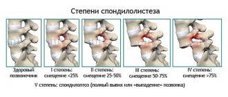
Degeneration of intervertebral discs leads to mechanical damage to the structure of the spinal column. Age-related changes include, for example, flattening of the lumbar deflection (lordosis), and pathological disorders include spondylolisthesis - displacement of the vertebral bodies relative to neighboring segments, practically “falling out” of one of them forward or backward.
With such a slippage, pinching of the spinal cord or its nerves inevitably occurs.
Scoliosis is also a spinal deformity - its curvature in the lateral planes with increased pressure on the spinal nerves and the development of symptoms of radiculopathy.
[node:field_similarlink]
Causes and risk factors
There are a large number of reasons that can provoke changes in the vertebrae and discs.
Among them the following stand out::
- Infectious diseases: vertebral tuberculosis, pyogenic infection, brucellosis, tertiary syphilis (now extremely rare).
- Injuries: fractures, sprains, dislocations and subluxations of the vertebrae.
- Hereditary factor.
- Diseases of an autoimmune and allergic nature.
- Poor nutrition (various unbalanced strict diets, improper vegetarianism, etc.).
- Lack of physical activity.
- Overstrain of untrained muscles.
- Prolonged stay in uncomfortable positions.
- Frequent short overloads of the neck, for example, when driving a car during sudden acceleration or braking.
- Hypothermia.
Among the risk factors, it is worth noting the following:
- poor nutrition;
- insufficient personal hygiene;
- smoking;
- lack of physical activity;
- physical activity without preliminary warm-up;
- sports training without the correct regimen and frequency;
- extreme sports;
- driving a car without a headrest;
- work associated with a forced position of the head for a long time;
- elderly age;
- regular stress.
Consequences
Dorsopathy of the cervical spine can provoke the following complications:
- Vertebral artery syndrome;
- Myelopathic syndrome;
- Vegetovascular dystonia.
The most dangerous consequence of neck dorsopathy is myelopathy and vertebral artery syndrome
Vertebral artery syndrome is a spasm of the cervical artery, which leads to deterioration of blood supply, cerebral ischemia, and stroke.
Myelopathic syndrome is a set of phenomena that accompany compression of the spinal cord. Possible paralysis of the limbs, dysfunction of the intestines and urinary organs, loss of sensitivity below the zone of compression of the brain.
Vegetative-vascular dystonia involves impaired regulation of the peripheral nervous system, which is responsible for the functioning of internal organs. It makes itself felt by cardiac arrhythmia, asthma attacks, increased nervous excitability, and thermoregulation disorders.
MRI
- MRI remains the imaging modality of choice for the cervical spine due to its complete imaging safety for the evaluation of the cervical spine.
- Advantages of MRI include: assessment of soft tissue morphology (eg, discs, muscle nerves, spinal cord), visualization of cerebrospinal fluid, non-invasiveness, and no radiation exposure to the patient.
- Modern MRI machines with high magnetic fields and new software products provide faster and more detailed visualization.
- Unfortunately, some MRI sequences depict larger-than-actual pathology and do not detect some abnormalities. Other disadvantages of MRI include high cost, the inability of claustrophobic patients to tolerate the procedure, the need for long periods of patient immobilization, and poorer visualization of bone structures compared to CT (MSCT).
- In addition, MRI is worse at differentiating compression of the root by disc herniation from the effect on the root by spondylous osteophyte.
EMG (ENMG).
Electrodiagnostic studies continue to be the standard methods for assessing neurological function of the cervical spine. The advantages of these methods are the low cost and safety of these tests.
Nerve conduction studies (NCG) and electromyography (EMG) provide the ability to assess the extent of root and peripheral nerve damage.
Needle EMG can detect acute, subacute and chronic radicular signs of motor nerve fiber pathology.
Laboratory diagnostics
. Necessary for differential diagnosis of inflammatory infectious or systemic conditions.
Treatment of dorsopathy with therapeutic anti-inflammatory patch NANOPLAST forte
The therapeutic pain-relieving anti-inflammatory patch NANOPLAST forte has shown high effectiveness in the treatment of many types of dorsopathy.
Thanks to the simultaneous influence of two physiotherapeutic factors - deep soft warming heat of infrared radiation and the influence of a magnetic field with specially selected characteristics - this innovative drug allows you to relieve pain and inflammation, improve blood circulation in the affected area, and reduce the dose of painkillers and anti-inflammatory drugs.
Therapeutic pain-relieving anti-inflammatory patch NANOPLAST forte can be used both in the complex treatment of dorsopathy and in monotherapy of many types of dorsopathy . The patch is applied to the cervical, thoracic or lumbar spine, depending on the type of disease and location of pain.
To relieve acute symptoms in the treatment of dorsopathy, a therapeutic patch is used for 3 to 5 days. The duration of the course of treatment is from 9 days. It is usually recommended to use the treatment patch in the morning for 12 hours, but it can also be used at night.
High efficiency, unique composition, long-term (up to 12 hours!) therapeutic effects, ease of use and affordable price make NANOPLAST forte the drug of choice in the treatment of most types of dorsopathy of various etiologies.
Read more about NANOPLAST forte