Spinal listhesis is a condition in which there is a horizontal displacement forward, backward, left or right relative to the underlying vertebra. Vertebral listhesis is a serious disease that poses a threat to the life and health of the patient. What happens with this shift? First of all, regardless of the trajectory of the displacement, a sharp narrowing of the spinal canal occurs. With total compression of the spinal cord, paralysis of part of the body is possible. And if the vertebral body in the cervical region is displaced, death is possible due to cardiac and respiratory arrest, since these systems function thanks to the support of the autonomic nervous system, controlled through the structures of the spinal cord.
If you discover such a problem, you should seek medical help as soon as possible and carry out the treatment prescribed by your doctor. Spinal listhesis can be treated by a vertebrologist, neurologist, osteopath and chiropractor. These specialists have sufficient competence to conduct a full examination and develop an individual course of therapy for each patient. A general practitioner in a city clinic most likely will not even think that the patient’s neck or lower back pain is associated with vertebral listhesis. It is easier for him to diagnose osteochondrosis and prescribe symptomatic treatment to relieve discomfort.
In Moscow, you can make a free appointment with a vertebrologist, neurologist, chiropractor or osteopath in our clinic. During the initial free examination, the doctor will be able to make a preliminary diagnosis. Then he will give you individual recommendations on how to conduct examinations and what will be required to treat the identified pathology.
If you are worried about lower back or neck pain, excessive muscle tension, clicking and other extraneous sounds when performing movements, do not hesitate, book a free appointment right now. Then it may be too late.
Causes of spinal listhesis
Potential causes of spinal listhesis include a variety of negative influences. The main ones are:
- traumatic impact that destroys the structure of the supporting structure and fixation (sprains of the ligamentous and tendon apparatus, deformation of the paravertebral muscles, cracks and fractures of the joints between the vertebrae);
- degenerative dystrophic changes in the cartilaginous tissues of the intervertebral discs, as a result of which they lose their height and ability to fix the vertebral bodies, often found in areas L4-L5, L5-S1, L3-L4;
- destruction and instability of the facet and facet intervertebral joints;
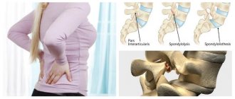
- isthmic violation of the integrity of various tissues of the spinal column - can be congenital or acquired;
- dysplasia of cartilage and bone tissues, including those provoked by necrosis against the background of a septic and aseptic inflammatory process;
- tumor etiology - as a tumor grows, it can displace healthy tissue in order to free up space for itself;
- curvature of the spine - compensatory listhesis occurs in order to maintain the balance of the supporting mechanism.
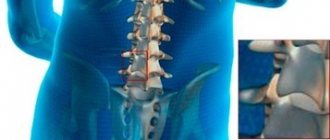
To understand the reasons for the development of vertebral listhesis, you need to have at least a general understanding of the anatomy of the spinal column. First of all, it is a supporting structure that maintains the balance of the body. Secondly, the spine is the seat of the spinal cord, the most important structural part of the autonomic nervous system, responsible for the functioning of the entire body. If the functioning of the autonomic nervous system is disrupted, the immune system malfunctions, digestion stops, cardiac and respiratory arrest may occur, arms and legs cease to obey (paralysis occurs), etc.
The spinal column consists of individual vertebral bodies with arcuate processes. It is they, together with the vertebra, that form the oval opening of the spinal canal. The vertebrae are separated from each other by cartilaginous intervertebral discs.
Each disc consists of an annulus fibrosus, a dense membrane, and a nucleus pulposus, an internal corpus pulposus. This structure does not have its own circulatory network and therefore can receive nutrition and fluid only through the process of diffuse exchange with the surrounding muscles.
If diffuse nutrition is disrupted, the height of the intervertebral disc decreases, the vertebrae become unstable and can move relative to each other.
The spinal column is secured by a system of ligaments:
- the posterior and anterior longitudinal ones begin at the back of the head and end at the coccyx;
- short intervertebral ones provide attachment of one vertebra to another;
- interspinous ligaments connect the spinous processes of the vertebrae;
- The transverse ligaments connect the transverse processes to each other.
Despite such a number of ligaments, they all have one big drawback - they lack elasticity. Therefore, when the height of the intervertebral disc decreases or after stretching, they can no longer fully fix the position of the vertebral bodies.
Also, the facet and facet joints of the spine take an active part in fixation. They connect the vertebral bodies and provide flexibility and mobility. Most often, joints are destroyed in the loaded lumbar region, less often in the cervical region. Very rarely, displacement of the vertebral bodies occurs in the thoracic region. The exception is cases of advanced scoliosis, in which lateral (lateral) vertebral listhesis occurs.
The need for surgical intervention
Spondylolisthesis tends to progress and its degree increases over time; on control radiographs after a few years, the displacement and slipping of the vertebra becomes larger and the anatomy becomes very hostile, which creates great difficulties for the surgeon during the operation, and the patient develops additional secondary changes in the spinal canal, affecting functions of the nervous system - the roots are more compressed, not only pain in the lower back, but also in the legs, numbness, and possible genital disorders.
Thus, it is logical to undergo surgical treatment already in the early stages of spondylolisthesis, so as not to bring the situation to a critically irreversible point.
To better understand the pathology, it is better to illustrate it with specific examples from practice.
Patient Z., whose isthmic, also known as congenital or true listhesis, was combined with discitis, inflammation of the disc.
The image clearly shows the zone of non-fusion (zigzag with a fountain pen).
Her photo, the intervertebral disc is almost completely absent, there is a vacuum effect, the vertebrae are sclerotic, all this did not allow us to carry out a des cage.
Intraoperative radiograph demonstrating the displacement of the vertebrae, pins determining the position of the screws in perspective.
Screws were inserted, reduction was performed, i.e. reduction of listhesis and its rigid fixation.
Control CT scans after surgery demonstrate reduction of listhesis, correct placement of fixators, and adequacy of decompression.
After the operation, the patient “rose to her feet” 3 days later, after regression of pain, she walked a lot around the department and was discharged with a good functional result.
An example of degenerative, secondary (due to osteochondrosis) spondylolisthesis.
An older patient P. with spondylolisthesis at the same level as the previous patient.
Compensatory thickening of the ligamentum flavum and the development of spondyloarthrosis led to the development of secondary narrowing of the spinal canal—spinal stenosis, manifested by neurological disorders.
MRI of patient Z. before the intervention.
The axial projection shows a clear demonstration of multifactorial narrowing of the spinal canal due to the mentioned reasons.
The patient underwent decompression of the spinal canal, restoration of the spinal axis - reduction of listhesis, and spinal fusion (Fusion) with a cage and TPF system.
Postoperative CT scan, lateral view showing reduction of vertebral displacement.
CT axial view after surgery demonstrating complete decompression with removal of two affected facet joints.
The patient was discharged with a good functional result.
Listhesis of the lumbar spine
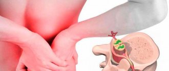
Lumbar listhesis is the most common localization of this pathology. This is due to the fact that the lumbar intervertebral discs bear the maximum shock-absorbing load during any body movement. If a person leads a sedentary lifestyle, is engaged in heavy physical labor or sedentary work, or is overweight, then the muscular frame of the lumbar region cannot cope with its functions. There is constant spastic muscle tension. In this state, they completely stop carrying out diffuse exchange of fluid with the intervertebral cartilaginous discs. Metabolites accumulate in the tissues of the fibrous ring, it becomes dehydrated and begins to collapse. This creates the prerequisites for the development of listhesis of the lumbar spine, which in the future will lead to disruption of the innervation of the internal organs of the abdominal cavity and lower extremities.
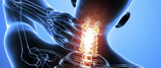
Listhesis of the cervical spine
Cervical listhesis is much less common due to the fact that there is no significant shock-absorbing load on this section. In most cases, cervical vertebral listhesis is associated with traumatic exposure. These are sudden turns of the head, emergency braking when driving a car, falling from a height with a hard landing, side impacts, etc.
There are congenital listhesis of the cervical spine, which can lead to paralysis, assimilation of the first vertebra by the occipital bone, etc. Correction of congenital dysplasia of intervertebral discs and joints must begin as early as possible. This will avoid poor posture and prevent delayed psychomotor development of the child.
In an adult, listhesis of the cervical spine can lead to serious pathologies of the cardiovascular system and brain. With prolonged incorrect position of the vertebra, posterior vertebral artery syndrome is formed. This can lead to depression, decreased mental performance, chronic fatigue syndrome, increased blood pressure, etc.
Symptoms of spondylolisthesis
The main complaints with listhesis are aching pain in the affected area, aggravated by physical activity or being in one position for a long time, limited movement; it is necessary to maintain an even spine through “effort of will.” However, depending on which vertebra is displaced, i.e. which part of the spinal cord is under pressure, specific symptoms may develop, often misdiagnosed as a separate disease.
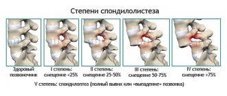
Degrees of vertebral listhesis
The degree of listhesis depends on how many degrees of displacement occurs. The higher this parameter, the more serious the danger to the patient’s health.
Currently, the following degrees of vertebral listhesis are accepted in the medical classification:
- 1st degree – deviation from the central axis by 25%;
- 2nd degree – deviation by 50%;
- 3rd degree – displacement up to 75%;
- Grade 4 – complete displacement or slipping of the vertebra.
Only the first and second degrees can be successfully treated with conservative methods. In the third, a planned surgical operation is required to restore the fixing ability of the ligamentous and tendon apparatus. In the fourth degree, emergency surgery is required to restore the patency of the spinal canal.
Content:
- Degrees of spondylolisthesis
- Symptoms of spondylolisthesis
- Diagnosis of spondylolisthesis
- The need for surgical intervention
- Rehabilitation after surgery
As you know, there are several types of displacement, or spondylolisthesis. They are classified according to etiology - the cause of the disease, namely
- Congenital, isthmic, true
- Degenerative, secondary, false
- Traumatic
- Neoplastic and some other rare forms
Types of listhesis
When considering the types of listhesis that occur, it is first of all worth talking about stable and plastic displacement. Stable displacement is characterized by the fact that the vertebra is fixed in its new place and does not move in the opposite direction. With the plastic type of displacement, the vertebra is not fixed by anything and moves freely. This is a more dangerous condition that requires immediate medical attention.
In addition, the following types are distinguished:
- anterior listhesis (antelisthesis) – anterior displacement;
- posterior scalene listhesis (antelisthesis) – displacement occurs in a stepwise manner in the posterior direction;
- lateral vertebral listhesis (laterolisthesis) – displacement to the right or left.
Scalene listhesis mainly occurs in the lumbar spine. Anterior listhesis is most often diagnosed in the cervical spine.
Treatment of listhesis of the lumbar spine
Listhesis can be treated with conservative methods, but only if the condition does not threaten the patient’s life. If he is already developing paralysis of the body, intestines and other internal organs, then it is better to agree to surgery. And after it, carry out comprehensive rehabilitation in order to strengthen all the tissues of the spinal column.
Conservative treatment of spinal listhesis is aimed primarily at restoring the normal height of the intervertebral disc, which does not fully fix the vertebra. If destruction of the intervertebral joint is detected, the doctor must take care of eliminating this problem.
In our manual therapy clinic, treatment of listhesis of the lumbar spine or any other is carried out using the following techniques:
- osteopathy – allows you to restore the physiological position of the vertebral body;
- massage improves the condition of the ligamentous and tendon apparatus, accelerates the circulation of blood and lymphatic fluid, thereby improving the diffuse nutrition of the cartilage tissue of the intervertebral disc;
- kinesiotherapy and therapeutic exercises increase muscle tone and improve their contractility, thereby restoring fixing and holding ability;
- reflexology allows you to activate the body’s hidden reserves and start the process of accelerated regeneration of damaged tissues;
- physiotherapy speeds up the healing process.
If you need effective and safe treatment for listhesis of the lumbar spine or cervical spine, you can make a free appointment with a vertebrologist at our manual therapy clinic. During the consultation, the doctor will give individual recommendations on restoring the integrity of the spinal column.
Diagnostics
At the “Medical Center YES!” A neurologist-vertebrologist performs diagnostics based on an examination of the patient, examination and description of the neurological status. The doctor can assess the patient’s condition by the volume of certain movements, the presence of deformities, and manifestations such as muscle spasms and functional blocks. To diagnose the disease, clarify the nature of neurological symptoms, localize the lesion and select the optimal treatment tactics, the doctor conducts a comprehensive neurological examination of the patient, using laboratory diagnostics and prescribing instrumental examination methods:
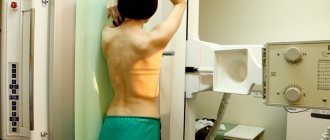
X-ray examination, CT, MRI, electroneuromyography.