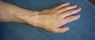
Often young women are perplexed: where did the strange lump appear on their hand?
?
Although painless, but quite large (from 0.5 to 6 cm in diameter), it cannot go unnoticed due to its location. So what is hygroma
- cancer or a purely aesthetic defect?
Although hygroma on the arm is a tumor-like formation, it, contrary to misconceptions, is not an oncological disease
, and also
never degenerates into cancer
. This disease is quite widespread (up to 50% of all neoplasms on the hands) and can be successfully treated. On the other hand, this subcutaneous formation can cause discomfort or even interfere with normal blood circulation in the hands.
Hygroma
(or, as it is also called,
ganglion
) is a capsule-bag that is dense to the touch, filled with a viscous and jelly-like protein liquid mixed with fibrin. The hygroma noticeably protrudes over the ligaments and tendons and practically does not move under the skin. Most often, synovial cysts are observed in women aged 20 to 30 years (⅔ of the total number of patients). Children are least likely to suffer from it.
In what cases should you worry about treating hygroma and can it be prevented?
Hygroma - formation in the form of a tumor
Causes of hygroma
The exact causes of hygroma on the hand have not yet been established. It is believed that joint injuries can lead to the formation of small cavities, which are filled with serous fluid and then merge into one large or several small cysts. Doctors identify several factors that contribute to the onset of the disease and the growth of education:
- tenosynovitis
(inflammation of the tendon sheath) or bursitis (inflammation of the joint capsule) with a subacute course; - hand injury
(bruise, sprain, fracture, ligament rupture); - chronic trauma to articular and periarticular tissues
(ligaments, tendons, cartilage lining), for example, due to repetitive professional movements; - previous hand surgeries
; - diseases of the musculoskeletal system localized in the hands
; - the need to write or draw a lot by hand
(especially if the position or selection of stationery is incorrect); - congenital weakness of the tissues that form the joint capsule
(in cases where the tendency to hygromas is inherited between family members).
It has been established that athletes (tennis players, golfers and others), musicians (pianists, violinists and others), as well as people whose work involves stress on their hands (painters, turners, loaders, typists, bakers, etc.) are most often susceptible to hand hygroma. hobby). Sewing, embroidery, beadwork and similar hobbies can also increase the risk of developing a hygroma.
When is surgery needed?
Most people with hygroma want a clear answer to this question. However, the doctor cannot answer it. A synovial cyst is not a dangerous disease; it has a chance to go away without any treatment; surgery (like any intervention) has its own possible complications and risks. Whether to operate on a hygroma or not is a patient’s informed decision. When a person understands the situation and is ready to share responsibility for the decision made with the doctor, then one can choose the time and place to perform the intervention.
How is the operation to remove hygroma performed?
I will describe the basic principles using the example of the most common dorsal hygroma on the wrist.
- The operation is outpatient. There is no need to stay in the hospital.
- Local anesthesia. Anesthesia means unnecessary risks, as well as excessive preoperative examination. I use a mixture of lidocaine and epinephrine, this avoids discomfort from the tourniquet, reduces the likelihood of a toxic reaction to the anesthetic and prolongs the effect of pain relief to 4-5 hours.
- Transverse access. Longitudinal access leaves rough, unsightly scars.
- It is imperative to excise the fragment of the joint capsule from which the hand hygroma grows. Only the skin is sutured.
- I don't use immobilization. For the first 2-3 days, a thick soft bandage is applied, which limits movement and makes the early postoperative period as comfortable as possible. Prolonged immobilization after surgery can lead to stiffness, but does not affect the likelihood of relapse.
- The skin heals in about 2 weeks.
Hygroma symptoms
In approximately 35% of cases, a synovial cyst may be asymptomatic
, except for the compaction under the skin.
Therefore, the main symptom of hygroma is considered to be the appearance of a lump
(one or several)
in the area of the wrist joint
,
fingers or hand
;
extremely rarely - on other parts of the body. Typically, such a lump reaches 1, 2 or 3 centimeters in size, but an advanced synovial cyst can be up to 6 cm in diameter. In the scientific literature there are references to even larger seals
! But there is usually no need to worry about its size, if it does not cause severe inconvenience, because the hygroma grows very slowly and does not form a truly large tumor-like body.
Synovial cyst
has a characteristic location: on the back of the wrist or ankle joint, as well as the hand or foot.
In addition to the elastic dense lump, patients may experience:
- difficulty moving the affected area;
- dull, usually aching, pain in the hand;
- deterioration of sensitivity in the hand;
- weakness of muscles and ligaments;
- discomfort that increases with physical activity on the hands (especially the supporting one);
- crawling sensation, tingling or burning sensation, as well as other symptoms of compression of nerve endings or blood stagnation;
- increased sensitivity of the skin in the area of the lump.
Special mention should be made of hygroma, in which the growing lump is located under the ligament
, and therefore
does not form a distinct protrusion above the surface of the skin
. In this case, patients complain of:
- constant or periodic pain (pressing or dull), which can radiate to the hand, fingers, forearms;
- pain when pressing;
- roughening, peeling of the skin over the formation;
Unlike other similar diseases, hygroma on the arm, as a rule, does not cause pain on palpation (sometimes it can only be slightly painful). It is characteristic that the skin over the lump does not undergo any changes - it remains elastic and retains its normal color, and does not “grow” onto the tumor. The formation, as a rule, can be moved from side to side, but it does not roll under the skin. One of the main differences between a hygroma and a real tumor is that it can shrink and increase in size. As a rule, hygroma loses volume during the rest period and swells after exercise.
Symptoms of hygroma may appear or intensify in women after the birth of a child.
What modern methods of getting rid of hygroma exist?
Removal of this bubble with a gel-like liquid is possible both from the skin and from the joint. There is a thin camera and special instruments that allow you to look inside the wrist joint through small punctures of 2-3 mm. This procedure is called arthroscopy from the words “arthro” joint and “scopy” to look. Through these punctures you can not only look into the joint, but also perform useful manipulations inside, for example, remove a hygroma. The advantages of the arthroscopic method are smaller scars on the skin, faster rehabilitation, and a relatively lower likelihood of relapse.
Diagnosis of hygroma
To make an accurate diagnosis, you need to visit a doctor - an orthopedist, surgeon or rheumatologist. Diagnosis of hygroma requires a comprehensive study of the clinical picture, taking into account localization, external signs, and also, if necessary, the results of laboratory or instrumental studies. Typically, a patient examination begins with palpation of the neoplasm, examination and questioning of the patient, as well as checking joint mobility. In some cases, this is enough, and sometimes additional examination of the lump is required to exclude lipoma, fibroma, atheroma or traumatic cyst. If the hygroma is in the palm area, tumor formations in the bone or cartilage tissue should be excluded
To exclude a malignant origin of the formation, the doctor may prescribe a puncture (fluid sampling) from the cyst for histological and/or cytological examination, which is carried out using a syringe.
In the case of an atypical location of the lump, an X-ray examination or ultrasound is performed to make a diagnosis. Ultrasound examination of the cyst helps determine the density of its contents, the presence of veins and other vessels nearby that it can compress, as well as the uniformity of the lump or the presence of several nodules. In some cases, the patient may be recommended an MRI (to determine the structure of the walls, the presence of vessels in the hygroma, and the consistency and composition of the filling of the cyst).
It is important to diagnose hygroma in the early stages
Possible complications of hygroma
Although hygroma is a disease with a positive prognosis and a relatively low risk of complications, it can lead to pathologies that require prompt surgical intervention
. First of all, this concerns painful or rapidly growing formations that can compress blood vessels and soft tissues and impair their trophism. This condition directly damages the bone and cartilage tissue in the hands, leading to starvation and degenerative changes in the wrist and interphalangeal joints, deterioration of muscle and ligament function, as well as fine motor skills of the fingers.
With an “unfortunate” location, the hygroma of the hand can compress veins and nerve bundles
, disrupting the conduction of nerve impulses in the hands, causing aches and severe pain after exercise. There is also a risk of traumatic crushing of the tumor.
In the worst case scenario, a large synovial cyst can cause:
- thrombophlebitis (inflammation of the venous wall with the formation of a blood clot, which leads to blockage of the vessel), venous stagnation (accompanied by swelling and cyanosis of the tissues around the hygroma);
- rupture of the hygroma’s own capsule, as well as blood vessels with hemorrhage;
- pinched nerves and disruption of the normal innervation of the hand, which is accompanied by numbness in the fingertips and tingling;
- bursitis and even purulent tendovaginitis;
- suppuration and sepsis if the wall of the hygroma bursts;
- contracture (persistent loss of mobility in the hand, finger or other affected parts of the body);
- re-development of benign formations (single or, more often, multiple) after injury or removal of hygroma.
If you encounter mechanical crushing of a hygroma on your hand, do not try to remove its contents yourself.
!
Treat the affected area with an antiseptic and apply a sterile bandage, then contact your surgeon within a few hours
.
Due to damage to the skin over the formation, infection of the tendon ganglion
(hygromas). The development of complications and the onset of the inflammatory process is indicated by:
- increase in general body temperature (up to 38-40°C);
- impaired mobility in the joint;
- changes in the structure of the skin over the hygroma (loss of elasticity, redness, roughness, peeling);
- soreness.
Treatment of hygroma in this case should be carried out by an orthopedist-traumatologist or surgeon. It is not worth leaving a crushed hygroma without treatment: after the capsule wall heals, it will fill with liquid again, and new formations may appear around it.
What is it?
The word is formed from two parts: hygro- means liquid, the ending -oma is added if we are talking about a tumor. In principle, this term describes quite well the essence of what is happening, that is, a neoplasm filled with liquid. When this ball or lump is isolated, the surgeon receives a thin translucent shell with gel-like or liquid contents.
There are also other terms for hygroma:
- synovial cyst
- ganglion
- ganglion
- tendon ganglion
- ganglion cyst
- synovioma
- bursitis
With one amendment or another, all these words can be considered synonyms.
Types of hygroma
As we mentioned above, hygroma can occur in the form of a single formation, delimited from other tissues by a membrane. It is also possible for several cysts to appear, fused or independent of each other.
However, most often the disease is classified according to the location of the lump. So, doctors distinguish the following types of it:
- hygroma of the wrist joint
; - hand hygroma
; - hygroma on fingers
.
Much less common:
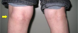
- hygroma of the knee joint
(formed in the knee fossa, also known as Becker’s cyst); - hygroma of the ankle joint
; - hygroma of the foot
(located on the dorsal, i.e., upper, side of the foot, usually at its outer edge).
The tendon ganglion, located in the wrist and hands, accounts for up to 88% of the total number of cases of the disease. The ankle joint and foot bothers up to 11% of patients who suffer from hygroma. A synovial cyst in the neck, shoulder, shoulder blade or knee occurs in only 1% of patients. It is extremely rare for patients to see a doctor with formations in the bones, muscles or spine.
Hygroma of the wrist
Hygroma of the wrist joint
- the most common type of synovial cyst. It becomes clearly visible in a bent position of the wrist joint (for visual diagnosis, you need to move the hand forward and back, and then left and right). Often, hygroma of the wrist joint is located on the front or back side of the joint, but it can also grow on the lateral surfaces or at the very base of the palm.
If the lump is located under the ligament, it may not cause any concern and may not even cause any aesthetic discomfort.
As a rule, the consistency of hygroma of the wrist joint is felt as soft or springy, “rubbery”.
Hygroma of the hand
Tendon ganglion in the hand area
(hygroma of the hand) usually occurs on the fingers (from the side of the palm), the palm itself, the back of the hand, or closer to the wrist joint. Typically, hand hygroma does not bother the patient until it reaches a large enough size and does not interfere with everyday activities.
Hygroma on the finger
Synovial cyst on fingers
often occurs on their back side, in the area of the distal phalanx (i.e., the last one on which the nail is located). Often it is located in close proximity to the cuticle or nail fold.
The skin over the hygroma on the finger, as a rule, looks thinned, stretched, and its natural pattern is smoothed out. Under the skin, as a rule, a rounded formation can be easily felt, which does not cause pain, except in case of injury. It is quite dense to the touch, so it can be perceived as a bone or cartilage growth.
Hygromas on the palmar side of the phalanx are usually larger than on the back, and can spread to the entire phalanx and even occupy the adjacent one. They can be quite painful due to compression of the nerves that run along the sides of the fingers. This type of hygroma often prevents patients from performing household chores.
Ganglions also appear at the proximal phalanges
(near the base of the finger). Such cysts are also quite painful, despite their small (about the size of a pea) size. Pain with a hygroma on the finger usually occurs when trying to tightly grasp a hard object (for example, the handle of a shovel, a rolling pin, etc.).
Hygroma of the lower extremities
Synovial cysts of the lower extremities are typically localized on the back of the foot or fingers, as well as on the front of the ankle joint. Such hygroma rarely bothers patients with pain. As a rule, discomfort occurs when the foot is compressed by tight shoes, the foot is rubbed or stuffed, and also when the lump is located in close proximity to the nerve.
Hygroma of the lower extremities
Treatment of hygroma
Although sometimes a hygroma on the arm can be left without treatment, due to the risk of complications and aesthetic discomfort, doctors usually recommend its removal. Sometimes a synovial cyst can resolve spontaneously if the factor that provokes it (for example, occupational stress) is eliminated in a timely manner - then the synovial fluid will simply stop flowing into the capsule.
The indication for treatment of hygroma is its multi-chamber nature, rapid growth of formation, development of the inflammatory process, pain syndrome, and limited mobility in the joint.
The preferred treatment for hygroma is surgical removal.
, since puncture or aspiration of the tumor is usually ineffective or poses a risk of complications.
Thus, conservative treatment of wrist hygroma is characterized by recurrence of the disease in approximately 80-90% of cases
, while for surgical treatment the same figure is only
8-20%
.
Surgery
Doctors do not recommend delaying surgical treatment of hygroma, since a large, albeit benign, formation soon begins to displace blood vessels, muscles, ligaments and nerves. Their non-physiological position significantly complicates the operation and may have consequences for the patient
.
Removal of a hygroma on the arm is usually carried out under local anesthesia with a tourniquet and lasts no more than half an hour. The operation is performed using a scalpel or laser equipment. In advanced cases, large or unusual formations may require general anesthesia. When carrying out the procedure, it is important to remove all degenerative cells without exception (otherwise the altered tissue may again develop into a cyst), wash the capsule, and also bandage the so-called. the mouth of the hygroma, which is associated with the joint and feeds the lump with synovial fluid. The drainage tube for fluid drainage is removed 1-2 days after the procedure, and postoperative sutures after complete healing of the incision are removed on days 7-10. After this, it is recommended to immobilize the operated area with a splint and a pressure bandage for several weeks and avoid serious stress on it for 30-60 days.
.
Recently, surgeons have given preference to endoscopic removal of wrist hygroma, in which the capsule and its contents are removed through a small incision a couple of centimeters in size. Rehabilitation after such an intervention occurs much faster.
Conservative treatment of hygroma
As a conservative treatment for hygroma of the hand, crushing it (without penetration and formation of a wound) is occasionally used, followed by the application of a compressive bandage. This method should never be used on infected synovial cysts due to the risk of sepsis, but it can be used to treat sterile lumps. The main disadvantage of this approach is the rapid recurrence of the disease: as soon as the burst edges of the capsule grow together, it will again begin to fill with serous fluid.
A somewhat more successful technique is aspiration puncture of the formation, in which its contents are sucked out through a small puncture, and glucocorticoid solutions and sclerotization preparations are injected in its place. This allows the capsule to be filled with connective tissue instead of liquid. This method is also indicated for administering antibiotics and treating infected hygroma.
Conservative techniques do not eliminate degenerative cells
, which provoke the formation of hand hygroma and are therefore considered ineffective.
There are several methods for treating hygroma
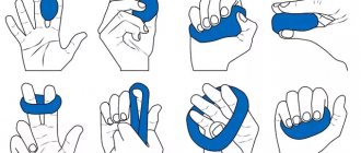
Physiotherapy
A small cyst that does not grow, does not become inflamed and does not cause pain can often be treated conservatively using physiotherapeutic techniques. They help reduce the volume of formation and ensure the outflow of its contents, slow down cell growth. Although physiotherapy cannot completely cure hygroma, in combination with chondroprotectors
it helps keep the tumor under control without surgery.
Sometimes physiotherapy is the optimal solution for non-standard location of the lump
, in which excision can be a rather traumatic procedure.
This type of treatment helps relieve compression of nerve endings, eliminate pain and inflammation, improve tissue regeneration, and relax the muscles around the tumor.
In modern physiotherapy for hygroma of the wrist and other joints, the following is used:
- electrophoresis
; - ultra high frequency therapy
; - phonophoresis with hydrocortisone
; - magnetotherapy
; - paraffin applications
; - mud baths and wraps
; - UV therapy
.
During the entire course of physiotherapy, and also, as prescribed by the doctor, after it, a tight, compressive bandage is applied to the area affected by hygroma, which prevents further accumulation of fluid in the capsule.
If the tumor is too large or continues to grow, there are signs of inflammation, the attending physician, together with the patient, usually decides on surgical intervention.
Drug treatment
For the medicinal treatment of hygroma, a wide range of anti-inflammatory drugs are used, which are applied to the surface of the cone or injected directly into its cavity.
However, anti-inflammatory therapy provides only a temporary and symptomatic effect
,
and therefore used in combination with other techniques
.
In case of a purulent process inside the cyst, and also as a preventive measure before surgery to remove it, the attending physician selects a specialized antibiotic for the patient - usually in the form of injections.
But the most effective medicine for the treatment of tendon ganglion are chondroprotectors
.
They are helping:
- establish metabolic processes in the tissues adjacent to the hygroma, strengthen muscles and ligaments, prevent the destruction of synovial cartilage;
- improve the quality of synovial fluid - as a result, the joint begins to produce it in smaller volumes, and the nutrition of the hygroma is reduced;
- slow down further growth of tumor formation;
- relieve symptoms of inflammation in the area of hygroma;
- significantly reduce pain (many patients note a reduction in pain by 50-80%);
- cope with discomfort from the tendon ganglion, which cannot be operated on due to its small size.
Chondroprotectors do not allow the joint on which the hygroma occurred to “starve” due to poor supply of nutrients. They also relieve swelling and restore normal mobility in the fingers, hands and wrist joint. In addition, they have a systemic effect on the body, improving the condition of the musculoskeletal system and preventing the appearance of new hygromas, as well as the recurrence of old ones. Due to the almost complete absence of side effects, chondroprotectors can be taken to prevent synovial cysts even before they appear
,
during treatment of a neoplasm
, as well as
after surgery
.
Chondroprotectors stimulate tissue repair after surgery and prevent the re-formation of modified cells. The standard course lasts from 3 to 6 months per year. Compared to other drugs in this series, Artracam
, the course of which lasts 1.5 months, and then can be repeated after 2 months if necessary. It is made exclusively from natural ingredients - seafood.
Drug Artracam
is one of the most effective chondroprotectors for hygroma of the wrist and hand.
Puncture
Hygroma puncture is a quick and safe way to get rid of it. Also, a puncture can be considered as a diagnostic procedure: if you get a gel, it means it’s definitely a hygroma. The probability of relapse after puncture is about 80-90%. Injecting any medications into its cavity does not reduce the likelihood of relapse. It is worth remembering that the entry of steroid hormones (they are sometimes used in the hope of influencing the possible return of hygroma) into the subcutaneous fat or articular cartilage can cause very serious irreversible destruction of these structures.
Prevention of hygroma and its complications
For standard prevention of hygroma of the wrist and other joints, you should:
- avoid injury and hypothermia;
- promptly treat arthritis, arthrosis and other connective tissue diseases;
- minimize the load on the joint, incl. monotonous (from time to time take a break from work or monotonous activities to warm up);
- pay attention to hereditary and metabolic diseases that can negatively affect the health of the musculoskeletal system (gout, diabetes);
- follow safety precautions and exercise rules when playing sports;
- Avoid self-medication for injuries and other lesions in the hands and wrists;
- Monitor your body weight and maintain a low-calorie diet rich in vitamins, minerals and amino acids.
If in your family or for your occupation there is a high predisposition to hygromas and other diseases of the musculoskeletal system, you should think about additional measures. Rheumatologists recommend an annual course of prophylaxis with chondroprotective drugs. They help strengthen connective tissue and improve its metabolism, preventing the development of degenerative cells. Artracam is ideal for this purpose.
- due to the high bioavailability of glucosamine sulfate, pleasant taste and convenient sachet form.
Take care of yourself!
Images designed by Freepik
Can it resolve?
Medicine knows cases when hygroma resolves. This happens both as a result of treatment and by itself. Cases of resorption of cones are quite rare, however, they are possible. This happens in several cases:
- If the cause is occupational activity, the lump may disappear when the load on the affected joint is reduced.
- In the initial stages, hygromas sometimes disappear on their own, without intervention. But you need to take into account that the joint is still affected, and the disease can recur at any time.
- Symptoms of the disease decrease or disappear with non-surgical treatment (ointments, physiotherapy).
Hygromas resolve in the initial stages, and there is a high chance of relapse. Only surgery can completely get rid of the disease.
Hygromas cause pain and can cause numbness or an abscess.