Synovioma is one of the most dangerous tumors, which very often metastasizes to adjacent bones, lymph nodes or some other organs. Such tumors are classified as sarcoma diseases. The disease is a cellular tumor that is formed from the soft tissue of the joint membranes, synovial bursae and tendon sheaths.
This pathology originates when cells in tissues begin to mutate and change, cell proliferation occurs (tissue proliferation by cell multiplication by division). Such neoplasms most often occur on the fingers and toe joints. In most cases, they are unilateral, affecting only one of the limbs. Very often, the presence of synovioma is detected during medical examination or when visiting a traumatologist and surgeon.
To date, the exact reasons for which synoviomas occur have not been identified. However, it is believed that the formation of such tumors is often influenced by heredity and genetic predisposition. In addition, factors such as long-term exposure of the body to food or industrial carcinogens (toxic substances), severe trauma to skeletal bones, heavy physical activity and several other root causes can affect the condition.
Benign synovioma
Benign tumors (giant cell synoviomas) are observed very rarely. Usually found in people under 45 years of age. People older than this age rarely get sick. With a benign neoplasm, severe pain is not observed. The tumor, as a rule, does not grow, does not penetrate adjacent tissues and does not impair the motor functions of the joints. It is located in a capsule and may not progress for many years. Very often, synovioma of the hand occurs on the fingers. In most cases, this type of tumor responds well to treatment.
Treatment is surgical; the tumor and adjacent tissues are excised. But there are also relapses when the tumor is in a hard-to-reach place and cannot be completely removed during surgery. In the benign form, such tumors are classified as “Benign neoplasms” and have the ICD-10-D10-D36 code.
Forecast
Due to the slow growth and “maturity” of tumor cells, the prognosis is favorable in most cases. However, relapses of synovioma can occur in approximately 40% of cases, especially when the tumor location is difficult to access for tissue excision and the desire to preserve limb function as much as possible after surgical treatment.
At Euroonko, experienced oncologists treat synoviomas. The clinic is equipped with all the necessary equipment and medications to provide care in accordance with modern treatment standards.
Book a consultation 24 hours a day
+7+7+78
Leading clinics in Israel
Assuta
Israel, Tel Aviv
Ikhilov
Israel, Tel Aviv
Hadassah
Israel, Jerusalem
Malignant synovioma
Malignant synoviomas (synovial sarcomas) are classified in the “Oncology” group and their code is O-3. The main risk group includes children and adolescents under 18 years of age, and is more common in males. In children, with timely treatment of the pathology, the prognosis is usually favorable. The tumor can develop on the fingers, elbow and shoulder joints. Slightly less commonly, these tumors affect the knees, feet and fingers of the lower extremities. There are very rare cases when synovioma is located inside the joint.
For a long time (up to two years or more), the disease can occur without obvious signs. This is the so-called latent stage, during which it is difficult to detect a tumor and make a diagnosis.
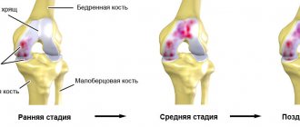
Synovial sarcoma of soft tissues is a shapeless neoplasm that reaches quite large sizes. It progresses very quickly, actively grows, metastases appear in the bones and various organs (lungs, lymph nodes, skin, abdominal organs). With such a tumor, the motor function of the joints is impaired, and severe pain is observed. The synovioma tissue contains modified bone and cartilaginous elements. Unlike benign synovioma, a malignant tumor is not enclosed in a capsule and tends to actively penetrate into adjacent tissues. Treatment is surgical, the tumor is excised to the extent of healthy tissue. In severe cases, limbs are amputated. If necessary, chemotherapy and radiation therapy are used.
Types of synovioma
First of all, it should be noted that synoviomas, based on the nature of their growth, the aggressiveness of the clinical picture and the tendency to metastasize, are divided into benign and malignant. In clinical practice, tumors are often encountered that are in one way or another associated with the synovial vagina, and then we need to talk about a benign form of synovioma. If the oncological process develops in soft tissues, and not necessarily localized near the joints, and progresses quickly, then a malignant form must be suspected - synovial sarcoma.
The existence of benign synoviomas is disputed by a number of authors. Some scientists argue that all diseases from this group have a malignant course, and it does not depend on the degree of maturity of the tumor. In a number of cases, biopsy material taken from a pathological focus reveals atypical elements in the form of giant cells, which gives grounds to divide benign synoviomas into 2 types: without giant cells and giant cell (according to the WHO classification, the latter condition corresponds to nodular tenosynovitis and does not apply to blastomas ).
G. Gailer purposefully studied synoviomas, who described cases of the benign course of the tumor. He gave as an example hard nodules that were localized near the joints and were surrounded by a clear capsule. Microscopically, they were a cluster of fibroblast-like and epithelial-like cells (the so-called biphasic structure), which most closely resembled mature synoviocytes.
Citing the example of a microscopic picture of a tumor, the scientist still argued that whether a process is benign should be judged not only by the results of microscopy, but also by the clinic, and suggested monitoring such patients for at least 5 years.
Classification of synovioma
Currently, in rheumatology, synoviomas are classified based on tumor characteristics. According to the classification of the World Health Organization, synoviomas are divided according to the type of cellular structure into two types: monophasic and biphasic synoviomas. Monophasic synovioma is very difficult to detect, since synovial differentiation (changes in size, shape, function and activity of the cell) is weak. Such neoplasms consist of one type of cell (large light cells of the spindle-shaped type).
In biphasic synovioma, differentiation is clear. Therefore, it is easier to detect and diagnose. Such tumors respond very well to treatment. They consist of two types of cells: sarcomatous and epithelial. In addition, the neoplasm contains cavities similar to synovial ones.
Based on the tissue structure, tumors are divided into fibrous and cellular. Fibrous synovioma is formed from fibers that are similar in structure to fibrosarcoma tissue. Cellular consists mainly of glandular tissue with papillomas and cysts.
Based on the cell structure, synoviomas are divided into the following types:
- Fibrous;
- giant cell;
- Alveolar;
- Adenomatous;
- Histoid;
- Mixed.
Causes of synovioma
The pathogenesis of the disease lies in the disruption of the formation of normal synovial cells during their reproduction due to atypical transformations. The following conditions may be the possible etiological factors:
- unfavorable environmental conditions;
- mutational defects encoded in the genetic code that are inherited;
- exposure to radiation, ultraviolet radiation;
- consumption of foods that contain carcinogenic substances;
- traumatic damage to joints;
- long-term exposure to carcinogens of physical and chemical origin.
Symptoms
One of the first symptoms of synovioma development is the appearance of a tumor, which can most often be detected by touch. Typically, such neoplasms are of medium density, but they can also be hard. The appearance of pain is a significant symptom of the development of a malignant process. Synovioma of the elbow, shoulder or knee joint limits their mobility. In cases where the tumor compresses the nerve, numbness of the limbs may occur. There are also general signs of the disease that do not depend on the location of the tumor. The patient feels unwell, feels weak and constantly tired, loses appetite, loses weight sharply, body temperature rises, and lymph nodes enlarge.
What is it and what causes it?
This pathology is also called giant cell synovioma, its definition is as follows: it is a tumor, the formation of which is associated with abnormal absorption of a free DNA molecule by the body cell, as well as the proliferation of structural elements of the synovial membrane of the joints. The pathological focus can also be localized in the mucous bursae and tendon sheaths. If metastasis occurs, the process occurs through the lymphogenous route.
Interesting! If we take malignant neoplasms of soft tissues, then synovioma is the most common. It occurs in 32.7% of cases.
Sometimes a benign synovioma occurs, which is distinguished by the presence of a capsule, beyond which the pathologically formed cells do not spread anywhere. The disease occurs equally in both men and women at different ages. If you look at a cross-sectional photo of a synovioma, the formation resembles fish meat, distinguished by a whitish color. It is also characterized by many cracks and cysts, hemorrhages and even areas of necrosis. Often the cavities of the neoplasm are filled with mucus, very similar to synovial fluid. By the way, due to its cystic structure, the tumor has a soft consistency. If calcification occurs, it becomes harder.
The reliable causes of many diseases have not been identified to date; this also applies to benign synovioma of the hand. But through ongoing research, scientists have been able to identify a number of factors that increase the likelihood of developing a painful condition. These include:
- genetic predisposition to cancer;
- serious injuries;
- internal organ transplantation;
- negative effects of carcinogens, ionizing radiation;
- immunosuppressive course of recovery for cancer.
Attention! Representatives of the age group from 14 to 20 years are at increased risk. Statistics show that synovioma of the knee or ankle most often develops.
Diagnostics
The success of treatment for synovioma depends on how quickly this tumor is detected and the correct diagnosis is made. In modern clinics equipped with the latest modern equipment, an accurate diagnosis is made. Errors, as a rule, are excluded (after all, synovioma of the knee joint, for example, is often mistaken for bursitis).
The necessary comprehensive diagnostics are prescribed:
- X-ray examination of the affected limb in different projections determines the size and location of the neoplasm (on X-rays, synovioma looks like a darkened spot with a lobular shape or blurred edges);
- Magnetic resonance and computed tomography (layer-by-layer scanning determines the size of the tumor and the spread of metastases);
- Biopsy (sampling of material for examination under a microscope to determine the malignancy or benignity of the tumor);
- Radioisotope scanning (strontium is used, the accumulation of which in the affected area helps to determine the degree of tumor germination and its localization);
- Angiography, which detects an increase in the network of vessels in the area of the neoplasm;
- Genetic material testing;
- Blood tests (general and biochemical);
- Ultrasound of the peritoneum (detects the presence of distant metastases);
- Scintigraphy of skeletal bones (determines distant tumor growth in bone tissue).
In some cases, other types of examination may be required (lung x-ray, lymph node puncture, ultrasonography of the peritoneal area, MSCT). This depends on the stage and degree of progression of the malignant process.
Synovioma prognosis
When a benign formation in the joint area is diagnosed, after surgery to excise it, patients are slightly at risk of complications and relapses, that is, they have a relatively favorable prognosis.
Malignant synovioma is dangerous with a high probability of metastasis. More than fifty percent of patients, after undergoing specific therapy, again sought medical help with a relapse of the disease. In this case, an important aspect is timely diagnosis and treatment, which, however, does not guarantee a complete recovery.
Treatment
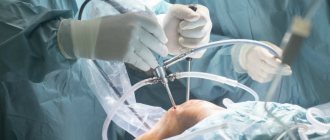
The most effective method of treating synovioma is complete surgical removal of the tumor. Depending on the complexity of the disease, the degree of surgical intervention during the operation varies. For small tumors, 3-5 cm of healthy tissue surrounding the tumor is excised. If the tumor is advanced and has reached a large size, then fairly large areas of adjacent soft tissue, bones and lymph nodes are excised. In particularly severe cases, the limb is amputated.
Radiation therapy is used before and after surgery. Before surgery, it is needed to stop the growth of the tumor and prepare it for excision. After surgery, it helps prevent complications and re-development of the tumor.
Chemotherapy is used in cases where the tumor is diagnosed late and has extensive metastases. Chemotherapy treatment is often used to increase the effectiveness of other methods or in cases where the tumor is at a late stage of development and it is impossible to operate on it.
Do you want to know the cost of cancer treatment abroad?
Synovioma in children
The disease practically never occurs in childhood. If diagnosed, the tumor could arise as a result of the carcinogenic effects of various factors on the woman’s body during pregnancy or directly on the child after his birth. Hereditary predisposition and the presence of cancer pathology in close relatives play a significant role. The symptoms and principles for diagnosing synovioma of the tendons, fingers, shoulders and other types are similar to those in adults. Treatment is selected by the doctor taking into account the child’s age, stage of cancer, and individual characteristics of the body.
Prevention
Currently, there are no special measures to prevent such neoplasms, since there is no exact explanation of the reasons for the onset of synovioma development. However, if you follow some simple recommendations, you can reduce the likelihood of their occurrence. It is necessary to avoid any injuries to the limbs, protect yourself from exposure to radiation and harmful substances, and follow safety and protection rules when contacting chemicals. Persons with a genetic predisposition to such diseases must undergo regular medical examinations, laboratory and instrumental examinations.
Symptoms of the disease
The neoplasm can develop for more than 2 years without showing signs. The pathology is discovered by chance during a routine medical examination. It is easy to find new growths in open areas. The benign membrane passes into the capsule and is easily treated, therefore, after detection, a course of therapy is carried out.
Synovioma on x-ray
Synovial sarcoma grows quickly and is accompanied by severe pain. Motor functions become difficult and then stop. The patient's health deteriorates sharply. The following symptoms appear:
- Body temperature remains at 37-39 degrees;
- Iron deficiency develops in the blood - anemia;
- The lymph nodes are noticeably enlarged;
- The victim experiences rapid fatigue;
- A person suddenly loses weight.
In the damaged area, active proliferation of malignant processes into other organs occurs, and a transformation of an atypical type is noticeable on the bone tissue. All this leads to immobilization of the joint.