L.G. Turbina, Doctor of Medical Sciences, Professor, MONIKI, Moscow
Pain in the back and limbs, not associated with inflammatory damage to the peripheral nerves, in our country is traditionally classified as vertebrogenic, implying spinal osteochondrosis as the etiological factor. However, studies of the last decade have shown that osteochondrosis is only one of the causes of such pain, but not the main one.
The causes of back pain can be divided into vertebrogenic and non-vertebrogenic.
Vertebrogenic causes of pain in the back and limbs:
- Disc herniation
- Spondylosis
- Osteophytes
- Sacralization or lumbalization
- Arthrosis of the intervertebral (facet) joints
- Ankylosing spondylitis
- Spinal stenosis
- Spinal segment instability with spondylolisthesis
- Vertebral fractures
- Osteoporosis
- Vertebral tumors
- Ankylosing spondylitis
- Functional disorders of the spine
Nonvertebrogenic causes of back pain:
- Myofascial pain syndrome
- Psychogenic pain
- Referred pain in diseases of internal organs
- Intra- and extramedullary tumors
- Metastatic lesions
- Syringomyelia
- Retroperitoneal tumors
Let us consider in detail the etiology, pathogenesis, clinical picture, diagnosis and treatment of the most common vertebrogenic and non-vertebrogenic pain.
Vertebrogenic pain
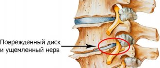
The causes of vertebrogenic back pain are often degenerative-dystrophic processes: osteochondrosis and spondyloarthrosis. With osteochondrosis, the intervertebral disc is primarily affected, resulting in reactive changes in the bodies of adjacent vertebrae, tissues of the facet joints and ligaments.
The process is primarily localized in the nucleus pulposus of the intervertebral disc, which becomes less elastic due to loss of moisture. Under the influence of mechanical stress, the nucleus pulposus can sequester and protrude towards the annulus fibrosus of the disc.
Over time, cracks form in the annulus fibrosus. A disc with an altered nucleus and fibrous ring can prolapse into the lumen of the spinal canal (disc prolapse), and masses of the nucleus pulposus penetrate through the cracks of the fibrous ring, forming disc herniations. The described processes in one spinal segment lead to reactive changes in adjacent vertebrae and intervertebral joints, resulting in disruption of the kinematics of the entire spinal column. In addition, the process may involve the ligamentum flavum, which becomes denser over time and puts pressure on the root or membranes of the spinal cord. Over the years, the process can stabilize due to disc fibrosis, but it never reverses.
Congenital bone anomalies, excessive physical activity and other reasons that contribute to the wear of cartilage tissue lead to the development of spinal osteochondrosis and aggravation of its course.
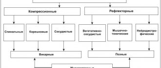
Depending on which structures of the spinal column are involved in the process in each specific case, either compression or reflex syndromes predominate in the clinical picture (see table).
Compression syndromes develop if altered structures of the spine deform or compress the roots, blood vessels or spinal cord. Reflex vertebrogenic syndromes arise as a result of irritation of various structures of the spine, which has powerful sensory innervation. It is believed that only the bone tissue of the vertebral bodies and the epidural vessels do not contain nociceptive receptors. Afferent impulses from the irritated elements of the spinal column through the dorsal root and spinal structures close on the motor neurons of the anterior horn, causing muscle-tonic reactions at the appropriate level. However, it should be remembered that the division of vertebrogenic syndromes into compression and reflex is very arbitrary, since reflex syndromes can occur in their pure form or accompany compression manifestations.
Based on localization, vertebrogenic syndromes are distinguished at the cervical, thoracic and lumbosacral levels.
Osteochondrosis of the lumbar spine - causes, mechanism of occurrence and development
No clear cause of osteochondrosis has been identified; the disease is considered multifactorial. The most popular theory is that the cause of spinal osteochondrosis is considered to be constant muscle overstrain or muscle hypotension, non-physiological muscle tension, which leads to overload of the vertebral segments, deterioration of their blood supply and, ultimately, to degeneration of the intervertebral discs and joints, which results in osteochondrosis.
The cause of premature aging of intervertebral discs can also be endocrine and metabolic disorders, hereditary predisposition to the development of osteochondrosis, autoimmune disorders, and microtraumas. The negative role of visceral, that is, associated with internal organs, pathologies in the development of spinal osteochondrosis has also been proven. Unfavorable heredity plays a significant role in the development of the disease. The contribution of hereditary factors to the appearance of lumbar osteochondrosis is about 60%, the rest is attributed to factors of adverse external influences on the spine.
Factors contributing to the development of osteochondrosis in both the lumbar and other parts of the spine are very numerous. The main ones are: incorrect posture, physical inactivity, muscle strain in the lumbar region as a result of constant carrying of heavy objects or sudden excessive load, hormonal changes, endocrine and somatic diseases, disruption of the normal blood supply to spinal tissues, emotional stress, age-related changes, poor nutrition. Under the influence of these factors, the processes of dehydration and circulatory disorders, arthrosis and subluxations in small joints, bone growths, prolapsed discs - hernias, gradually increase in the structures of the spine.
The most obvious causes of lumbar osteochondrosis are most often considered to be a sedentary lifestyle and back injuries. Therefore, the risk group primarily includes those people whose profession is directly related to stress on the back. This is a very wide range of professions - salespeople and waiters, surgeons and teachers - everyone who is forced to spend most of their working time standing. The risk group includes builders and loaders, as well as athletes whose occupation places heavy stress on the spine. In addition, drivers, various office workers, programmers suffer from osteochondrosis of the lumbar region - that is, those who have a mostly sedentary job and who suffer from physical inactivity.
Compression syndromes of the cervical localization
At the cervical level, not only roots and vessels, but also the spinal cord can be subjected to compression. Compression of blood vessels and/or the spinal cord is manifested by a clinical syndrome of complete or, more often, partial transverse lesion of the spinal cord with mixed paresis of the arms and lower spastic paraparesis.
Root compression can be clinically divided into:
- root C3 – pain in the corresponding half of the neck;
- root C4 – pain in the area of the shoulder girdle, collarbone. Atrophy of the trapezius, splenius and longissimus muscles of the head and neck. Possible cardialgia;
- root C5 – pain in the neck, shoulder girdle, lateral surface of the shoulder, weakness and atrophy of the deltoid muscle;
- root C6 – pain in the neck, scapula, shoulder girdle, radiating along the radial edge of the arm to the thumb, weakness and hypotrophy of the biceps brachii muscle, decreased reflex from the tendon of this muscle;
- root C7 - pain in the neck and scapula, spreading along the outer surface of the forearm to the II and III fingers, weakness and atrophy of the triceps brachii muscle, decreased reflex from its tendon;
- root C8 – pain from the neck spreads along the inner edge of the forearm to the fifth finger of the hand, decreased carporadial reflex.
Vertebral artery syndrome due to cervical osteochondrosis
Typically, vertebral artery syndrome develops over several months or even years. Accordingly, clinical manifestations do not arise acutely, but increase until they lead to a significant deterioration in the patient’s quality of life. Unfortunately, at this stage of development, cervical osteochondrosis and vertebral artery syndrome are very difficult to treat and surgical assistance is often required to restore blood supply to the posterior parts of the brain. Many patients learn about the existing problem only after they develop a stroke (acute cerebrovascular accident) with subsequent paralysis of the body.
We strongly recommend that you study in detail the first signs and classic symptoms of this dangerous disease. If they are detected, immediately consult a neurologist or vertebrologist.
Vertebral artery syndrome against the background of cervical osteochondrosis can be triggered by degenerative dystrophic changes in the cartilaginous tissue of the intervertebral discs. The posterior vertebral arteries are tightly adjacent to the vertebral bodies and any changes in their configuration entail a violation of the patency of the blood vessels. It can be partial or complete.
Potential reasons for the development of this pathology:
- degenerative dystrophic destruction of the fibrous ring of the intervertebral disc;
- sclerosis of articular endplates;
- deformation of the facet and facet joints;
- arthrosis of uncovertebral joints;
- instability of the position of the vertebral bodies against the background of protrusion of the intervertebral discs;
- hernial protrusions of the nucleus pulposus through a violation of the integrity of the fibrous ring of the intervertebral disc;
- spondylosis and spondyloarthrosis;
- Ankylosing spondylitis and other types of rheumatoid ankylosis;
- formation of bone growths on the vertebral bodies and spinous processes;
- spasms of the muscles of the neck and collar area, including fibromyalgia;
- cicatricial deformation of the lateral and longitudinal ligaments of the spine after stretching or microscopic ruptures;
- myositis and other types of muscle fiber damage;
- curvature of the cervical spine, including the “widow’s hump” in the area of 6-7 vertebrae.
When identifying potential causes, the division of the vertebral artery sections into intracranial and extracranial should be taken into account. The first is damaged due to displacement and subluxation of the first cervical vertebra. The second may be subject to compression due to degeneration of the cervical vertebrae. It passes through a canal formed by openings on the sides of all cervical vertebrae. In the area of the first two vertebrae on the outside there is no protection of the arteries other than the skin. Therefore, any external pressure (tight clothing, incorrect head position during sleep) will lead to compression of the lumen of the cerebral blood vessels.
Constriction of the vascular bed can be a reflex, occurring due to irritation of Frank's sympathetic nerve or damage to the radicular nerves extending from the spinal cord through the foramen in the vertebral bodies.
With vertebral syndrome, segments of the artery passing in the area of 4-5 vertebrae can be damaged. Less commonly, pathology is diagnosed in the area of the 6-7 vertebrae.
Cervical reflex syndromes
Clinically manifested by lumbago or chronic pain in the neck area with irradiation to the back of the head and shoulder girdle. On palpation, pain is detected in the area of the facet joints on the affected side. Sensitivity disorders, as a rule, do not occur.
It should be noted that the cause of pain in the neck, shoulder girdle, and scapula can be a combination of several factors, for example, reflex pain syndrome due to spinal osteochondrosis in combination with microtrauma of the tissues of the joints, tendons and other structures of the musculoskeletal system. Thus, with glenohumeral periarthrosis, many researchers note in such patients damage to the C5-C6 discs, as well as injury to the shoulder joint, or myocardial infarction, or other diseases that play the role of triggers.
Clinically, with glenohumeral periarthrosis, pain in the periarticular tissues of the shoulder joint and limitation of movements in it are noted. Only pendulum-like movements of the shoulder in the sagittal plane are possible (frozen shoulder syndrome). The adductor muscles of the shoulder and periarticular tissues are painful on palpation, especially in the area of the coracoid process and the subacromial zone. Sensory disorders are not determined, tendon reflexes are preserved, sometimes somewhat animated.
Reflex cervical syndromes include the anterior scalene muscle syndrome. The anterior scalene muscle connects the transverse processes of the middle and lower cervical vertebrae with the first rib. When this muscle is involved in the process, pain occurs along the anterior outer surface of the neck, radiating along the ulnar edge of the forearm and hand. When palpating the anterior scalene muscle (at the level of the middle of the sternocleidomastoid muscle, somewhat laterally), its tension is determined, and in the presence of muscle trigger points, pain distribution zones are reproduced in it - shoulder, chest, scapula, hand.
Vertebrogenic neurological complications in the thoracic spine with osteochondrosis are rare, since the bone frame of the chest limits displacement and compression. Pain in the thoracic region more often occurs in inflammatory (including specific) and inflammatory-degenerative diseases (ankylosing spondylitis, spondylitis, etc.).
In medical practice, the first place in terms of treatment is taken by lesions of the lumbar and lumbosacral spine.
Symptoms
According to the clinical course, two stages of vertebral artery syndrome are distinguished: functional and organic.
The functional stage of vertebral artery syndrome is characterized by a certain group of symptoms: headaches with some autonomic disorders, cochleovestibular and visual disturbances. Headache can have various forms, both acute throbbing and constant aching or sharply intensifying especially when turning the head or prolonged static load. The headache may spread from the back of the head to the forehead. Disturbances in the cochleovestibular system can manifest as paroxysmal dizziness (swaying instability) or systemic dizziness. In addition, some hearing loss may occur. Visual disturbances can manifest as darkening of the eyes, a feeling of sparks, sand in the eyes.
Prolonged and prolonged episodes of vascular disorders lead to the formation of persistent foci of ischemia in the brain and the development of the second (organic) stage of vertebral artery syndrome . In the organic stage of the syndrome, symptoms of both transient and persistent hemodynamic disorders of the brain appear. Transient hemodynamic disturbances are manifested by symptoms such as dizziness, nausea, vomiting, and dysarthria. In addition, there are characteristic forms of ischemic attacks that occur during turning or tilting the head, during which attacks of falling with preserved consciousness may occur, so-called drop attacks, as well as attacks with loss of consciousness lasting up to 10 minutes (syncope episodes). Symptoms usually regress in a horizontal position and are thought to be due to transient ischemia of the brainstem. After such episodes, general weakness, tinnitus, and autonomic disturbances may occur.
Based on the type of hemodynamic disturbances, several variants of vertebral artery syndrome are distinguished (compressive, irritative, angiospatic and mixed forms).
Vessel narrowing in the compression variant occurs due to mechanical compression on the artery wall. In the irritative type, the syndrome develops as a result of reflex spasms of the vessel due to irritation of sympathetic fibers. In the clinic, most often, combined (compression-irritative) variants of vertebral artery syndrome are encountered. In the angiospastic variant of the syndrome, there is also a reflex mechanism, but it arises from irritation of receptors in the area of the motor segments of the cervical spine. In the angiospastic variant, vegetative-vascular disorders predominate and the symptoms are not so strongly associated with head turns.
Lumbar compression syndromes
Upper lumbar compression syndromes are a relatively rare location.
Compression of the LII root (LI-LII disc) is manifested by pain and loss of sensitivity along the inner and anterior surfaces of the thigh, and decreased knee reflexes.
Compression of the LIV root (LII-LIV disc) is manifested by pain along the anterior inner surface of the thigh, decreased strength and subsequent atrophy of the quadriceps femoris muscle, loss of the knee reflex.
Compression of the LV root (LIV-LV disc) is a common location. It manifests itself as pain in the lower back with irradiation along the outer surface of the thigh, the anterior surface of the leg, the inner surface of the foot and big toe. Hypotonia and wasting of the tibialis muscle and decreased strength of the dorsal flexors of the thumb are noted.
Compression of the SI root (LV-SI disc) is the most common location. It manifests itself as pain in the buttock, radiating along the outer edge of the thigh, lower leg and foot. The strength of the triceps surae muscle decreases, sensitivity in the areas of pain irradiation is impaired, and the Achilles reflex fades.
Lumbar reflex syndromes
Lumbago - acute pain in the lower back (lumbago). Develops after physical activity. Manifests itself with sharp pain in the lumbar region. The antalgic posture and tension of the lumbar muscles are objectively determined. Neurological symptoms of loss of function of the roots or nerves of the lumbosacral region, as a rule, are not detected.
Lumbodynia is chronic lower back pain. It manifests itself as dull aching pain in the lower back. Palpation determines the pain of the spinous processes and interspinous ligaments and facet joints (at a distance of 2-2.5 cm from the midline) in the lumbar region. Movement in the lumbar region is limited. Sensory disorders are not defined.
Piriformis syndrome
The piriformis muscle begins at the anterior edge of the upper sacrum and attaches to the inner surface of the greater trochanter of the femur. Its main function is hip abduction. The sciatic nerve passes between the piriformis muscle and the sacrospinous ligament. Therefore, when the piriformis muscle is tense, compression of the nerve is possible, which occurs in some cases with lumbar osteochondrosis.
The clinical picture of piriformis muscle syndrome is characterized by sharp pain in the subgluteal region radiating along the posterior surface of the lower limb. Adduction of the hip causes pain (Bonnet test), the Achilles reflex is reduced. The pain syndrome is accompanied by regional autonomic and vasomotor disorders, the severity of which depends on the position of the body - pain and autonomic disorders decrease in the supine position and intensify when walking.
Coccydynia – pain in the sacral area. A polyetiological clinical syndrome that may be caused by discopathy of the first coccygeal disc, causing reflex tension of the pelvic floor muscles, or ligament pathology. No sensory disorders are detected. A rectal examination reveals areas of tenderness in the muscles involved (usually the levator ani muscle).
Differential diagnosis of compressive and reflex vertebrogenic syndromes
| Compression | Reflex |
| The pain is localized in the spine, radiating to the limb, right up to the fingers or toes | The pain is local, dull, deep, without irradiation |
| Pain intensifies with movement in the spine, coughing, sneezing, straining | Pain intensifies with load on the spasmed muscle, its deep palpation or stretching |
| Regional vegetative-vascular disorders are characteristic, often depending on body position | Regional autonomic-vascular disorders are not typical |
| Symptoms of loss of function of compressed roots are determined: sensory disturbance, muscle wasting, decreased tendon reflexes | There are no symptoms of loss |
Treatment of vertebrogenic pain syndromes
In the acute period of the disease, when the pain syndrome is severe, the main task of the doctor is to relieve pain. To successfully complete this task you must:
- Create peace for the spine. To do this, place a shield under the mattress or place the patient on a special orthopedic mattress. For 5-7 days, the motor mode is limited, and the patient is allowed to stand only in an immobilizing belt or corset and only when physiologically necessary. The rest of the time, bed rest is indicated. The expansion of the motor regime is carried out carefully; the recommended movements should not cause pain.
- Drug treatment should be structured taking into account all links in the pathogenesis of pain. The source of pain in compression syndromes is pathologically altered structures of the spinal column, which either irritate tissue nociceptors or compress the spinal roots. In reflex syndromes, the source of pain can be both the spine itself and reflexively spasmed muscles that form tunnel syndromes. In addition, with chronic (lasting more than 3 months) or recurrent pain, depressive, anxiety, hypochondriacal and other affective disorders develop. The presence of such disorders must be actively identified and treated, since they have an extremely negative impact on the course of the disease.
- Non-drug treatment. Physiotherapy, manual therapy, kinesitherapy, etc. are widely used in the treatment of vertebrogenic pain syndromes.
- Surgery. Used when conservative treatment is ineffective for 4 months or there are signs of spinal cord compression with dysfunction of the pelvic organs, sensory conduction disorders or damage to the central motor neuron (in the presence of pyramidal signs).
Treatment
Treatment of vertebral (vertebral) artery syndrome consists of two main areas: improving hemodynamics and treating diseases that lead to compression of the vertebral arteries.
Drug treatment
Anti-inflammatory and decongestant therapy is aimed at reducing perivascular edema resulting from mechanical compression. Drugs that regulate venous outflow (troxerutin, ginkgo biloba, diosmin). NSAIDs (Celebrex, lornoxicam, celecoxib)
Vascular therapy is aimed at improving blood circulation in the brain, since hemodynamic disturbances occur in 100% of patients with this syndrome. Modern diagnostic methods make it possible to evaluate the effectiveness of treatment with these drugs and the dynamics of blood flow in the vessels of the brain using ultrasound examination. The following drugs are used for vascular therapy: purine derivatives (trental), vinca derivatives (vincamine, vinpocetine), calcium antagonists (nimodipine), alpha-blockers (nicergoline), instenon sermion.
Neuroprotective therapy
One of the most modern areas of drug treatment is the use of drugs to improve energetic processes in the brain, which allows minimizing neuronal damage due to episodic circulatory disorders. Neuroprotectors include: cholinergic drugs (citicoline, gliatiline), drugs that improve regeneration (Actovegin, Cerebrolysin), nootropics (piracetam, Mexidol), metabolic therapy (mildronate, Thiotriazoline, Trimetazidine)
Symptomatic therapy includes the use of drugs such as muscle relaxants, antimigraine drugs, antihistamines and others.
Treatment of degenerative diseases includes non-drug treatment methods, such as exercise therapy, physiotherapy, massage, acupuncture, and manual therapy.
In most cases, the use of complex treatment, including both drug and non-drug treatment, can reduce symptoms and improve blood circulation in the brain.
Surgical treatment methods are used in cases where there is severe compression of the arteries (disc herniation, osteophyte) and only surgical decompression can achieve a clinical result.
Drug treatment
- Analgesics, anti-inflammatory non-steroidal drugs, anesthetics. To relieve pain, the use of analgesics metamizole sodium (Analgin), paracetamol, tramadol (Tramal) and non-steroidal anti-inflammatory drugs (NSAIDs) enterally and parenterally is indicated. The use of NSAIDs is pathogenetically justified, since drugs in this group have an analgesic effect, and also, due to their effect on cyclooxygenase (COX-1 and COX-2), inhibit the synthesis of prostaglandins, which prevents the sensitization of peripheral nociceptors and the development of neurogenic inflammation.
Of the drugs in this group, the following have proven themselves well: diclofenac, which is available in the form of tablets of 50 and 100 mg, rectal suppositories and solutions for parenteral administration. The drug ketorolac (Dolac) has a powerful analgesic effect, which is recommended to be administered for severe pain syndromes 30 mg intramuscularly for 3-5 days, and then switch to tablet forms, prescribing 10 mg 3 times a day after meals for no more than 5 days.In addition to those listed above, you can use other drugs in this group: meloxicam (Movalis), lornoxicam (Xefocam), ketoprofen (Ketonal), etc. But it should be remembered that most NSAIDs are contraindicated for gastric and duodenal ulcers, with a tendency to bleeding . If the patient is diagnosed with the above diseases, even in remission, the listed NSAIDs are contraindicated. In such cases, the drugs of choice are selective COX-2 inhibitors, which do not have such a significant effect on the gastrointestinal tract. These drugs include celecoxib (Celebrex), a selective COX-2 inhibitor. It should be prescribed at a dose of 200 mg 3 times a day after meals for 7-10 days.
To reduce pain, paravertebral blockades can be performed with an anesthetic (procaine, lidocaine, etc.) in combination with corticosteroids (50 mg hydrocortisone, 4 mg dexamethasone, etc.). Blockades using anesthetics and corticosteroids are recommended to be carried out once every 3 days. In most cases, 3-4 blockades are sufficient for a course of treatment (elimination of acute pain).
- Vascular agents. Considering the mandatory participation of the vasomotor component in the pathogenesis of vertebrogenic syndromes, especially those of a compression nature, it is necessary to introduce vasoactive drugs into the treatment complex. The choice of drug depends on the presence of concomitant vascular disease and the severity of vasomotor disorders. In mild cases, oral administration of vasodilators (nicotinic acid preparations or their analogues) is sufficient. If the patient is diagnosed with severe compression radiculopathy, parenteral administration of drugs that normalize both the arterial inflow and venous outflow of pentoxifylline (trental) is necessary.
- Psychotropic drugs. Patients with chronic pain need correction of affective disorders. To carry out adequate correction of psychoaffective disorders, their diagnosis is necessary (consultation with a psychotherapist or psychodiagnostic testing). In case of predominance of anxiety-depressive and depressive disorders, the prescription of antidepressants is indicated. Preference is given to drugs that have, along with an antidepressant, anxiolytic effect: amitriptyline - from 25 to 75 mg/day. for 2-3 months, tianeptine (Coaxil), mianserin (Lerivon), etc. If the patient has predominant hypochondriacal disorders, tricyclic antidepressants should be combined with antipsychotics that do not cause extrapyramidal disorders, tifidazine (Sonapax) - 25-50 mg/day. , sulpiride (eglonil) - 25-50 mg/day.
Vertebral lumbar pain: multifactorial origin, symptomatology, treatment principles
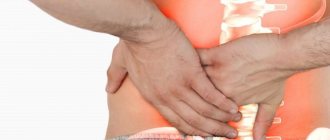
Back pain, familiar to almost every person, is most often associated with damage to the lumbosacral spine. The development of chronic vertebral pain, including lumbar pain, is facilitated by heavy physical exertion and, conversely, by the lack of adequate exercise, unfavorable meteorological factors (especially permanent ones associated with unsatisfactory working and living conditions), congenital or acquired pathology of the musculoskeletal system and spinal column, and also the presence of excess weight and osteoporosis. In terms of its prevalence in our country, chronic vertebral pain is one of the mass public health problems.
Lumbar vertebral pain, in addition to primary damage to the spine, can have an “extravertebral” origin - due to secondary involvement of the osteochondral and nervous structures of the spinal column.
Main factors and clinical forms of spinal lesions
The primary factors of damage to the spine - in particular, its lumbosacral region - include focal or widespread vertebral changes associated with independently occurring pathology of the spinal column. Among them, the main factor is the dystrophic vertebral process (osteochondrosis of the spine).
Secondary damage factors are associated with the presence of an extravertebral pathological process, which also leads to the development of focal or widespread changes in the spine. Among them, osteoporosis and metastatic lesions of the spine are of greatest clinical importance.
The first factor (osteoporosis) is highly prevalent among middle-aged, elderly and senile women. At the same time, spinal osteoporosis most often occurs without the development of neurological disorders, and therefore its clinical manifestations are rarely a reason for neurological observation. The second factor (metastatic lesions) is many times higher than the incidence of primary spinal tumors. In some cases, vertebral disorders of metastatic origin come under the supervision of a neurologist even before the diagnosis of the underlying disease is made. The diagram - factors and clinical forms of primary and secondary spinal lesions - is presented on
.
Clinical manifestations of pathology of the lumbosacral spine
In neurological practice, differentiation of forms of vertebral lesions begins with the definition of vertebral syndrome, taking into account the characteristics of which the underlying disease is established. Clinical manifestations of pathology of the lumbosacral spine represent 3 groups of vertebral syndromes (
):
- actually painful;
- radicular monoradicular;
- polyradicular.
Pain (reflex) syndromes
Painful (reflex) syndromes of the lumbosacral region, not accompanied by focal neurological symptoms, can manifest themselves:
- lumbodynia - acute, subacute or chronic pain in the lumbosacral region (Fig. 3), in some cases - lumbago (sharp, sudden lumbar pain - “lumbago”);
- lumboischialgia - lumbar pain radiating along the dermatome of the sciatic nerve - n. ischiadicus (Fig. 3);
- coccydynia—pain in the coccyx area (Fig. 3).
Radicular syndromes (radiculopathies)
Radicular syndromes (radiculopathies) caused by damage to the lumbosacral spine are less common than lumbar pain syndromes. The presence of radiculopathy is indicated by symptoms of loss of sensitive, reflex and motor functions of a certain spinal root.
Manifestations of lumbosacral radiculopathy:
- pain in the lumbar region, radiating to the leg (down to the foot);
- hyperesthesia or paresthesia (tingling sensation, crawling “goosebumps”) - mainly in the area of pain;
- hypoesthesia/hypalgesia - mainly in the distal innervation of the root (outer/inner edge of the foot);
- asymmetry (due to decreased) or absence of Achilles and knee reflexes;
- decreased muscle strength—mainly in the extensor and flexor muscles of the toes.
The most common forms of lumbosacral radiculopathies are associated with damage to the fifth lumbar (L5) and first sacral (S1) spinal roots. Clinical differences between these radiculopathies relate to the areas of localization of pain and sensory disorders, as well as the presence of the Achilles reflex, which disappears with S1 radiculopathy (Fig. 4).
Clinical features of spinal osteochondrosis
In the vast majority of cases, the development of lumbar pain and radicular syndromes is caused by spinal osteochondrosis, especially often affecting the joints of the two lower lumbar vertebrae and the base of the sacrum (intervertebral discs LIV-LV, LV-SI).
The main clinical manifestations of spinal osteochondrosis, which limit the patient's motor activity to varying degrees, include recurrent or chronic vertebral pain, painful spinal mobility, as well as monoradicular neurological disorders.
The course of clinical manifestations of spinal osteochondrosis is most often cyclical - with alternating periods of exacerbation and remission (complete or partial). Exacerbations are usually seasonal (autumn and spring). The highest frequency of exacerbations of the disease occurs in the fifth decade of life. In most cases, the development of exacerbations of vertebral pathology is predictable - in case of violation of the regime that limits physical activity and excludes cooling.
The most characteristic clinical feature of spinal osteochondrosis as a disease with a chronic, long-term course is the inevitability of a gradual “subsidence” of vertebral pain (usually at the turn of the 5th–6th decades of life). This feature of spinal osteochondrosis is due to the transition of the current degenerative process to the final stage, stabilizing the position of the bone and soft tissue structures of the spinal column. In this regard, regression of vertebral pain is often accompanied by an even greater limitation of spinal mobility. Pain and limitations in the patient’s daily physical activity that persist after 50 years are most often associated with a previous injury, another form of spinal damage, or osteoarthritis of the hip joint.
The most unfavorable form of manifestation of lumbar osteochondrosis is the development of discogenic compression of the structures of the spinal canal, in particular the cauda equina, which threatens severe neurological complications and disability of the patient. Compression of the spinal canal structures may also be associated with secondary forms of spinal damage ().
Signs of acute compression of the spinal canal structures (including the cauda equina):
- the occurrence of bilateral weakness and numbness of the legs, numbness in the perineum;
- retention of urine and feces;
- with compression (compression-ischemic) damage to the spinal cord—spontaneous reduction of pain, followed by a feeling of numbness of the pelvic girdle and limbs.
Clinical features of secondary spinal lesions
Symptomatic vertebral pain caused by secondary damage to the spine, at the very beginning of its development, can occur similar to manifestations of spinal osteochondrosis. The presence of this pain often becomes the reason for physiotherapy, which can further aggravate the manifestations of the underlying disease.
Establishing the symptomatic nature of vertebral pain is facilitated by:
- a thorough analysis of the patient’s complaints during “chronization” and increasing intensity of vertebral pain, as well as its atypical manifestations;
- clinical examination of the patient if the treatment is insufficiently effective.
Atypical vertebral manifestations (characteristic of secondary spinal lesions):
- vertebral pain:
–more often occurs in patients over 40, especially 50 years old; – gradually increases; – intensifies with movement, persists or intensifies at rest; – daytime, as well as nighttime, including “awakening”; – accompanied by local vertebral pain - “soreness” or sharp pain within one or two adjacent vertebrae when pressing and “tapping” the spinous processes;
- the effectiveness of non-narcotic analgesics: in usual therapeutic doses - short-term, gradually decreasing;
- the presence of extravertebral pain - paravertebral, abdominal, lower abdomen or groin area;
- combination with somatic disorders:
– increased body temperature; – general weakness, loss of appetite, loss of body weight; – changes in laboratory parameters—acceleration of ESR, anemia, leukocytosis.
Principles of treatment
Analgesics
The main analgesic drugs for eliminating vertebral pain are non-steroidal anti-inflammatory drugs (NSAIDs). NSAIDs, like other painkillers, are usually self-administered by patients when pain intensifies and returns. However, long-term use of analgesics, which increases the risk of complications of drug therapy, requires the use of NSAIDs under medical supervision.
Today, in the arsenal of a practicing physician there is a wide range of NSAIDs, which, according to their mechanism of action, belong to “non-selective” anti-inflammatory drugs - blocking the enzyme cyclooxygenase (COX) and “selective” - blocking the COX-2 isoenzyme. These drugs differ significantly in the ratio of advantages and disadvantages, respectively, in the severity and duration of therapeutic effects and the side effects caused. Probably more preferable for patients with vertebral pain (in terms of availability, effectiveness and lower likelihood of side effects) are: among the “non-selective” NSAIDs - diclofenac, ibuprofen and ketoprofen, and among the “selective” - meloxicam.
Methods of using NSAIDs:
Diclofenac - orally 100-150 mg/day (regular tablet forms 25-50 mg, retard form - 100 mg); intramuscularly or subcutaneously; rectally; locally.
Ibuprofen - orally 1200 mg/day; locally.
Ketoprofen - orally 150–300 mg/day (regular tablet form 50 mg, retard form 150 mg); intramuscularly; rectally; locally.
Meloxicam—orally 7.5–15 mg/day (once); intramuscularly; rectally.
The general rule for NSAIDs is to take the oral form during or immediately after a meal with plenty of water.
The duration of NSAID use depends on the severity and duration of vertebral pain. For acute back pain, short-term (several days) use of NSAIDs is sufficient. In the presence of intense, especially radicular pain, the period of use of the same NSAID is usually at least 3–4 weeks.
The most likely side effects of NSAIDs are related to their effects on the gastrointestinal tract. A lower incidence of gastric and intestinal dyspepsia, as well as gastrointestinal bleeding, is observed with the use of meloxicam.
B vitamins
The use of neurotropic B vitamins is a common method in clinical practice for the treatment of patients with damage to the peripheral nervous system, including neurological manifestations of spinal osteochondrosis. To carry out the so-called vitamin therapy, the method of alternately administering solutions of thiamine (vitamin B1), pyridoxine (vitamin B6) and cyanocobalamin (vitamin B12) - 1-2 ml intramuscularly with daily alternation of each drug - for 2-4 weeks was traditionally used. The disadvantages of this scheme have long been known - small doses and frequent injections lead to low compliance.
Currently, the multicomponent drug “Milgamma” is more often used, each ampoule of which contains 100 mg of thiamine and pyridoxine and 1000 mcg of cyanocobalamin, as well as lidocaine, which provides a local anesthetic effect when administered intramuscularly. Milgamma, which has an antinociceptive (probably serotonergic) effect, is used for acute, recurrent and chronic vertebral pain. Due to the established influence of Milgamma on the processes of regeneration of nerve fibers and the myelin sheath, this drug is especially widely used in vertebral and extravertebral forms of damage to the peripheral nervous system.
Milgamma compositum - for oral administration, includes benfotiamine (a fat-soluble form of vitamin B1 that retains its pharmacological activity after absorption in the gastrointestinal tract) and pyridoxine. The presence of these two components ensures the effectiveness of further therapy (within 6 weeks, after a course of use of the drug "Milgamma".
Treatment regimen:
Milgamma - 2 ml intramuscularly, daily, for 10 or 15 days.
Milgamma compositum - orally, 1 tablet 3 times a day, for 6 weeks.
Non-drug treatments
In case of exacerbation of vertebral pain and its reverse development, along with drug treatment, physiotherapy (including massage), acupuncture and manual therapy are carried out alternately and in different sequences. At the same time, gradual motor activation, which does not increase the severity of pain, should be carried out, which is an effective method of “self-help” for the patient, and, without exacerbation, a method of preventing chronic vertebral pain.
Surgical methods of treatment
Surgical methods for treating vertebral pathology can be planned or emergency. The planned procedure for surgical intervention is determined by a relatively stable clinic of vertebral pathology, requiring radical removal or accessible for surgical intervention.
The purpose of such operations (at the lumbar level) is:
- decompression of the spinal roots in case of discogenic compression or spinal canal stenosis;
- removal of a tumor of the spine, spinal cord, spinal membrane or root, which is not accompanied by signs of increasing compression.
Most spinal surgeries are performed for chronic or frequently recurring low back pain. The main argument in favor of choosing surgical treatment is usually the “exhaustion” of the entire arsenal of conservative methods available for a given case.
When making a decision to undergo surgical treatment - radical, but much more expensive - it is necessary to take into account:
- a certain likelihood of resumption of vertebral pain (including in the area of other vertebrae) after surgery;
- the possibility of gradual spontaneous weakening and even complete regression of vertebral pain without surgical intervention;
- a significant dependence of the results of surgical treatment, even methodically successful operations, on the premorbid status of the patient: postoperative persistence and resumption of vertebral pain is typical for patients with hypochondriacal and depressive disorders, alcohol addiction, as well as those with concomitant somatic pathology.
The need for emergency surgical intervention arises with the acute development of neurological disorders caused by compression of the spinal cord and cauda equina. In the absence of radical treatment, against the background of a further increase in spinal and polyradicular symptoms, the development of irreversible neurological disorders is possible. However, an emergency surgical operation can eliminate acute compression of the spinal cord, its vessels and the cauda equina, and ensure the restoration of motor, sensory and pelvic functions.
Yu. V. Grachev, Doctor of Medical Sciences V. I. Shmyrev, Doctor of Medical Sciences, Professor of the Scientific Research Institute of Advanced Promotion of the Russian Academy of Medical Sciences, MC Administrative Center of the President of the Russian Federation, Moscow
Non-drug treatment of vertebrogenic pain syndromes
Physiotherapy plays an important role in the treatment of pain syndromes. In the acute period of the disease, preference is given to the use of physical factors that reduce pain, improve regional hemodynamics, especially the outflow of blood from the area of compression, and relieve muscle spasm. At the first stage, diadynamic currents, microwave fields, magnetic therapy, ultraviolet irradiation, and acupuncture are used. As the pain subsides, physiotherapy is prescribed to improve tissue trophism and increase the range of movements (laser therapy, massage, light therapy, kinesitherapy). During the recovery period, it is recommended to actively involve the patient in the treatment process: expand the motor mode, strengthen the muscle corset, etc.
It should be remembered that complete comprehensive treatment of patients with vertebrogenic lesions of the nervous system allows one to achieve complete and long-term remission. During the period of absence of pain, it is necessary to recommend an active lifestyle, physical exercise (without significant vertical and “twisting” loads on the spine), and recreational swimming.
Literature
- Belova A. N., Shepetova O. N. Guidelines for the rehabilitation of patients with movement disorders. M., 1998. 221 p.
- Kukushkin M. L. Pathophysiological mechanisms of pain syndromes//Pain. 2003. No. 1. P. 5-13.
- Podchufarova E. V., Yakhno N. N., Alekseev V. V. et al. Chronic pain syndromes of lumbosacral localization: the significance of structural musculoskeletal disorders and psychological factors // Pain. 2003. No. 1. P. 34-38.
- Shmyrev V.I. Treatment and rehabilitation program for patients with dorsalgia. Guidelines. M., 1999. 28 p.
- Yakhno N. N., Shtulman D. R. Diseases of the nervous system. T. 1. 2001
Treatment of lumbar osteochondrosis with therapeutic anti-inflammatory patch NANOPLAST forte
The therapeutic analgesic anti-inflammatory patch NANOPLAST forte has shown high effectiveness in the treatment of many types of osteochondrosis, including lumbar osteochondrosis.
Thanks to the simultaneous influence of two physiotherapeutic factors - deep soft warming heat of infrared radiation and the influence of a magnetic field with specially selected characteristics - this innovative drug allows you to relieve pain and inflammation, improve blood circulation in the affected area, and reduce the dose of painkillers and anti-inflammatory drugs.
Therapeutic pain-relieving anti-inflammatory patch NANOPLAST forte can be used both in the complex treatment of lumbar osteochondrosis and in monotherapy. The patch is applied to the lumbar spine, to the buttocks, depending on the type of disease and location of pain.
To relieve acute symptoms in the treatment of lumbar osteochondrosis, a therapeutic patch is used for 3 to 5 days. The duration of the course of treatment is from 9 days. It is usually recommended to use the treatment patch in the morning for 12 hours, but it can also be used at night.
High efficiency, unique composition, long-term (up to 12 hours!) therapeutic effects, ease of use and affordable price make NANOPLAST forte the drug of choice in the treatment of osteochondrosis of the lumbar spine.
Read more about NANOPLAST forte