March 1, 2019
103803
0
3.4 out of 5
The human spine is the basis of the musculoskeletal system. At the same time, it not only performs a supporting function and provides the ability to walk upright, but also represents a fairly flexible axis of the body, which is achieved due to the mobility of the vast majority of its individual parts. In this case, the anterior part of the spine participates in the formation of the walls of the thoracic and abdominal cavities. But one of its most important functions is to ensure the safety of the spinal cord that runs inside it.
Features of the structure of the spine
The human spine is formed by 31-34 vertebrae lying on top of each other, between the bodies of which there are peculiar cartilaginous formations - intervertebral discs. In addition, adjacent vertebrae are connected to each other by joints and ligaments. In general, in the spine one can distinguish 122 joints of different sizes and structures, 365 ligaments and 26 cartilaginous joints, but there are only 52 true joints.
Most vertebrae have a similar structure. They have:
- body - the main part of the vertebra, which is a spongy bone close to a cylindrical shape;
- arch - a semicircular shaped bone structure located at the back of the vertebral body and attached to it by two legs;
- articular, transverse and spinous processes - have different lengths and extend from the vertebral arch, forming the spinal canal together with the body and arch, and the articular processes of adjacent vertebrae form true joints, called facet or facet joints.
Spongy bone is a special type of bone tissue that is highly durable. Inside, it has a system of bone crossbars diverging in different directions, which ensures its increased resistance to multidirectional loads.
Formed by the posterior part of the vertebral bodies, arches and processes, the vertebral foramina clearly coincide with each other and create a single spinal canal, where the spinal cord is located, conventionally divided into segments. On average, in an adult, its cross-sectional area is about 2.2-3.2 cm2, but in the cervical and lumbar regions it has a triangular shape, while in the thoracic region it is round.
At the level of each vertebra, spinal roots extend in pairs from the corresponding segments of the spinal cord. They pass through natural openings formed by the processes of the vertebrae. Blood vessels providing nutrition to the spinal cord are also located here.
Changing the position of the spine is carried out with the help of muscles attached to the vertebral bodies. It is thanks to their contraction that the body bends, and relaxation leads to the restoration of the normal position of the vertebrae.
Sections of the spine and structural features of the vertebrae
The spine is divided into 5 sections: cervical, thoracic, lumbar, sacral and coccygeal. At the same time, no matter how strange it may be, in fact, in different people, the spine can be formed by a different number of vertebrae. This:
- 7 cervical vertebrae – C1-C7;
- 12 chest – T1—T12;
- 5 lumbar – L1-L5;
- 5 sacral – S1-S5;
- 2-5 coccygeal.
The sacral and coccygeal vertebrae are connected motionlessly.
The cervical spine has the greatest mobility. It has 2 vertebrae, the structure of which is very different from the rest, since they must ensure the connection of the spinal column with the bone structures of the head, and also create the opportunity for turns and tilts of the head. The thoracic region is the least mobile. It has direct connections with the ribs, which provokes the appearance of corresponding anatomical features of the vertebrae of this section. Overall, it provides organ protection and support to the body. The lumbar spine is distinguished by massive vertebrae that bear the main weight of the body. The sacrum, formed by 5 fused vertebrae, helps maintain the vertical position of the body and takes part in distributing the load. The last part of the spine, the coccyx, serves as the attachment point for ligaments and other anatomical structures.
There are also developmental anomalies in which there is a change in the number of vertebrae. Normally, during embryonic development, the 25th vertebra should fuse with the sacrum. But sometimes this does not happen, which leads to the formation of the 6th lumbar vertebra. In such cases, they speak of the presence of lumbarization. There are also opposite cases, when not only the 25th, but also the 24th vertebrae fuses with the sacrum. As a result, 4 vertebrae remain in the lumbar region, while the sacrum is formed by the 6th. This is called sacralization.
The vertebrae of different parts of the spinal column have different sizes and shapes, but all of them are covered on the front, back and sides with a thin layer of dense tissue, perforated by vascular canals. The cervical vertebrae are the smallest, while the first of them, the atlas, has no body at all. As the serial number increases, the size of the vertebral bodies increases and reaches a maximum in the lumbar region. The fused sacral vertebrae bear the entire weight of the upper body and connect the spine to the pelvic bones and lower limbs. The coccygeal vertebrae are the remnants of a rudimentary tail and are small bone formations that have extremely poorly developed bodies and are completely devoid of arches.
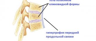
Normally, the height of the vertebral bodies is the same over the entire area, with the exception of the 5th lumbar vertebra (L5), the body of which is wedge-shaped.
The spinous processes present in almost all vertebrae, tiledly covering each other, extend from them at different angles in different parts of the spine. Thus, in the cervical and lumbar regions they are located almost horizontally, and at the mid-thoracic level, which corresponds to 5-9 thoracic vertebrae, they are located at rather sharp angles. At the same time, the processes of the upper and lower thoracic vertebrae occupy an intermediate position.
The spinous processes, as well as the transverse ones, are the base to which the ligaments and muscles that move the vertebrae are attached. The articular processes of adjacent vertebrae form facet joints. They create the ability to bend the spine back and forth.
Thus, the vertebral bodies are connected by intervertebral discs, and the arches are connected by intervertebral joints and ligaments. The anatomical complex formed by the intervertebral disc, two adjacent intervertebral joints and ligaments is called the spinal motion segment. In each individual segment, the mobility of the spine is small, but the simultaneous movement of many segments provides a sufficient level of flexibility and mobility of the spine in different directions.
Normally, the spine has 4 physiological curves, which provide a weakening of shocks and concussions of the spine during movement. Thanks to this, they do not reach the skull and ensure the safety of the brain. There are:
- cervical lordosis;
- thoracic kyphosis;
- lumbar lordosis;
- sacrococcygeal kyphosis.
Lordosis is a curvature of the spine that is convex toward the front of the body, and kyphosis is a curve in the opposite direction.
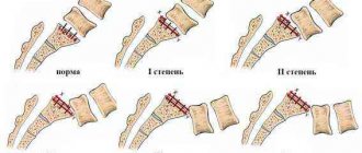
Due to the presence of physiological curves, the human spine has an S-shape. But normally they should be smooth and not exceed permissible values. The presence of pronounced angles or the location of the spinous processes at different distances from each other is a sign of a pathological increase in kyphosis or lordosis. In the lateral or frontal plane, any bends or tilts should normally be absent.
Moreover, the degree of physiological bending is not a constant value even for an absolutely healthy person. The fact is that the angle of inclination depends on the age of the person. Thus, a child is born already having physiological curves of the spine, but they are much less pronounced. The degree of their manifestation directly depends on the age of the child.
In a horizontal position of the body, physiological bends straighten out a little, and in a vertical position they are more pronounced. Therefore, in the morning after sleep, the length of the spine increases slightly, the curves are less pronounced, and in the evening the situation changes. Moreover, when the load increases, the magnitude of the bends increases proportional to the applied load.
All vertebrae are different sizes. Moreover, their width and height progressively increase with distance from the head. The dimensions of the intervertebral discs correspond to the vertebral bodies and are present between almost all of them. Such a cartilaginous layer, which acts as a shock absorber and ensures mobility of the spine, is absent only between the 1st and 2nd cervical vertebrae, i.e., the atlas and axis, as well as in the sacrum and coccyx.
There are a total of 23 intervertebral discs in the adult human body. Each of them has a pulpous nucleus, called the pulposus, and a surrounding tough fibrous membrane, called the annulus fibrosus. The intervertebral disc passes into a fairly thin plate of hyaline cartilage, which covers the bone surface.
Ligamentous apparatus
The spine is equipped with a powerful ligamentous apparatus formed by a large number of different ligaments. The main ones are:
- The anterior longitudinal ligament is formed by fibers and bundles of different lengths, which are firmly attached to the vertebral bodies and much more loosely to the corresponding intervertebral discs. It runs along the anterior and lateral surfaces of the vertebral bodies. This ligament originates from the occipital bone and passes through the entire spinal canal up to the 1st sacral vertebra.
- The posterior longitudinal ligament also originates from the occipital bone and covers the posterior surface of the vertebral bodies down to the lower part of the sacral canal. Its thickness is greater than that of the anterior similar ligament, and at the same time it is more elastic due to the presence of a larger number of elastic fibers. Unlike the anterior one, it firmly fuses with the intervertebral discs, but is more loosely attached to the bony bodies of the vertebrae. Therefore, in places of contact with cartilaginous plates it is thicker in cross section, and at the point of attachment to the vertebrae it takes on the appearance of a narrow strip. The lateral parts of the posterior longitudinal ligament form a thin membrane that delimits the venous plexuses of the vertebral bodies from the dura mater of the spinal cord, thereby protecting the spinal cord from compression.
- Ligamentum flavum – located between the vertebral arches, closing the gaps and forming the spinal canal. They are formed from elastic fibers, but with age they tend to become denser, that is, ossify. The ligamentum flavum resists excessive forward flexion and extension of the spine.
There are also interspinous, intertransverse and supraspinous ligaments that connect the corresponding processes. But the legs of the arches are not connected by ligaments, which is why intervertebral foramina are formed, through which the spinal roots and blood vessels emerge.
Connection of the spine to the skull
The spinal column is connected to the skull through:
- paired atlanto-occipital joints;
- median atlantoaxial joints;
- lateral atlantoaxial joints.
The atlanto-occipital joints are formed at the point of contact of the protruding parts (condyles) of the occipital bone with the upper articular fossae of the 1st vertebra of the cervical spine, called the atlas. Both atlanto-occipital joints are surrounded by wide articular capsules and strengthened by 2 membranes: anterior and posterior. These joints have physiological restrictions on mobility: flexion up to 20°, extension not exceeding 30°, head tilt to the side within 15-20°.
By the way, it is through the posterior atlanto-occipital membranes, which are wider, that the vertebral arteries, which are responsible for the blood supply to the vertebrobasilar area of the brain, pass.
The median atlantoaxial joint has a cylindrical shape and includes 2 separate joints, which are formed by the posterior and anterior articular surfaces of the tooth of the 2nd cervical vertebra, a fossa on the posterior side of the arch of the 1st cervical vertebra, and a fossa on the anterior surface of the transverse ligament. Both tooth joints have separate articular cavities and capsules. The vertebral tooth is connected to the foramen magnum by a corresponding ligament, at the same time it has 2 strong pterygoid ligaments, which begin on its lateral surfaces and are attached to the condyle of the occipital bone, thereby preventing excessive rotation of the head. Therefore, rotations in the joint are only possible by 30-40° in each direction.
The lateral atlantoaxial joint is a paired combined multiaxial low-moving joint, the formation of which involves the lower articular fossa of the C1 vertebra and the upper articular surfaces of the axial vertebra. Each joint has a separate capsule and is additionally strengthened by the cruciate ligament of the atlas. It originates from the apex of the tooth and ends at the front of the foramen magnum.
Spinal cord
The spinal cord is one of the parts of the central nervous system. It is a long, delicate cylindrical cord, slightly flattened from front to back, from which the nerve roots branch. It is the spinal cord that is responsible for transmitting bioelectric impulses from the brain to every organ and muscle and vice versa. It is responsible for the functioning of the sense organs, contraction when the bladder is filled, relaxation of the sphincters of the rectum and urethra, regulation of the functioning of the heart muscle, lungs, etc.
The spinal cord is located inside the spinal canal, and its length in an adult is 45 cm in men and 41-42 cm in women. Moreover, the weight of such an anatomical structure, which is so important for the human body, does not exceed 34-38 g. Thus, the length of the spinal cord is less than the length of the spinal canal. It starts from the medulla oblongata, which is the lower part of the brain, and thins out at the level of 1 lumbar vertebra (L1), forming the conus medullaris. The so-called filum terminale departs from it, the lower part of which consists of the spinal membranes and is ultimately attached to the 2nd coccygeal vertebra.
In men, the apex of the conical point of the spinal cord is localized on the border of the lower edge of L1, and in women - in the middle of L2. From this moment on, the spinal canal is occupied by the lumbosacral roots, extending from the last segments of the spinal cord, which forms a large nerve formation - the cauda equina. Its constituent nerve roots emerge at an angle of 45° from the corresponding intervertebral foramina.
In newborn children, the spinal cord ends at the level of L3, but by the age of 3, its cone is already at the same level as in adults.
The spinal cord is divided by longitudinal grooves into two halves: anterior and posterior. Its central part is formed by gray matter, and the outer layers are formed by white matter. In the central part of the spinal cord there is a canal that contains cerebrospinal fluid. It communicates with the fourth ventricle of the brain. In adults, this canal is closed in some parts or along the entire length of the spinal cord. Gray matter is formed by the bodies of neurons, i.e., nerve cells, and in cross section resembles a butterfly in shape. As a result, it contains:
- The anterior horns contain motor neurons, also called motoneurons. Like any other neurons, they have long processes (axons) and short branches (dendrites). The axons of motor neurons transmit impulses to the skeletal muscles of the arms, legs and torso, provoking their contraction.
- Posterior horns - the bodies of interneurons are located here, which connect sensory neurons with motor neurons, and also take part in the transmission of information to other parts of the central nervous system.
- Lateral horns - neurons that create the centers of the sympathetic nervous system are localized in them.
On average, the diameter of the spinal cord is 10 mm, but in the region of the cervical and lumbar spine it increases. In these places, so-called thickenings of the spinal cord are formed, which is explained by the influence of the functions of the arms and legs. Therefore, in the cervical spine its transverse size is 10-14 mm, in the thoracic spine - 10-11 mm, and in the lumbar spine - 12-15 mm.
The spinal cord is bathed in cerebrospinal fluid, or cerebrospinal fluid. It is designed to act as a shock absorber and protect it from various damages. In this case, the cerebrospinal fluid is the most filtered blood, devoid of red blood cells, but saturated with proteins and electrolytes, the vast majority of which are sodium and chlorine. Thanks to this, it is completely transparent. Liquor is formed in the ventricles of the brain at a rate of approximately 0.5 liters per day, although on average its volume in the canal does not exceed 130-150 ml. Therefore, even with significant losses of cerebrospinal fluid, its losses are quickly compensated by the body. A small part of the cerebrospinal fluid is absorbed by the blood and lymphatic vessels of the spinal cord.
Spinal cord membranes
The spinal cord is surrounded by 3 membranes: the hard outer membrane, the arachnoid membrane, separated from the first by the subdural space, and the internal one, called the pia mater. The latter is adjacent directly to the spinal cord and is separated from the membrane occupying the middle position by the subarachnoid space. Each of the spinal membranes has its own structural features and performs specific functions.
Thus, the hard shell is a kind of connective tissue case for this sensitive and important nervous structure, densely intertwined with blood vessels and nerves. It consists of collagen fibers and has 2 layers, the outer one fits tightly to the bone structures of the spine and, in fact, forms the periosteum, and the inner one forms the dural sac of the spinal cord. The dura mater is additionally strengthened by multiple bundles of connective tissue, which connect it to the posterior longitudinal ligament, and in the lower parts of the spine they form the filum terminale (filum terminale of the spinal cord), which is ultimately attached to the periosteum of the coccyx. The hard shell has different thickness in different areas, which ranges from 0.5 to 2 mm. It reliably protects the spinal cord from most external influences and runs from the foramen magnum down to 2-3 sacral vertebrae, i.e., it covers the delicate spinal cord along its entire length.
In addition, this shell has cone-shaped protrusions. They are designed to form a protective layer for the nerve roots extending at the level of all vertebrae, and therefore exit with them into the intervertebral foramina.
The dura mater is delimited from the wall of the spinal canal by the epidural space. It contains fatty tissue, spinal nerves and numerous blood vessels responsible for the blood supply to the vertebrae and spinal cord.
The subdural space mentioned above separates the dura mater and the arachnoid membrane of the spinal cord. Essentially, it is a narrow gap filled with thin bundles of connective tissue fibers. In this case, the subdural space ends blindly at the S2 level, but has a free connection with a similar space inside the cranium.
The arachnoid membrane is a delicate, transparent anatomical structure formed by multiple trabeculae (cords), which does not have a rigid fixation system with the dura spinal membrane. They are connected to each other only at the intervertebral foramina.
The arachnoid membrane is separated from the soft membrane by a subarachnoid (subarachnoid) space in which cerebrospinal fluid circulates, and also connective tissue cords pass that unite these membranes with each other. The subarachnoid space communicates with the fourth ventricle of the brain, which ensures continuous circulation of cerebrospinal fluid.
The third membrane of the spinal cord is located in the closest proximity to it and has many blood vessels that provide blood delivery to the spinal cord. It is connected to the arachnoid membrane by a significant number of connective tissue bundles.
Spinal roots
As already mentioned, the entire spinal cord is divided into segments. Moreover, it is shorter than the spinal canal, so there is a discrepancy between the serial number of its segments and the positions of the vertebrae. Thus, the upper cervical segments fully correspond to the position of the vertebral bodies. A shift in numbering is already observed in the lower cervical and thoracic segments. They are one vertebra higher than the corresponding vertebrae. In the central part of the thoracic spine, this difference already increases by two vertebrae, and in the lower part - by 3. Therefore, it turns out that the lumbar segments of the spinal cord are at the level of the bodies of the 10th and 11th thoracic vertebrae, and the sacral and coccygeal segments correspond to 12 thoracic and 1 lumbar vertebrae. But the spinal roots always exit through the intervertebral foramina at the level of the discs corresponding in number.
A pair of nerve roots depart from each spinal segment: anterior and posterior. There are 31 pairs in total. They originate from the lateral surface of the spinal cord and penetrate the dural sac, which forms a protective sheath for them. When leaving it, the spinal roots pass through the hard shell, which has special protrusions in the form of funnel-shaped pockets designed specifically for them. Thanks to this, the spinal roots can bend physiologically, but there is no risk of folding or stretching.
Each dural funnel-shaped recess has 2 openings through which the anterior and posterior nerve roots pass. Moreover, they are delimited by parts of the hard and arachnoid membranes. They are firmly fused with the roots, so leakage of cerebrospinal fluid beyond the subarachnoid space is excluded.
The anterior and posterior roots unite at the level of the intervertebral foramina, forming the spinal nerves. But the posterior intervertebral foramen thickens, forming the so-called ganglion. The anterior and posterior roots join together immediately after the ganglion to form the spinal nerve. Each has several branches:
- Posterior – responsible for innervation of deep muscles, skin of the back and neck.
- Anterior - takes part in the formation of the cervical, brachial, lumbar and sacral plexuses. In this case, the anterior branches of the thoracic nerves form the intercostal nerves.
- Meningeal - ensures the transmission of bioelectric impulses to the dura mater of the spinal cord, as it returns to the spinal canal through the vertebral foramina.
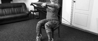
Necessary measures for correct posture
All people with pathological curvature of the spinal column must be monitored by an orthopedist-traumatologist. In turn, the doctor must prescribe all possible procedures aimed at eliminating the diseases: massage, physical therapy, orthopedic supplies, manuals, manual therapy, physiotherapy, etc. are prescribed. In severe cases, surgical treatment is indicated.
As measures aimed against the development or progression of scoliosis, kyphosis and lordosis, it is necessary to carry out comprehensive measures:
- sleep on a hard bed on an adjustable mattress, in a position on your back or stomach;
- wearing orthopedic shoes or inserts in the form of insoles with an anatomical structure;
- physical labor: swimming, athletics, tourism and others;
- strict adherence to the daily routine (getting up and going to bed at the same time, eating);
- giving up bad habits (smoking, alcohol abuse, drug addiction). Other bad habits include avoiding certain incorrect positions, such as sitting cross-legged or standing on one leg;
- self-monitoring of correct posture during sedentary work, at a school desk, at home on a chair, etc.;
- wearing special equipment, corsets and other accessories in case of violation of the normal curvature of the spinal column to fix it;
- uniform distribution of the load on the spine when carrying bags, backpacks, briefcases.
Parents need to monitor the health of their children at any age. It is from adolescence that pathological curves of the spine most often form. If you do not notice this in time, the diseases will progress, bringing a lot of discomfort and health problems.
Blood vessels
The blood supply to the spine is realized through fairly large arteries that pass either in close proximity to the vertebral bodies or along them. The arteries of the cervical vertebral bodies originate from the subclavian artery, the thoracic vertebrae are supplied by the intercostal arteries, and the lumbar vertebrae are supplied by the lumbar arteries. As a result, the spine is actively supplied with blood at all levels, and the pressure in the vessels is at fairly high levels. But if the bone structures have a direct blood supply, then the intervertebral discs are deprived of this. Their nutrition is carried out through the diffusion of substances during compression/straightening of the disc during physical activity.
The lumbar and intercostal arteries are located along the anterolateral surfaces of the vertebral bodies. In the area of the intervertebral natural foramina, posterior branches branch off from them, which are responsible for feeding the soft tissues of the back and dorsal parts of the vertebrae. In turn, spinal branches depart from them, which deepen into the spinal canal, where the blood vessels are again divided into 2 branches: anterior and posterior. The anterior branch is larger in size and is located transversely to the anterior part of the vertebral body, and on the posterior surface it unites with a similar vessel on the opposite side of the body. The posterior branch extends along the posterolateral surface of the spinal canal and connects with a similar artery on the opposite side.
Thus, the spinal arteries form an anastomotic network that covers the entire spinal canal and has transverse and longitudinal branches. Numerous vessels responsible for feeding the vertebral bodies and spinal cord are diverted from it. The arteries penetrate into the vertebral bodies near the midline, but they do not pass into the intervertebral discs.
The spinal cord has 3 blood supply basins:
- Cervicothoracic, where the first 4 segments are fed from the anterior spinal artery, formed by the fusion of 2 vertebral arteries, the next 5 segments have absolutely independent nutrition, and the blood supply is provided by 2-4 large radicular-spinal arteries, branching from the vertebral arteries, the ascending and deep cervical arteries.
- The intermediate (middle) thoracic basin, including segments T3-T8, is supplied exclusively by one single artery located at level 5 or 6 of the thoracic root. Due to such anatomical features, there is a high risk of developing severe ischemic lesions in this part of the spinal cord.
- Lower thoracic and lumbosacral basin - blood supply is provided by one large anterior radicular artery.
As for the venous system, the spine has 4 venous plexuses: 2 external, localized on the anterior surface of the vertebral bodies behind the arches, and 2 internal. The largest venous plexus is the anterior intravertebral plexus. Its large vertical trunks are interconnected by transverse branches. It is firmly fixed to the periosteum along the posterior surface of the vertebrae by a large number of jumpers. The posterior venous intravertebral plexus can easily move because it does not have strong connections with the vertebral bodies. But at the same time, all 4 venous plexuses of the spine are closely interconnected by numerous vessels that penetrate the vertebral bodies, as well as the yellow ligaments. In general, they form a single whole and extend from the base of the skull to the tailbone.
Venous blood is drained through the system of the superior and inferior vena cava, into which it enters from the vertebral, intercostal, lumbar and sacral veins. All intervertebral veins exit through the corresponding openings of the spine. At the same time, they are firmly attached to the periosteum of the bony edges of the foramina.
The spinal cord itself has 2 venous blood outflow systems: anterior and posterior. In this case, the veins of the surface of the organ are united by a large anastomotic network. Therefore, if it is necessary to ligate one or more veins, the likelihood of developing spinal disorders is close to zero.
How is the numbering done?
The numbering of the vertebrae of the ridge is always carried out from top to bottom, increasing in number. For convenience, each department is designated by a Latin letter, based on its Latin name:
- cervical (cervical) − C1-C7, the occipital part of the skull is conventionally considered the zero vertebra, which means C0;
- the thoracic (thoracic) region can be designated in three ways, namely Th1-Th12, or T1-T12, or D1-D12;
- lumbar (lumbar) – L1-L5;
- sacral (sacral) – S1-S5;
- coccygeal – Co1-Co5.
Age and gender characteristics of the spine
The length of the spinal column in newborns does not exceed 40% of the total height. But during the first 2 years of life, its length almost doubles. All this time, all parts of the spine are growing at a high speed, but mainly in width. From 1.5 to 3 years, the growth rate decreases, especially in the cervical and upper thoracic regions. At about 3 years of age, active growth of the lumbar and lower thoracic spine begins. From 5 to 10 years, a phase of smooth, uniform growth in all parameters begins, followed by a phase of active growth, lasting from 10 to 17 years. After this, the growth of the cervical and thoracic regions slows down, but the growth of the lumbar region accelerates. The entire process of development of the spinal column is completed at 23-25 years of age.
Thus, in an adult man, the length of the spine is on average 60-75 cm, and in a woman - 60-65 cm. Over the years, degenerative changes occur in the intervertebral discs, they flatten and cease to fully cope with their functions, and physiological bends increase. As a result, not only various diseases arise, but also the length of the spinal column decreases in old age by about 5 cm or more.
Thoracic kyphosis and lumbar lordosis are more pronounced in women than in men.
Thus, the human spine has a complex structure, a dense network of nerves and blood vessels. This largely explains the difficulty of performing surgical interventions on it and the possible risks. Therefore, today all efforts are aimed at finding the least invasive methods of performing operations that involve minimal tissue trauma, which sharply reduces the likelihood of developing complications of varying severity.