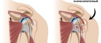
The joint is surrounded by a capsule, synovial bursa, tendons, muscles, and ligaments. With an inflammatory process in the entire periarticular area, periarthritis can be diagnosed. Consequently, this term means that all periarticular tissues are simultaneously damaged, after which the inflammatory process begins to develop in the joints that are located with the source of inflammation. Periarthritis is a rheumatic disease.
Under the influence of this disease, damage occurs to all tissues and interosseous joints that relate to the musculoskeletal system. In most cases, glenohumeral periarthritis is detected in women over forty-five years of age. Men are less likely to be affected by this disease.
The occurrence of glenohumeral periarthritis occurs in more than 75% of all registered cases. This happens due to the fact that the tendons, which are located in the place where the joint is attached, are constantly subject to functional overstrain. It is somewhat less common that the muscle tendon that is attached to the joint of the elbow, wrist, and hand is affected. It is extremely rare for an inflammatory process to occur in the tendons and muscles of the leg joint.
When diagnosed with glenohumeral periarthritis, a person experiences a deterioration in their quality of life and an increased risk of partial or complete disability. It is necessary to know what causes the development of the disease and what the symptoms look like. This will help you contact an experienced specialist in time so that complications do not begin to appear.
How does glenohumeral periarthritis manifest?
Humeral periarthritis can have a simple form, acute, or chronic. When the disease develops after injury, the first signs appear after a few days. Given this, it can be difficult for the patient to name the reason that led to the development of the disease.
General signs of a simple form of the disease include:
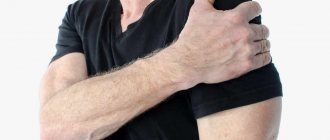
- A person complains of mild pain localized in the shoulder.
- The appearance of painful sensations is observed when performing certain movements.
- There is no pain at rest.
- When you try to rotate your arm when the elbow joint is involved, or overcome resistance, the pain syndrome increases.
- Mobility is slightly limited. This manifests itself in difficulty raising the arm up.
- In some cases, pain disturbs a person even at night. This applies to those who are used to sleeping on their back.
- Palpation determines the presence of painful points, the localization of which is the anterior outer surface of the shoulder.
- In most cases, after a few weeks the simple form of glenohumeral periarthritis resolves on its own.
If the disease progresses unfavorably, there is a constant load on the affected area, or repeated injury occurs, then the simple form transforms into an acute one.
The acute form of glenohumeral periarthritis is characterized by the appearance of the following symptoms:
1. Sudden onset of painful sensations followed by increased intensity. This occurs because calcifications migrate from the short tendon into the bursa. Pain in this case is a consequence of excessive physical exertion.
2. The localization of pain is not only the shoulder, but also the neck and arms.
3. Increased pain occurs at night.
4. The occurrence of severe pain is provoked by rotational movements of the joint, as well as attempts to move the arm behind the back.
5. A decrease in pain intensity occurs when the hand is on the chest. The arm should be bent at the elbow.
6. Development of swelling of the frontal surface of the shoulder.
7. Violation of the general condition of a person (increase in body temperature, deterioration in sleep, performance).
8. From a blood test it can already be determined that the erythrocyte sedimentation rate has increased, and from an x-ray - the presence of calcifications.
In the absence of appropriate treatment for acute glenohumeral periarthritis, the risk of its transition to the chronic stage increases. It can be recognized by the following signs:
- Low-intensity pain is felt in the shoulder.
- Movement of the shoulder joint provokes increased pain.
- At night, the patient may complain of shoulder pain.
- When you rotate your hand or make a sudden movement, the pain will be shooting in nature.
If the chronic form of periarthritis is ignored, the development of ankylosing periarthritis may occur. This will last for several years. Symptoms of this form of the disease are expressed as follows:
- The density of the tissues that surround the joint increases.
- Complete immobilization of the shoulder.
- The appearance of sharp and unbearable pain when raising the arm.
This form is the final stage of development of glenohumeral periarthritis. If we talk about the percentage, then this occurs in 25% of all cases.
Inspection
When examined, there are usually no external changes. During palpation, painful points are determined along the anterior, lateral or posterior surface of the shoulder joint at the attachment points of the rotator cuff muscles. Movement in the shoulder joint is painful. Characterized by greater pain during active (which the patient himself makes) movements, while passive movements (which the doctor makes with the patient’s limb) are less painful. Simple functional tests can help determine which rotator cuff muscles are inflamed.
If the supraspinatus tendon is affected, abduction of the shoulder is painful. The appearance of pain only at the beginning and at the end of movement indicates inflammation of the serous bursa located under the acromial process of the scapula (subacromial bursitis). Nowadays this condition is more often called impingement syndrome.
Pain during external rotation of the shoulder (trying to comb one's hair) indicates damage to the tendons of the infraspinatus and teres minor muscles.
Placing the arm behind the back (internal rotation of the shoulder) indicates inflammation of the subscapularis tendon.
Flexion at the elbow joint and supination of the forearm, reminiscent of turning a key in a lock (the doctor resists this movement), indicates damage to the tendon of the head of the biceps.
With painful passive movements in the shoulder joint, as in the photo below, you can think about damage to the shoulder joint itself (arthrosis, arthritis).
Clarifying which muscle is affected can be useful when carrying out blockades.
How to diagnose the disease
If a person experiences pain in the shoulder area or limited mobility, it is recommended to immediately contact an experienced specialist. To diagnose glenohumeral periarthritis, the patient needs to visit a therapist. After examining the patient, the therapist can give a referral for examination by a specialist (surgeon, neurologist, rheumatologist, orthopedist).
An external examination and history taking is complemented by an assessment of the motor activity of the shoulder joint. Also, palpate the area where the inflammatory process occurs. To clarify the diagnosis and determine the cause of this disease, the patient is referred for an X-ray examination. It is necessary to examine the diseased joint and cervical spine. In addition, ultrasound and magnetic resonance imaging may be needed.
Diagnostic measures involve a blood test. In the presence of an acute form of the disease, an increased erythrocyte sedimentation rate and c-reactive protein are detected. Other forms of glenohumeral periarthritis are not detected by blood tests.
If surgical intervention is necessary, the patient may be referred for an invasive diagnostic procedure (arthrography, arthroscopy). It should be borne in mind that such a disease can easily be confused with another pathology that manifests itself with similar symptoms. this means that it is advisable to carry out a differential diagnosis of arthritis, arthrosis, thrombosis of the artery located under the collarbone.
Causes
Periarthrosis is the result of the development of an inflammatory process or degenerative-dystrophic changes in the shoulder joint, which often accompany each other. Therefore, most often it occurs against the background of other pathologies of the musculoskeletal system, which explains the higher incidence of development in older people.
In general, the disease can result from:
- receiving microtrauma to muscles and tendons, which is facilitated by excessive loads on the shoulder, regular performance of repetitive movements (raising/lowering, adduction/abduction of the arm), which is due to the characteristics of work or sports;
- injuries to the soft tissues of the shoulder, leading to the formation of a hematoma, swelling in the area where the blow was received;
- prolonged hypothermia, leading to spasm, i.e. narrowing of the lumen, blood vessels of the skin and reflex spasm of the vessels of the periarticular tissues;
- maintaining a sedentary lifestyle and prolonged stay in a static position, which causes impaired blood circulation, including in the area of the shoulder joint;
- neuroreflex spasms of muscles and small blood vessels, leading to malnutrition of soft tissues (ischemia), which can be observed with myocardial infarction, angina pectoris, as well as osteochondrosis of the cervical spine;
- disturbances in the nutrition of the soft tissues of the shoulder as a result of the inclusion of neurotrophic mechanisms, which is observed in diabetes mellitus, tuberculosis, obesity, Parkinson's disease and leads to the development of ischemia with subsequent death of areas of soft tissue and their replacement with coarse connective tissue;
- congenital anomalies of the shoulder joint, including dysplasia and arthropathy, as well as developmental disorders.
Effect of drugs during treatment
To relieve the inflammatory process and painful syndrome, the patient may be prescribed the following medications:
1. Non-steroidal anti-inflammatory drugs. This refers to taking ketorol, nimesil, dikloberl, etc. Thanks to the effects of these drugs, pain is eliminated and inflammation is relieved from the affected muscles.
2. Painkillers (baralgin, analgin).
3. Muscle relaxants (mydocalm). This is a group of medications that relax muscles and help relieve spasms by reducing muscle tone.
4. Chondroprotectors (structum). Thanks to such drugs, the physiological activity of the joints improves, intra-articular fluid decreases, and swelling is eliminated. As a result, the pain disappears and there is a therapeutic effect.
If the pain is not relieved by the above medications, the patient may be prescribed a subscapular nerve block. The injection is given into the subacromial space. The blockade will be carried out twice throughout the entire treatment period. At least three weeks must pass between the first and second blockade.
Before agreeing to the blockade, you should consult with an experienced doctor about contraindications and side effects.
For glenohumeral periarthritis, a novocaine blockade can be administered. After administration of the drug, you can observe the manifestation of immediate results. In most cases, novocaine is combined with a glucocorticoid. Thanks to this, the inflammatory process is reduced, pain is eliminated, and swelling is relieved. It must be taken into account that the use of hormonal drugs can negatively affect the patient’s immune system. This means that such medications must be used strictly as prescribed by a doctor and under his close supervision.
1.General information
Humeral periarthritis is a disease caused by inflammatory processes in the periarticular space of the shoulder.
A diagnostically significant sign that makes it possible to make just such a diagnosis is the presence of completely healthy elements in the joint itself. With periarthritis, only the muscles, ligaments, tendons, periarticular bursa, and synovial capsules are affected - the structures that provide mobility of the arm in the shoulder.
Periarthritis should be distinguished from periarthrosis, in which the inflammatory process is a consequence of an existing systemic degenerative disease. In patients with periarthrosis, as a rule, pain and functional impairment manifest themselves multiple times - symmetrically, as well as in joints of different groups. With periarthritis, it is possible to identify the factor that caused the pathology of one glenohumeral joint. In general, a person can be completely healthy, have excellent metabolism, hormonal levels, and a vascular system, and the doctor, based on the results of the diagnosis, rules out any trophic disorders or degeneration.
A must read! Help with treatment and hospitalization!
Conducting physical therapy
In order for a speedy recovery to occur, you should use therapeutic exercises. A big plus is doing exercises in water. Swimming and hydrokinesotherapy are an integral part of the recommended complex during glenohumeral periarthritis. Exercising in the pool normalizes muscle tone, relieves excess tension, and increases the mobility of the damaged joint.
If we talk about the goals of the gymnastics complex, we can highlight:
- Blood flow is normalized.
- Tissues are enriched with oxygen.
- Stagnation is eliminated.
- Muscles are strengthened.
- Metabolic processes are normalized.
It should be remembered that physical therapy is contraindicated if glenohumeral periarthritis is in the acute stage or if there is severe pain in the joint.
Exercise therapy Jamaldinova
Muslim Jamaldinov, the director and leading specialist of Dr. Popov’s rehabilitation center, is known to many today. The healing program he created is effective even with severe pain and loss of mobility. The secret of such versatility lies in pre-warming the muscles with massage.
As the basis for his methodology, Muslim Jamaldinov took Popov’s set of exercises for glenohumeral periarthritis, reworked it and supplemented it with his discoveries.
The result is a soft, completely non-traumatic system for restoring the affected joint:
- Bend the limbs at the elbows and in this position “shrug” the shoulders.
- Sitting on a bench and placing your hands on your hips, try to reach your opposite knee with your shoulder.
- Connecting your fingers into a “lock”, make circular movements with your hands in front of you.
Jamaldinov’s therapeutic exercises for shoulder periarthritis can relieve stress from bone structures, relax muscles, reduce pain and restore range of motion.
