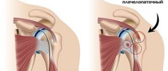
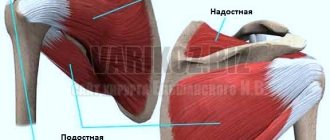
Calcific shoulder tendinitis occurs when calcium builds up in the tendons of the shoulder. The tissues around the calcium deposits become inflamed, resulting in severe pain. This disease is quite common and most often occurs in people over 40 years of age. Calcific tendonitis occurs in the rotator cuff tendons. The rotator cuff is made up of several tendons that connect the muscles around the shoulder to the humerus. Calcium deposits usually form on the rotator cuff tendon, called the supraspinatus tendon.
There are two different types of calcific shoulder tendonitis: degenerative calcification and reactive calcification. The wear and tear processes of aging are the main cause of degenerative calcification. As we age, the blood supply to the rotator cuff tendons decreases, causing the tendons to weaken. The wear process is accompanied by microtears of tendon fibers. And in damaged tendons, simultaneously with regeneration, processes of calcification deposition occur.
Reactive calcification is different from degenerative calcification. The mechanism of development of this type of calcification is not completely clear. This type of calcification is not associated with degenerative changes and is much more likely to cause shoulder pain than degenerative calcific tendonitis. The development of reactive calcific tendonitis is believed to occur in three stages. In the initial stage of calcification, changes occur in the tendons, which create conditions for the formation of calcifications. During the calcification stage, calcium crystals are deposited in the tendons. But at this stage, calcifications are resorbed (reabsorbed) by the body. It is at this stage that pain is most likely to appear. In the post-calcification period, the body repairs the tendon and the damaged tissue is replaced with new tissue. The mechanism that triggers the absorption of calcifications into the body is not clear, but once this occurs and the tissue begins to regenerate, the pain usually decreases or disappears completely.
Causes
No one knows what exactly causes calcific tendinitis. Physical stress, aging or a combination of both leads to degenerative calcification. Some researchers suggest that calcium deposits are formed due to tissue hypoxia and insufficient oxygen supply to the tendon tissues. Others believe that pressure on the tendons can lead to their damage, resulting in the deposition of calcium deposits.
The mechanism of formation of reactive calcification has not been completely studied. Typically, this type of calcific tendinitis occurs in younger patients and occurs without any apparent cause.
Drug treatment
A course of exercise therapy, massage sessions and physiotherapeutic procedures will be ineffective if they are not combined with taking medications. Local and systemic medications are usually used. The first option involves applying gels or ointments to the affected area. For example, Nise gel. This is a drug for external use containing NSAIDs. It must be used at least 3 times a day.
If the disease is chronic, it is advisable to use irritating ointments. An example of this is Capsaicin. The ointment is applied in a thin layer no more than 2 times a day.
Long-term tendinitis requires a more serious approach to treatment. Patients are already prescribed local painkillers in the form of injections into the joint. They retain the analgesic effect for a long time. An example of drugs from this group is Lidocaine.
If the effectiveness of the above methods is low, they resort to joint blockade. The procedure is repeated once every 2-3 weeks. The hormones Prednisolone and Hydrocortisone are used in advanced cases. Their use can lead to a decrease in the elasticity of the tendons. If you have biceps tendinitis, this procedure is prohibited.
If the underlying cause of the disease is a bacterial infection, you cannot do without the help of antibiotics. Penicillins and cephalosporins are usually prescribed. Before antibiotic therapy, the patient must undergo a bacterial culture to determine sensitivity to certain drugs.
Diagnostics
To diagnose calcific tendonitis of the shoulder, your doctor will first take a medical history and perform a physical examination. Shoulder pain can be associated not only with calcific tendonitis, but also with other diseases. Therefore, to make an accurate diagnosis, instrumental studies are necessary. Radiography allows one to visualize the presence of calcium deposits in the tendons. But the most informative way to visualize ligaments and tendons and the presence of pathological changes in them is MRI (magnetic resonance imaging). Visualization of calcifications using radiography or MRI in dynamics allows you to determine treatment tactics (conservative or surgical treatment). Laboratory tests are necessary in cases where it is necessary to differentiate this disease from inflammatory diseases of connective tissue.
Calcific tendonitis of the shoulder
Anatomy of a disease
Which is accompanied by quite acute pain. The disease is called calcific tendonitis of the shoulder. This disease is in most cases age-related and very common, and the main risk group is people over forty years of age, but it also occurs in younger people.
Several tendons connect the muscles to the humerus, the so-called rotator cuff tendons. It is on these tendons that calcific tendinitis forms.
There are two types of calcific tendonitis of the shoulder: degenerative and reactive. The degenerative form of the disease mainly occurs in older patients. And younger patients are more often susceptible to the reactive form of the disease.
Causes of the disease
At the moment, there is no precise scientific basis for the occurrence of calcific tendinitis. And many experts find it difficult to name the causes of the disease. But based on many years of doctors’ observations of patients, we can assume that the cause of the degenerative form of the disease is the general aging of the body. Our tendons wear out and tissue destruction and micro-tears occur. The flow of fresh blood deteriorates. And deposits of calcium compounds form in damaged tissues.
The reactive form of the disease follows a completely different scenario and mainly in this form of the disease acute pain sensations appear. Experts suggest that the cause is physical damage to the tendons, and divide the course of the disease into three stages.
At the first stage, tendon damage occurs and, as a result, favorable conditions for calcium deposits are formed.
At the second stage, deposits begin to gradually dissolve in the body. And it is this stage that is accompanied by pain.
And finally, at the last stage, our body restores damaged organs, replacing damaged fibers with new ones.
Until now, experts have not established the conditions for triggering mechanisms for tendon restoration and resorption of calcium salts. And there is no clarity about the causes and mechanisms of the disease itself.
Symptoms
At the onset of the disease, it is very difficult to identify any symptoms. Pronounced symptoms begin to appear already at the stage of salt dissolution, as discussed above. Pain, stiffness of movement of the shoulder joint and limited trajectory of raising the arms. Here are the main symptoms that should make you think about contacting a specialist.
Diagnosis of calcific tendonitis
Pain in the shoulder area can be caused by various reasons. Including inflammatory processes that are in no way related to calcific tendonitis. For this reason, more precise laboratory tests are often required. And, if there is a suspicion of calcific tendinitis, the specialist will definitely refer you for an x-ray. Deposits of calcium salts will be clearly visible on x-rays. Another method for detecting calcium deposits is magnetic resonance imaging (MRI). After carefully studying the data from X-rays or magnetic resonance imaging, the treating specialist will decide on treatment methods. Or he will refer you for additional laboratory tests for a more detailed study of the inflammatory processes of connective tissues in the shoulder joint.
Treatment
In most cases, conservative treatment can be used. This will relieve pain and localize inflammatory processes. At this stage, rest and the use of anti-inflammatory drugs are recommended. For severe pain, injections can be used to relieve pain and reduce inflammation.
When the body begins to dissolve calcium deposits, the pain can be especially severe. The treating specialist can decide on a procedure called lavage. Two punctures are made in the area where calcium compounds are formed. And through these holes, using saline solution, they try to remove some of the deposits. During this procedure, calcium deposits may break up, and the resulting particles can be removed with a needle. The lavage procedure helps to significantly relieve pain and speed up the recovery period for the tendons. If the procedure is unsuccessful, when it was not possible to break up the deposits, this will help relieve excess pressure on the tendons. Which will have a beneficial analgesic effect.
Also, with a conservative approach to treatment, physiotherapeutic procedures are used. For example, ultrasound therapy. This helps relieve inflammation and relieve pain. But for ultrasound treatment to be effective and efficient, the treatment must be a course. Ultrasound therapy is the most modern method in the treatment of calcific tandinitis. The ultrasonic wave destroys deposits of calcium salts and thereby helps the body dissolve them. On average, a course of ultrasound therapy takes approximately six to eight weeks.
At the final stage of calcific tindinitis, therapeutic and recreational physical education is recommended. This will strengthen the weakened muscles of the shoulder joint and bring them into working condition. Improves blood flow and exchange in the area of inflammation. Exercising is a very important part of treatment. The rotator cuff muscles are strengthened and relieve excess pressure on calcium deposits. Helping shoulder joint stability.
And if all these methods do not help, you will have to resort to a last resort - surgical intervention. The most modern method of surgical treatment now is arthroscopic. This method can significantly reduce the recovery period after surgery. For example, it will be possible to remove the supports supporting the shoulder in about three days. And complete restoration of the functionality of the operated ligaments occurs within six weeks.
This is the least traumatic way to impact the joints. An arthroscope and saline solution are inserted through several microscopic incisions for better visibility of the surgical site. And then they destroy and remove calcium deposits from the affected areas. And one of the main advantages of the method is that the patient does not have to be hospitalized. All procedures can be performed on an outpatient basis.
And the very last option, open surgery.
In this operation, an incision is made in the affected area and calcium deposits and damaged tendons are removed. Then everything is stitched together. After surgery, the rehabilitation period will last much longer. Only after six weeks can you begin physical therapy procedures. And strictly under the supervision of specialists. Full recovery can take from twelve to twenty weeks. Author: K.M.N., Academician of the Russian Academy of Medical Sciences M.A. Bobyr
Treatment
Conservative treatment
The main goal of conservative treatment is to reduce inflammation and pain. Therefore, at the first stage, conservative treatment includes rest and taking NSAIDs (ibuprofen). Anti-inflammatory drugs can reduce the inflammatory process and reduce pain. If severe pain is present, corticosteroid injections may be prescribed. The use of steroids can effectively relieve swelling and inflammation for some time.
During the period of time when calcium deposits begin to be reabsorbed, pain can be especially severe. In such cases, it is possible to remove part of the calcium deposits using saline rinsing. solution through two punctures in the area of calcium deposits. This procedure is called lavage. Sometimes with this procedure it is possible to break the calcifications into pieces (they are removed with a needle). Removing deposits allows you to quickly reduce pain and achieve faster tendon recovery. Even when lavage does not remove calcium deposits, it can relieve pressure in the tendons, resulting in less pain.
Physiotherapy . Physiotherapy is one of the main components of the conservative treatment of calcific tendinitis. The use of a technique such as ultrasound helps reduce pain and inflammation. But the effect of using ultrasound is achieved only with a course of treatment (up to 24 procedures over 6 weeks). Shock wave therapy is currently the most modern method of conservative treatment of such diseases. The shock wave breaks down large calcium deposits, allowing the body to absorb them more quickly.
Exercise therapy is indicated at the stage of completion of reabsorption and allows you to restore muscle tone and improve blood supply to the structures of the shoulder. As a rule, an individual selection of exercises is carried out and exercise therapy is carried out for 4-6 weeks. Exercise is important to strengthen the rotator cuff muscles, as these muscles help control the stability of the shoulder joint. Strengthening these muscles can actually reduce the pressure on the calcium deposits in the tendon.
How does shock wave therapy treat calcifications?
By applying a shock wave to an area of tissue with calcification, we trigger a number of processes in the tendon. These processes lead to natural lysis and removal of calcification from the body. What are these processes? – Multiple increases in blood circulation at the site of exposure, and at the same time an increase in metabolic processes. – Activation of fibroclasts and macrophages, which eliminate (“eat”, dissolve) pathological calcium deposits. – Stimulation of connective tissue growth factors, which help restore the natural structure of the tendon, stimulates fibroblasts that synthesize collagen and other components of the tendon. – Powerful activation of lymph circulation at the site of exposure – calcification breakdown products are naturally removed. As a result, after some time, calcium deposits are resorbed and replaced by healthy tendon tissue. Inflammation goes away, pain disappears, and the full range of movement in the joint is restored. The man is healthy.
Results of treatment of calcifications in the shoulder using a method developed at the Avatage MC using shock wave therapy.
Example 1: Calcific tendinitis of the shoulder. Patient G., 56 years old. The duration of the disease is more than 1.5 years. R-graphy data before and after the course of treatment at the Avatage MC. The complete removal of calcification is visible in the second image.
Calcific tendonitis of the shoulder, before and after shock wave therapy treatment
Example 2. Calcification in the shoulder. Patient K., 53 years old. Calcific tendinitis of the shoulder. After 1 course of treatment, I completely got rid of shoulder pain and restored full range of motion in the shoulder joint.
Example 3. Calcific tendonitis of the shoulder, calcification in the supraspinatus muscle ligament. 7 UVT procedures were performed.
|
|
Example 4. Calcific tendonitis of the shoulder, the patient underwent 8 shock wave therapy procedures. After the course of treatment, the patient ceased to be bothered by pain, and the range of motion in the shoulder joint was completely restored.
|
|
Calcific tendonitis of the shoulder treatment in Zaporozhye, phone 061 222 87 00.
Surgery
If conservative treatment is ineffective and shoulder function deteriorates or there is persistent pain, surgical treatment is recommended. As a rule, surgical treatment is carried out using a minimally invasive atroscopic method, which allows the patient not to stay overnight in the surgical department. During an atroscopic operation, the surgeon visually determines the localization of calcium deposits in the tendons of the rotator cuff, removes them and washes this area; free calcium crystals, which can irritate the surrounding tissues, are also removed.
In rare cases, open surgery may be necessary. With this operation, access to calcium deposits is through an incision in the muscles of the ligaments with the removal of part of the tendons. After removing the deposits, the muscles and tendons are sutured.
Rehabilitation after shoulder surgery can take quite a long period of time. In the first 6-8 weeks after surgery, it is recommended to wear an orthosis and limit movements, then it is necessary to gradually begin to develop the joint and combine physiotherapy with exercise therapy. The volume of loads on the joint must be increased gradually and very carefully under the supervision of a physical therapy doctor. Exercises usually begin no earlier than 6 weeks after surgery. The exercises are aimed at improving the muscle strength of the shoulder girdle and rotator cuff muscles. Full recovery of shoulder function after surgery may take 3 to 4 months. After open surgery, recovery is much slower than with atroscopic resection.
Diagnosis of calcific tendonitis
The diagnosis can be made after a detailed clarification of complaints, collection of anamnesis, examination and physical examination, as well as after analysis of signs of calcification identified by radiography. The diagnosis of calcific tendonitis of the shoulder is confirmed by radiological examination in the presence of calcification of the tendons. This sign is not easy to detect. To detect it, photographs of the shoulder joint both in the direct projection with its neutral position, and in external and internal rotation. In the early stage, deposits can be dense and well defined. An MRI scan can also help in making a diagnosis.
Symptoms of the disease
The clinical picture is determined by the area of tendon damage. Most often, the patella suffers due to subluxations and sprains. After heavy loads, tension occurs at an early stage, followed by dull short-term pain at the bottom of the cup or in the tubercle of the tibia. As you progress, the sensations increase. There comes a time when every bend of the leg is difficult. Sometimes the entry of an infectious agent into the capsule causes suppuration, damage to the anterior cruciate bundle and nearby tissues. Symptoms and treatment for tendovaginitis of the knee joint differ little. In the affected area all the signs of the catarrhal process are present:
- edema;
- local increase in temperature;
- reduction in motor amplitude;
- creaking when walking.
When deep layers are affected, sharp sensations occur even with intense palpation of the medial tendon. In case of damage to the patellar retinaculum, the pathological syndrome increases. It is difficult for a person to climb stairs or get out of a chair. If pain in the leg bothers you when extending with deliberate resistance, this indicates a partial or total rupture of the connective tissue structures and tendinitis of the collateral ligaments. The chronic form is distinguished by its sluggish nature. Periodic problems are caused by recurrent inflammation, which loosens the density of bone tissue and provokes an aseptic or infectious catarrhal process.
CAUSES OF THE DISEASE
Tendonitis in most clinical cases occurs for two main reasons:
- • increased physical activity with excessive stress on the tendons;
- • age-related degenerative changes in the ligamentous apparatus, which are characteristic of people over the age of 40 years.
The development of the disease is facilitated by a number of anatomical features of the human body, in particular, different lengths of the lower extremities, disproportionate location of the patella, flat feet, curvature of the legs, etc.
The disease can be caused by various pathological conditions, including:
- • diseases associated with gross metabolic disorders in the body;
- • immunodeficiency states;
- • rheumatism;
- • infectious diseases of various etiologies (gonorrhea, streptococcal infection, chlamydia).
Tendinitis, as a secondary pathology, develops mainly in people with weakened immunity, in whom the body's resistance to infections is sharply reduced.
PREVENTION MEASURES
It is always easier to prevent any disease than to cure it later without consequences. And tendinitis is no exception to this rule. You can reduce the risk of developing tendon inflammation by following simple recommendations from specialists:
- refusal of uncomfortable high-heeled shoes;
- conducting small warm-ups throughout the day;
- contacting a professional trainer when performing strength exercises or intense training;
- reducing the load on the affected area;
- timely treatment of infectious diseases;
- strengthening the immune system;
- giving up bad habits and normalizing your diet.
Symptoms and diagnosis
The disease primarily manifests itself as pain. Unpleasant sensations intensify when making certain movements, the nature of which depends on the location of the inflammation. Some patients cannot raise their arm up, others cannot stretch it forward, and still others cannot throw their limb behind their head. Changes in body position may also worsen symptoms.
If left untreated, the pathological process progresses, pain occurs even with minor movements. The patient is unable to perform simple actions. In the later stage, pain is constantly present, even at rest. With calcific tendovaginitis, a crunching sound appears when moving. This is due to the appearance of areas of ossification in the tissues of the ligaments and joint capsule.
A preliminary diagnosis can be made based on patient complaints and clinical examination data. An orthopedist-traumatologist conducts a visual examination of the joint area, palpation, and also evaluates the volume of active and passive movements.
Differential diagnosis is carried out with damage to the rotator cuff, dislocations, fractures, arthrosis. These diseases can be distinguished by taking an anamnesis and clinical examination. If, with injuries to the rotators, the volume of passive movements is significantly greater than active ones, then with tenosynovitis of the tendon of the long head of the biceps, these indicators are approximately the same.
MRI will help make a final diagnosis, determine the volume and localization of inflammation, and identify secondary changes. This method has a high diagnostic value and helps to understand even complex situations.
FEATURES OF THE CLINICAL PICTURE
The symptoms of tendinitis depend on the location of the pathological process and the stage of its development. In the initial stages of tendon inflammation, pain occurs only after intense physical activity. Later, the pain begins to bother the person even at rest.
Pain can be localized directly in the area of the projection of the ligament or on the sides of it. They are often aching in nature, but can also be quite intense. Rarely pain is present on an ongoing basis. On palpation the pain intensifies. Acute inflammation of the tendons is accompanied by severe swelling, redness and impaired mobility in the affected joint.
Sometimes tendon tendinitis is accompanied by crepitus. As a result of the long course of the disease, small nodules are formed along the diseased ligament, which can be felt when touched. Over time, calcium is deposited in the thickness of such formations, which makes them very dense. If left untreated, partial or complete rupture of the tendon may occur.
The clinical picture of the disease largely depends on the stage of its course:
- acute phase - pain and discomfort in the area of the affected tendon bother the patient only after physical exertion and quickly disappear at rest;
- the subacute stage is characterized by a more pronounced pain syndrome even in a state of complete rest, which is accompanied by hyperemia of the skin, swelling of the soft tissues and limited mobility;
- Chronic tendonitis is accompanied by degenerative changes in the tendon with constant pain, decreased range of motion and persistent stiffness.
What will help soothe a stretched tendon, how to treat its inflammation and prevent complications of the pathological process? Only a qualified doctor can answer all these questions.
Causes and mechanism of development
The articular surface of the shoulder joint has a slight concavity, so the head of the humerus is fixed with the help of tendons. This anatomical feature allows movement in different directions, but makes the joint vulnerable.
Tenosynovitis is caused by injury or intense physical activity, which results in microtrauma. This is a trigger factor in the development of inflammation. Most often, in addition to the biceps tendon, the supraspinatus tendon is damaged, followed by other elements of the rotator cuff.
Prolonged immobilization of the hand or osteochondrosis of the cervical spine (due to limited mobility) leads to the development of degenerative changes that weaken the tendon and, in the presence of microtraumas, lead to the development of tedinitis.
The need for surgery
Surgical intervention is resorted to in particularly serious cases. Most often, these are situations when drug therapy for a long time does not bring positive results. In addition, the operation is prescribed at the third stage of the pathological process for the purpose of resection of part of the acromion - one of the areas of the scapula. There are several surgical options:
Advertising:
- Redressing
. The orthopedist takes the patient's hand and breaks the joint capsule using special movements. All manipulations are performed using anesthesia. - Open methodology
. Scar structures are excised through a small incision. - Arthroscopy
. This is the most common intervention option. Through several tiny punctures, the doctor inserts instruments for surgical manipulation into the affected area.
After the operation, patients are prescribed antibiotics to prevent the spread of infection and painkillers. Exercise therapy and physiotherapeutic procedures are required.
The recovery period depends on the severity of the pathological process and, as a rule, ranges from 1 to 3 months.
DIAGNOSIS OF THE DISEASE
Diagnosis of tendonitis is based on the collection of anamnestic data and evaluation of the results of examinations. Sometimes the doctor only needs to examine the patient and listen to his complaints. But in the vast majority of cases, to confirm the diagnosis, you need to undergo x-rays of the affected area, ultrasound, and even MRI. These methods will help determine the exact localization of the inflammatory process, assess the degree of its neglect and the nature of tendon degeneration.