Humeral periarthritis is a pathological condition in which inflammatory processes affect the periarticular tissues of the shoulder.
The following are susceptible to the disease:
- joint capsule;
- articular ligaments;
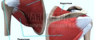
- muscles around the joint;
- tendons around the joint.
Most often, symptoms of glenohumeral periarthritis appear in older people, and treatment will be more successful the earlier it was started.
At CELT you can get a consultation with a traumatologist-orthopedic specialist.
- Initial consultation – 3,000
- Repeated consultation – 2,000
Make an appointment
Etiology of glenohumeral periarthritis
It is worth noting that this disease is quite common and both men and women are susceptible to it. The factors that stimulate its appearance are quite diverse:
- joint injuries (strong blows, falls on the shoulder or outstretched arm);
- intense and/or prolonged physical activity;
- diseases of the spine, poor posture;
- some surgical interventions (for example, breast removal).
Clinical manifestations of glenohumeral periarthritis
Symptoms of glenohumeral periarthritis are very diverse and depend on the form in which the disease occurs.
The initial form is characterized by the following clinical manifestations:
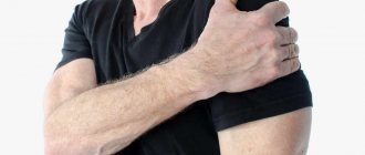
- mild pain in the shoulder that is felt when performing certain movements;
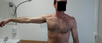
- limited movement in the joint, which is manifested by the inability to place the arm far behind the back or stretch it upward;
- Vivid pain sensations when trying to rotate a straight arm around an axis with resistance.
If treatment for glenohumeral periarthritis is not started at the onset of the disease, it will develop into an acute form, which is characterized by the following symptoms:
- sudden increasing pain in the shoulder, radiating to the upper limb and neck;
- increased pain at night;
- inability to rotate the upper limb around an axis;
- the presence of a slight swelling on the front surface of the shoulder;
- deterioration of the general condition of the body (fever, insomnia).
If treatment is not started at this stage, the disease becomes chronic, which is manifested by the following symptoms:
- frequent moderate pain localized in the shoulder;
- the appearance of acute pain during unsuccessful movements;
- a feeling of aching shoulders at night, leading to insomnia.
Material and methods
The clinical study included 42 patients with a diagnosis of “scapulohumeral periarthritis” aged from 50 to 60 years (average age - 55.8±3.03 years), of which 23 were men, 19 were women. Inclusion criteria were the presence of pain in the shoulder joint for a long time, limited movement in the affected joint and the absence of infectious diseases. The study did not include patients who took steroid drugs more than 5 times a year and who suffered from chronic diseases that could negatively affect the effectiveness and safety of GC use. All patients were randomized into 2 groups. Patients of group 1 (n=19) were administered HA (drug Armaviscon 1%, 20 mg/2 ml) once a week around the periarticular tissue of the shoulder joint (according to the method of local injection therapy for enthesopathies) [14]. The duration of the course was 3 injections. Group 2 included patients (n=23) who used GCS once for the entire course of treatment; only in the absence of a clinical effect, the drug was re-administered. The study included 5 visits: visits 0–2—randomization of the clinical group and start of therapy, visits 4–5—assessment of the effectiveness and safety of therapy. The interval between injections was 1 week. The following diagnostic tests were used to assess functional activity:
visual analogue scale (VAS) from 0 to 100 mm to assess pain during active and passive movements;
Questionnaire of disability in diseases of the shoulder girdle; the maximum score is 22 points, which corresponds to the most severe limitation of life activity due to shoulder pain [15];
Oxford Shoulder Questionnaire; the lowest value is considered to be 12 points; its increase to 60 points indicates a serious pathology of the shoulder joint [16].
To visualize the condition of the periarticular tissue of the shoulder joint, radiation methods were used, such as radiography of the shoulder girdle in standard projections and ultrasound examinations with a black-and-white scanner and a 5–10 MHz linear sensor, where the condition of the tissues of the shoulder joint was assessed in patients of both groups.
Statistical analysis
was carried out using standard methods using the Statistica software package, version 13, and Microsoft Excel 2010.
Classification of glenohumeral periarthritis
In medicine, glenohumeral periarthritis does not have an independent nosology, because there are many causes of dysfunction of the shoulder joint. According to the International Classification of Diseases, 10th revision, the following diseases can cause lesions of the shoulder joint:
- Adhesive capsulitis.
- Biceps tendonitis.
- Subacromial syndrome.
- Calcific tendinitis.
- Compartment syndrome and bursitis of the shoulder joint.
There are 3 forms of pathology: simple, acute, chronic. In rare cases, bilateral glenohumeral periarthritis has been observed.
How to diagnose the disease
If a person experiences pain in the shoulder area or limited mobility, it is recommended to immediately contact an experienced specialist. To diagnose glenohumeral periarthritis, the patient needs to visit a therapist. After examining the patient, the therapist can give a referral for examination by a specialist (surgeon, neurologist, rheumatologist, orthopedist).
An external examination and history taking is complemented by an assessment of the motor activity of the shoulder joint. Also, palpate the area where the inflammatory process occurs. To clarify the diagnosis and determine the cause of this disease, the patient is referred for an X-ray examination. It is necessary to examine the diseased joint and cervical spine. In addition, ultrasound and magnetic resonance imaging may be needed.
Diagnostic measures involve a blood test. In the presence of an acute form of the disease, an increased erythrocyte sedimentation rate and c-reactive protein are detected. Other forms of glenohumeral periarthritis are not detected by blood tests.
If surgical intervention is necessary, the patient may be referred for an invasive diagnostic procedure (arthrography, arthroscopy). It should be borne in mind that such a disease can easily be confused with another pathology that manifests itself with similar symptoms. this means that it is advisable to carry out a differential diagnosis of arthritis, arthrosis, thrombosis of the artery located under the collarbone.
Treatment of glenohumeral periarthritis
Humeral periarthritis responds well to treatment. Modern methods used by doctors at the CELT clinic allow our patients to completely get rid of any form of this disease. Of course, as already mentioned, it is advisable to start treatment as early as possible. The first thing our specialists focus their efforts on is eliminating the cause of the pathology:
- if there is displacement of the intervertebral joints, appropriate manual therapy procedures are prescribed;
- if there is a circulatory disorder in the shoulder, our specialists will prescribe medications that improve it;
- limiting physical activity
Treatment of glenohumeral periarthritis itself is as follows:
- taking non-steroidal anti-inflammatory drugs;
- use of local medicines (ointments, gels, compresses);
- laser therapy, magnetic therapy;
- periarticular injections;
- exercise therapy exercises.
Often, the term “humeral periarthritis” hides partial or complete ruptures of the rotator cuff tendons. In this case, to solve the problem, surgical intervention is necessary - arthroscopic repair of the rotator cuff tendons.
Effect of drugs during treatment
To relieve the inflammatory process and painful syndrome, the patient may be prescribed the following medications:
1. Non-steroidal anti-inflammatory drugs. This refers to taking ketorol, nimesil, dikloberl, etc. Thanks to the effects of these drugs, pain is eliminated and inflammation is relieved from the affected muscles.
2. Painkillers (baralgin, analgin).
3. Muscle relaxants (mydocalm). This is a group of medications that relax muscles and help relieve spasms by reducing muscle tone.
4. Chondroprotectors (structum). Thanks to such drugs, the physiological activity of the joints improves, intra-articular fluid decreases, and swelling is eliminated. As a result, the pain disappears and there is a therapeutic effect.
If the pain is not relieved by the above medications, the patient may be prescribed a subscapular nerve block. The injection is given into the subacromial space. The blockade will be carried out twice throughout the entire treatment period. At least three weeks must pass between the first and second blockade.
Before agreeing to the blockade, you should consult with an experienced doctor about contraindications and side effects.
For glenohumeral periarthritis, a novocaine blockade can be administered. After administration of the drug, you can observe the manifestation of immediate results. In most cases, novocaine is combined with a glucocorticoid. Thanks to this, the inflammatory process is reduced, pain is eliminated, and swelling is relieved. It must be taken into account that the use of hormonal drugs can negatively affect the patient’s immune system. This means that such medications must be used strictly as prescribed by a doctor and under his close supervision.
Prevention
The main rule is to avoid increased stress on the shoulder joints in order to avoid injuries. Additionally, diseases of the spine can provoke the development of pathology, so at the first symptoms you need to undergo a comprehensive diagnosis. Before physical activity, you need to thoroughly warm up your muscles and ligaments, and also avoid increased stress and severe stretching of the shoulder girdle.
Doctors at the CELT clinic will individually select a course of treatment for glenohumeral periarthritis and help you return to your usual lifestyle!
Results and discussion
Initially, clinical parameters in patients of both groups were comparable. Patients had limited movement and pain in the shoulder joint, the right side was predominantly affected (62%), the left side was less affected (38%). Active movements in the shoulder joint were limited due to pain in all patients, passive movements - only in 28 people. The dynamics of the severity of pain during active movements, assessed by VAS, are presented in Fig. 1.
In group 2, at visit 2, 12 patients required repeated administration of GCS due to the lack of a therapeutic effect.
Initially, the results of assessing disability in diseases of the shoulder girdle were 15.4±0.4 and 16.1±0.3 points in patients of groups 1 and 2, respectively. During treatment at visit 5, this indicator decreased to 3.2±1.4 and 7.8±1.4 points, respectively. In 15 patients of group 2, limitations in life activity associated with pain in the shoulder joint persisted to varying degrees. The Oxford Shoulder Questionnaire scores in patients of groups 1 and 2 were 51.2±0.23 and 51.6±0.2 points, respectively; at visit 5 these scores decreased to 15.2±0. 3 and 21.6±0.9 points, respectively. In both cases, the intergroup differences were statistically significant (p<0.05). When examining the shoulder girdle in both groups at visit 4, a decrease in the compaction of the shoulder muscles and a decrease in pain at the attachment points of the rotator cuff muscles were found.
Also, patients with severe limitations in joint mobility required additional courses of therapeutic physical education (PT). The effectiveness of the pharmacotherapy depended on the individual characteristics of the patients and the severity of tendo-periosteal reactions and osteoarthritis, which were observed on radiographs. During therapy, an increase in functional indicators was noted in the compared groups. Thus, in patients of the 1st group, the rehabilitation potential exceeded that of patients from the 2nd group; 18 (95%) patients gave a positive response to the therapy in the 1st group; only 1 patient of the 1st group remained in pain shoulder joint. During a similar observation period in group 2, in 8 (35%) patients, quality of life parameters did not reach the values of group 1 (Fig. 2).
Diagnostic imaging using ultrasound examination of the shoulder joint made it possible to quickly diagnose tendinopathy and monitor their dynamics. Thus, in patients of group 1, after the introduction of GC at visit 4, there was a decrease in foci of fibrosis of the supraspinatus and infraspinatus muscles, a decrease in muscle swelling, foci of chondrocalcinosis and narrowing of the joint space of the shoulder joint. In patients of group 2 at visit 4, a short-term decrease in fluid exudation in the periarticular tissue of the shoulder joint was noted; foci of fibrosis of the rotator cuff remained.
Also, after the administration of GCS, thickening of the capsule of the shoulder joint with foci of cystic formations was observed. After completion of treatment using GCs, patients in group 1 showed a more pronounced reduction in pain and restoration of kinematic activity of the shoulder joint than in patients in group 2, where GCs were used. In patients of group 1 at visits 3 and 4, significant differences in indicators of quality of life and functional activity of the shoulder joint remained. Patients of group 2 required additional therapeutic treatment, which took from several weeks to several months [17–19]. All patients were prescribed exercise therapy and mobilization of the affected joint until the range of motion was completely restored. Patients of group 2 were additionally prescribed post-isometric relaxation of the muscles of the shoulder girdle and arm.
The safety of therapy was also assessed. In patients of the 1st group, in 5 cases subcutaneous hematomas were recorded in the area of drug administration, in 1 case in the 2nd group a local reaction was noted, and in 1 patient the development of muscle weakness. Systemic reactions and changes in the hemogram were not observed in both groups.
Orthopedics and traumatology services at CELT
The administration of CELT JSC regularly updates the price list posted on the clinic’s website. However, in order to avoid possible misunderstandings, we ask you to clarify the cost of services by phone: +7
| Service name | Price in rubles |
| Appointment with a surgical doctor (primary, for complex programs) | 3 000 |
| Ultrasound of two symmetrical joints (except hip) | 4 000 |
| MRI of the shoulder joint (1 joint) | 7 000 |
All services
Make an appointment through the application or by calling +7 +7 We work every day:
- Monday—Friday: 8.00—20.00
- Saturday: 8.00–18.00
- Sunday is a day off
The nearest metro and MCC stations to the clinic:
- Highway of Enthusiasts or Perovo
- Partisan
- Enthusiast Highway
Driving directions
