Treatment of salt deposits in the shoulder joint can be done not only with medication. Complex therapy includes physiotherapeutic procedures and other alternative techniques, diet and massage are required. Folk remedies also help to cope with the disease. And specially selected exercises will return the joints to their former flexibility.
Causes of salt deposits in the shoulder and other joints
Salt deposition is not an independent disease. This term refers to the accumulation of crystalline compounds, often urates, in joints, tissues and body fluids. People with diseased kidneys or impaired metabolism primarily suffer from this pathology.
However, not every one of the listed risk groups experiences salt deposition. In order for the disease to begin to develop, exposure to provoking factors is necessary.
For example, these:
- unhealthy diet with a predominance of red meat, fat, game, sausages and smoked meats;
- alcohol abuse;
- passive lifestyle;
- stress, nervous breakdowns, overwork;
- hypothermia of the body;
- joint injury.
Often, salt accumulation is recorded in young patients with a genetic predisposition. Well, by old age, almost every second person suffers from a similar illness.
Signs of the disease
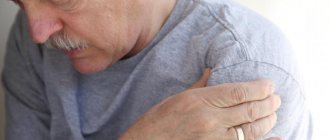
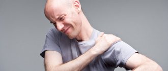
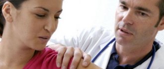
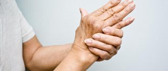
The first symptom of damage to the shoulder joint is pain. At first it is unexpressed and occurs only after excessive physical exertion or prolonged monotonous hand work. But the further the disease develops and the more the joint becomes deformed, the stronger the discomfort becomes.
Soon other signs of the disease appear:
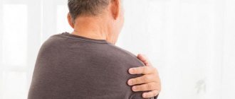
- swelling and redness of the shoulder;
- decreased physical activity;
- crunch in the joint;
- numbness of the limb;
- muscle weakness.
The process of joint destruction can continue for many years. In the final stages of the lesion, the pain becomes constant, disturbing the person even in a static position. The range of joint movements is limited - the patient cannot raise his arm to the side or in front of him.
Causes
No one knows what exactly causes calcific tendinitis. Physical stress, aging or a combination of both leads to degenerative calcification. Some researchers suggest that calcium deposits are formed due to tissue hypoxia and insufficient oxygen supply to the tendon tissues. Others believe that pressure on the tendons can lead to their damage, resulting in the deposition of calcium deposits.
The mechanism of formation of reactive calcification has not been completely studied. Typically, this type of calcific tendinitis occurs in younger patients and occurs without any apparent cause.
Possible consequences
Before talking about the effect of salts on human health, we should briefly define their role in the body. These complex substances are formed as a result of acid-base metabolism and are necessary in small quantities to protect tissues from oxidants. In addition, salts determine the performance of the excretory system and the stability of metabolic processes.
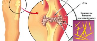
The consequence of urate accumulation is gout.
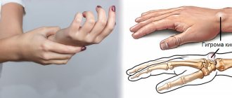
The localization of salts and the ailments they cause depend on the type of compounds. For example, calcium in the form of phosphates is most often deposited in the upper half of the body, causing osteochondrosis and calcific tendonitis of the shoulder.
Urates tend to accumulate in the small joints of the legs and arms, causing the development of gout. These same crystals form stones and sand in the kidneys, gall bladder and bladder. Oxalates (oxalic acid salts) can settle anywhere - on the surface of joints, in bones and blood vessels, in muscles and tendons, causing atherosclerosis, increased blood pressure and heart pathologies.
Diagnostics
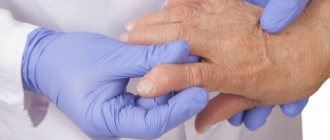
To diagnose calcific tendonitis of the shoulder, your doctor will first take a medical history and perform a physical examination. Shoulder pain can be associated not only with calcific tendonitis, but also with other diseases. Therefore, to make an accurate diagnosis, instrumental studies are necessary. Radiography allows one to visualize the presence of calcium deposits in the tendons. But the most informative way to visualize ligaments and tendons and the presence of pathological changes in them is MRI (magnetic resonance imaging). Visualization of calcifications using radiography or MRI in dynamics allows you to determine treatment tactics (conservative or surgical treatment). Laboratory tests are necessary in cases where it is necessary to differentiate this disease from inflammatory diseases of connective tissue.
How to treat salt deposits?
How to get rid of salts? It is known that patients are most often concerned about urate. They cause severe pain and immobilize a person for several days. Therefore, the primary goal of therapy is to relieve gout attacks, and then restore salt metabolism.
Drug treatment
Anti-inflammatory drugs from the group of non-steroids will help cope with acute pain:
- Nise;
- Nimesil;
- Indomethacin;
- Diclofenac;
- Naproxen.
These medications are available not only in tablet form, but also in the form of injections, as well as ointments for external use.
For unbearable pain in the foot, corticosteroids are prescribed:
- Methylprednisolone;
- Betamethasone;
- Ketazone.
Hormones are most often administered intramuscularly, then switching to tablets and ointments.
The special anti-gout drug Colchicine perfectly relieves pain and inflammation. Its use is started from the first hours of the attack, and when the pain decreases, it is stopped. The medication is toxic and in overdose can cause diarrhea, vomiting and abdominal pain.
Anti-inflammatory drugs are available in the form of tablets, injections and ointments
Indole and pyrazolone medications are good at relieving pain in the shoulder joint: Butadione, Reopirin, Phenylbutazone.
Start taking analgesics with maximum doses. As you feel better, the number of tablets is reduced to the average therapeutic value, observing the frequency of use. The use of capsules is supplemented with the application of anti-inflammatory ointments: Diclofenac, FullFlex, Piroxicam, Tiger Balm White.
To reduce the production of uric acid salts, the patient is prescribed uricodepressants:
- Orotic acid;
- Allopurinol;
- Milurite;
- Febuxostat;
- Thiopurinol.
To improve urate excretion, uricosuric medications are prescribed: Amplivix, Benemid, Flexen, Anturan.
All the drugs discussed have contraindications and cause unpleasant reactions if the dosage is incorrect, so it is recommended to use them only as prescribed by a doctor.
Physiotherapeutic procedures
What should the patient do after eliminating acute pain? How to continue treatment to reduce the incidence of gout attacks? The best assistant that can prolong the period of remission will be physiotherapy.
Its main tasks are to activate trophism and blood circulation, enhance the effect of medications, relieve swelling and start recovery processes.
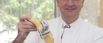
Ultrasound therapy
The technique is based on the pulsation effect acting at the level of cells and fibers of connective tissue. Micromassage is well complemented by the use of anti-inflammatory gels containing indomethacin or diclofenac.
Ultrasound deeply warms tissues and improves blood circulation.
Intense heat reduces swelling, removes effusion and reduces pain. The procedure is contraindicated in the presence of any local infection.
Magnetotherapy
This is one of the types of electrotherapy, which is based on the influence of a low-frequency magnetic field. Using the technique you can achieve the following results:
- improve blood circulation and metabolism in the shoulder joint;
- saturate the affected tissues with oxygen and other nutrients;
- stimulate lymph flow and reduce swelling;
- relieve inflammation and speed up joint recovery.
It is not advisable to carry out the procedure at elevated temperatures, during pregnancy, or if cancer is suspected.
Hirudotherapy
Treatment with leeches has been known since time immemorial. Even in Ancient Rus', they were placed to reduce pain and improve blood circulation in case of joint ailments. Hirudin contained in saliva stimulates peripheral circulation and cleanses the lymph flow of accumulated waste products.
When salts are deposited in the shoulder joint, leeches are placed around the affected joint for 2–3 hours every 14 days. Judging by the reviews, this is a very effective remedy - the patient feels noticeable relief immediately after the session, and the result lasts until the next visit to the hirudotherapist.
Treatment with leeches is effective for diseases caused by salt accumulation
If you do not want to use the “services” of live leeches, you can buy hirudin-based products at the pharmacy - Sophia cream, Girudovazin gel, Girudox balm, Piyavit dry powder. External products should be used with caution, as they are quite allergenic.
Cryotherapy
For diseases caused by salt deposits, cold treatment helps well. By irritating the nerve endings on the surface of the skin, cryoagents stimulate blood circulation and the metabolic process in the affected joint, increase local immunity, and restore water-salt metabolism.
The procedure perfectly removes inflammation and swelling, pain and hyperemia, and improves lymph flow. If the skin in the treatment area is damaged and there are metal objects in the joint, cryotherapy is prohibited.
Physiotherapy
The maximum effect in the fight against salt deposits is achieved by performing specially selected exercises. When choosing a treatment complex, it is recommended to pay attention to Dr. Popov’s gymnastics. It is aimed at reducing pain and restoring flexibility of the upper extremities, as well as post-isometric muscle relaxation.
The load on the joint and range of motion increases as the patient feels better. To prevent injuries and sprains, exercises are done under the supervision of a specialist.
At home, you can perform simple gymnastics, including swinging and rotating your arms, raising and lowering your shoulders, placing your limbs behind your back or moving them to the side. In this case, the intensity and amplitude of movements should increase gradually as the joint prepares to perform more complex exercises.
Massage
In combination with medications and other physiotherapeutic procedures, massage restores lymphatic drainage and tissue trophism, helps fight pain, improves joint mobility, and relaxes spasmodic muscles.
The procedure can be either manual or hardware. In the first case, stroking, kneading and rubbing movements are used. As the pain decreases, the intensity of the effect and the number of techniques are increased.
When salts are deposited, vibration massage using a manual device “Tonus” is very effective. You can do it at home, but only after the pain subsides.
Traditional medicine recipes
Folk remedies help with salt deposits in the shoulder joint. It is better to use them in combination with medications and physical therapy. During therapy, it is advisable to completely remove the load on the sore shoulder and try not to make sudden movements with your hand.
Bay leaves will help clear joints of salt.
You can use alternative medicine recipes both during an exacerbation and during a painless period.
So, how to cleanse joints of salts using folk remedies:
- Prepare a decoction of bay leaves. To do this, pour a pack of crushed raw materials with a liter of water, boil, cool and take before bed for 3 days;
- An equally popular recipe for cleaning joints from salts using rice. Being a natural adsorbent, soaked and boiled grain perfectly draws out urates and accelerates their removal from the bloodstream.
- At home, you can prepare a horseradish compress. For treatment, take fresh leaves, mashed and dipped in boiling water for 2-3 seconds. Warm greenery is applied to the shoulder and bandaged for several hours or left overnight.
Natural juices and drinks will help get rid of accumulated salt. Cranberry juice will not only restore the acid-base balance in the body and reduce the production of urates, but will also be an excellent source of vitamins. You can drink it without restrictions, but under one condition - if there are no gastrointestinal diseases.
Another, no less tasty and healthy drink is made from citrus fruits. For a cocktail, take orange, lemon or grapefruit. You can add a spoonful of strawberries to fresh juice.
A decoction of lingonberry leaves has a strong diuretic and cleansing effect. The drink removes salts from joints, cleanses the kidneys and bladder, disinfects and restores water-salt balance.
Compresses made from anti-inflammatory herbs help with the accumulation of salts: chamomile, thyme, calamus, calendula flowers. A napkin folded in several layers is moistened in the hot infusion, wrung out and placed on the shoulder. The top is covered with film and insulated.
Diet for salt deposits
One of the main components of therapy for salt deposition is a properly selected diet. The patient is recommended to drink plenty of fluids - this can be purified or alkaline mineral water, oatmeal jelly, rosehip decoction, green tea, but not black coffee.
To reduce the amount of purines ingested from food, exclude red meat, fat, sausages and smoked foods from the diet. Limit the consumption of sugar, sweets, yeast baked goods, legumes, spinach and sorrel.
But fermented milk products, vegetables, grains and fruits will only bring benefits. Preference is given to zucchini, cucumbers, watermelon, potatoes, and unprocessed rice. Twice a week you can eat fish or white meat, preferably rabbit.
Other localization of salt deposits in the body
In addition to the shoulder joint, salts can accumulate in other joints. Thus, the favorite place of urates is the first metatarsal joint of the right foot, although gout often affects the little toe or joints of the fingers.
A little less often, salts are deposited in the large joints of the lower extremities - the knee and hip. The elbow joint does not suffer as often, but it brings more suffering.
In the spinal column, the most attractive place for crystal deposition is the cervical region. This is most likely due to the high mobility of the segment.
OSTEOCHONDROSIS OF THE CERVICAL SPINE
Cervical osteochondrosis, or osteochondrosis of the cervical spine, due to morphological and functional characteristics, is characterized by various clinical manifestations and, often, the diagnosis is difficult due to the many “masks”. Clinical manifestations are directly dependent on the location of the lesion. Radicular (radicular) syndromes in cervical osteochondrosis are a manifestation of combined damage to often several roots.
Damage to the nerve root can be of the type of irritation, compression or conduction disturbance. With irritation and compression, the main clinical sign is pain, and with a conduction interruption - radicular paralysis. The pain is accompanied by widespread irritation, which leads to poor circulation, swelling and fibrosis of the tissue around the root.
The figure below (Fig. 2) shows the zones of innervation by nerves emanating from the cervical spine. Knowing these zones, it is easy to imagine the clinical picture of damage to one or another nerve root of the cervical spine.
Fig.2
If the C3 root (3rd cervical vertebra) is affected, pain occurs in the left or right half of the neck, a change in taste in the mouth, a feeling of swelling of the tongue, and difficulty moving food with it.
Irritation of the C4 root causes pain in the clavicle, shoulder girdle, atrophy and decreased tone of the posterior neck muscles, leading to an increase in the supraclavicular air cushion, which is a characteristic symptom of irritation of the above root.
When the C5 root is compressed or irritated, pain appears in the shoulder girdle and along the outer surface of the shoulder, and hypotrophy of the deltoid muscle.
When the C6 root is irritated, pain occurs in the neck and scapula, radiating along the outer surface of the shoulder, forearm and thumb, and hypotrophy of the biceps muscle appears.
Traumatization of the C7 root leads to pain in the neck, scapula with irradiation along the outer surface of the shoulder, into the dorsum of the forearm and to the 2nd and 3rd fingers of the hand.
Prevention of salt deposits in joints
Any disease, including salt accumulation, is easier to prevent than to cure. Unfortunately, a clear algorithm for preventing the disease has not yet been created. However, a healthy lifestyle, avoidance of alcohol, a balanced diet and moderate physical activity can protect the skeletal system from damage.
One of the best ways to prevent salt deposits in the shoulder is swimming.
If we talk specifically about the shoulder joint, you should avoid falling on outstretched arms, sudden jerks on the limb, excessive loads and prolonged monotonous movements. In the latter case, it is recommended to take frequent breaks, combining them with light exercises or shoulder massage.
Prevention of salt deposits is especially important for patients at risk for joint pathologies: athletes, people who have suffered an injury, the elderly or those with genetic inheritance.
SPINAL HERNIA
Intervertebral hernia is one of the most complex diseases of the musculoskeletal system. The cause of the disease can be excessive stress on the spinal column, injuries, incorrect posture, and age-related changes. Under the influence of these factors, the membrane of the spinal disc is destroyed and its contents enter the spinal canal, compressing the spinal roots or the spinal cord itself.
At an early stage of changes in the lumbosacral spine, they make themselves felt by aching pain in the lower back. If the disease is advanced, then the pain caused by the hernia depends on which roots it compresses, and clinically it can manifest itself as impaired movement in the legs, impaired urination and potency.
Treatment. Surgical intervention for this pathology gives quick results, but is fraught with postoperative complications and the development of diseases in other parts of the spine. It is indicated in only 10-12% of patients. The most effective method is a combination of laser and manual therapy. Laser therapy allows you to relieve swelling and inflammation in the hernia area, and manual therapy, which is carried out after a course of laser therapy, allows you to painlessly correct the hernia. There have been cases where hernias disappeared only after several courses of laser therapy.
Answers on questions
Will a honey massage help cope with salt deposits in the shoulder?
Yes, sure. This is a very effective treatment method, but to obtain a lasting result, at least 10 sessions are necessary.
The procedure is performed with warm honey, which is applied to a well-warmed joint. Then the sweet medicine is rubbed into the skin using patting and massaging movements. The total procedure time is at least 20 minutes.
It is recommended to do this massage at night. After washing off the honey, the shoulder is well insulated with woolen cloth for better warming.
How to treat salt deposits in the hymen joint with bee venom?
A bee sting is an ancient healing technique that perfectly helps with all kinds of joint ailments. In some countries, this method of treatment is even recognized as official medicine. There are no special contraindications to it, except that in some people a bee sting causes severe allergies and shock. Therefore, apitherapy should be carried out only under the supervision of a physician.
During treatment, you should not drink alcohol or visit a bathhouse or sauna, as all this will neutralize the effect of the poison. This therapy is also contraindicated for children.
Of course, not everyone will decide to have a bee land on themselves and get a painful sting, even under the supervision of a doctor. Ointments based on bee venom will help replace this dangerous procedure: Virapin, Apizartron, Apitrin. Apply them 1-3 times a day, thoroughly rubbing into the skin above the joint.
Is it possible to use dietary supplements for salt deposits in the shoulder joint and which ones? How effective are they?
Dietary supplements can be used as part of complex treatment, but not as an independent medicine. The most effective are Glucosamine and Chondroitin or their combination with the addition of methylsulfonylmethane (MSM).
These drugs relieve inflammation and pain well, improve the quality of synovial fluid and joint mobility. In addition, chondroprotectors protect cartilage tissue from corrosion by salts and subsequent destruction. It is recommended to take such medications for at least 5–6 months.
Types of shoulder bursitis
The most common types of bursitis of the shoulder joint are localized:
- Subacromial and supracromial.
- Subcoracoid.
- Subdeltoid.
Pathology is distinguished according to the characteristics of its course:
- Acute is an intense process with severe pain, which lasts from several days to a month and develops 3-5 days after an injury or other cause of inflammation.
- Subacute is a form of pathology in which pain intensifies with movement and is absent at rest.
- Chronic – less noticeable and obvious inflammation with a protracted course (up to one year).
- Recurrent - bursitis in a chronic form, which worsens periodically for various reasons (from constant injury to the joint to chronic infections in the body).
Conclusions about the nature of inflammation can also be drawn from how the composition of the synovial fluid has changed. There are different types of bursitis:
- Serous - the liquid becomes clear (sometimes with a slight cloudiness). It contains protein, leukocytes and synovial membrane cells.
- Hemorrhagic - exudate with blood.
- Purulent - inflammation caused by infection occurs rapidly and with severe pain. Antibiotics are required for treatment.