Bone exostosis is a pathological process that results in uneven thickening of tissue areas in the area of flesh and tubular bones. The tibia, fibula, femur, and humerus can be damaged. But the most famous case of osteochondral exostosis in adults is a heel spur. This degenerative disease develops against the background of planetary fasciitis. It causes a lot of suffering and is very difficult to respond to conservative treatment methods.
Osteochondral exostosis in children can be a consequence of congenital deformation, or it can begin with the onset of puberty. In the first case, risk factors that lead to disruption of the formation of embryonic tissues are important. Children of parents who smoke and drink alcohol often suffer. It is also important that any hormonal medications taken by the expectant mother during pregnancy are taken. With the acquired form of bone exostosis, hormonal imbalance is important. It can be a consequence of excessive release of sex hormones into the blood during puberty.
Delamination and degeneration of cartilage tissue occurs, and the growth points of hyaline cartilage open. Then its lattice structure is filled with deposits of calcium salts. The tissue acquires the properties of rough spongy bone. Over time, it becomes covered with a layer of synovial cartilage tissue. And the whole pathological process repeats itself again. Places of thickening of bone tissue with exostoses can increase in volume very significantly. This reduces motor activity and over time leads to the formation of serious contractures, the development of which will take more than one month.
Treatment of osteochondral exostosis at an early stage of development does not require surgical intervention. Therefore, if characteristic clinical signs appear, it is necessary to promptly consult a specialist. This is quite difficult to do, since in most cases, pathological growth of cartilage and bone tissue is discovered by chance during an examination for another orthopedic disease.
But there are indirect signs of trouble. It is important to pay attention to them and consult a doctor if they appear. They will be described in this material.
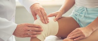
In Moscow you can get a comprehensive consultation with an orthopedist completely free of charge. You can make an initial free appointment with a doctor at our manual therapy clinic. During the consultation, the doctor will conduct an examination, make an accurate diagnosis and talk about the prospects and possibilities of manual therapy in your case of illness.
Reasons for the development of bone exostosis
There are various reasons for the development of bone and osteochondral exostosis. They are divided into factors of external and internal influence. Let's start getting acquainted with them with the factors of external negative impact - these are:
- injuries, including those without violating the integrity of bone tissue (fractures and cracks);
- exposure to negative temperatures on exposed areas of the body;
- increased physical activity while performing professional duties or while playing sports;
- squeezing (compression) of the limbs, including when a medical tourniquet is applied incorrectly to stop bleeding;
- wearing incorrectly fitted shoes and clothing.
In addition, there are internal factors of negative influence and there are many more of them:
- infectious processes and chronic inflammatory foci in any part of the human body (they provoke the nominal system to work in an enhanced mode, which can result in autoimmune deformation of cartilage tissue in large joints of the upper and lower extremities in the intervertebral discs);
- disruption of microcirculation processes of blood and lymphatic fluid in soft tissues;
- muscle fiber dystrophy due to a sedentary lifestyle;
- excess body weight (causes thickening of the vertebral bodies and condyles of the femur and tibia);
- degenerative dystrophic diseases of cartilage tissue throughout the body;
- Bechterew's disease (ankylosing spondylitis or articular form);
- rheumatoid inflammatory process;
- severe forms of allergic reactions in the human body;
- deforming osteoarthritis of large joints of the upper and lower extremities;
- violation of drinking rules;
- deficiency in the diet of certain vitamins and minerals;
- the process of osteoporosis in women in the menopausal phase.
In children of early infancy, the causes of exostosis of bone and cartilage tissue can be excess vitamin D in the body (if the rules for the prevention of rickets are violated), chondropathy, dysplasia injury, etc.
In adolescents, the most important factor in the destruction of bone and cartilage tissue is the process of lysis due to excess levels of sex hormones. The role of the pituitary and pineal glands is important. If the growth points of bone tissue are not closed in a timely manner, gigantism may occur. In any case of cartilaginous or bone exostosis in adolescents, a complete endocrine examination is required. It is important to do a series of specific blood tests to determine the levels of all important hormones. If parents notice too rapid growth of their child’s limbs between the ages of 9 and 14, they should visit an endocrinologist as soon as possible. In the early stages, hormonal deformations can be easily corrected using replacement therapy. In the later stages, this disease is no longer curable and inevitably leads to external deformities in the human body.
Symptoms of subungual exostosis
The formation of an osteochondral neoplasm under the nail bed leads to elevation of the free edge of the nail plate and its middle part, as well as various deformities. Gradually, the nail of the affected finger begins to curl. In some cases, its ingrowth (onychocryptosis) is also observed, which can lead to:
- development of the inflammatory process in the periungual fold;
- the appearance of pain:
- the appearance of purulent discharge;
- changes in the structure and color of the nail;
- development of infectious complications.
But even in the absence of onychocryptosis, subungual exostosis not only significantly worsens the appearance of the nail, but also causes unpleasant and downright painful sensations. In this case, the disease tends to progress over time and can also lead to complete loss of the nail.
Subungual exostosis has common features with various types of onychodystrophy, mycosis and ingrown nails. Therefore, to diagnose the causes of changes in the shape and condition of the nail plate, you should consult a podiatrist or orthopedist.
The formation of subungual exostosis may occur differently in different patients. In some cases, the osteochondral protrusion is small in size and does not appear in any way for years. In such situations, it may be accidentally detected on x-rays taken for completely different reasons.
But more often, subungual exostosis causes a person discomfort of varying severity, pain and limits the motor function of the affected toe. It looks like an enlargement of the anterior periungual fold, into which the growing nail rests. Sometimes this is accompanied by redness and swelling of the periungual area. Discomfort tends to increase during any physical activity and especially running or walking, as well as when wearing tight shoes and even slight pressure on the end of the big toe.
However, the main symptoms of subungual exostosis are:
- spontaneous peeling of the nail;
- deformation of the nail plate;
- increase in the size of the periungual fold;
- ingrown nail edges.
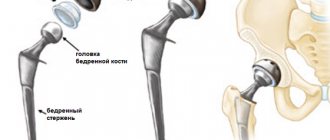
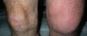
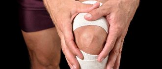
Bone exostosis of the knee joint
In adolescents, bone exostosis of the knee joint is relatively rare. In adult patients, solitary osteochondral exostosis of the tibia is observed, which leads to severe deformation of the bone articulation. This shape is easily identified even with the naked eye. During the initial examination and palpation, a dense, absolutely painless neoplasm can be detected under the skin. It may have a smooth or rough shape.
The second type is bone exostosis of the femur with numerous foci of deformation. The entire surface of the femur can be covered with multiple foci of calcium salt deposition. This type can occur in both adults and adolescents. It is predominantly asymptomatic. It can be detected randomly during an X-ray examination or during deep palpation of the femur.
Marginal bony exostoses of the calcaneus
Bone exostosis of the calcaneus is better known as a heel spur. This is a neoplasm that begins to develop from tendon and fascial tissue. The primary factor in the inflammatory reaction is a violation of the integrity of individual fibers of the plantar fascia. It begins to gradually thicken. Plantar fasciitis develops.
This pathology entails constant injury to the synovial cartilage tissue in the heel bone area. The cartilage begins to grow and marginal bone exostoses form, causing severe suffering to the patient when walking.
It is possible to cure bone exostoses on the heels without surgery only in the initial stages. Therefore, if you experience pain in the heels that develops after physical activity (for example, after walking long distances), you should immediately consult an orthopedist.
The doctor will order an x-ray of the heel bone. It will show the formation of marginal bone exostoses along the surface of the heel bone. These formations injure the surrounding soft tissues when walking. An inflammatory process occurs, which stimulates further growth of cartilage and bone tissue.
What is subungual exostosis
Subungual or subungual exostosis is a very common problem. This term refers to osteochondral growth in the area of the last or nail phalanx of the toe, or rather at its end. Thus, this is a benign neoplasm of a non-tumor nature. Initially, it is a cartilaginous growth that tends to ossify over time and injure soft tissues, cause pain and impede the growth of the nail plate. The disease was first described by Dupuytren back in 1817.
In the vast majority of cases, subungual exostosis forms on the first or big toe and is the body’s reaction to constant mechanical irritation.
What does it look like?
Exostosis itself forms under the soft tissues, so it may not appear for a long time. It forms on the surface of the bone and is a cartilaginous growth. But over time, it begins to ossify and transform into spongy bone, after which a thin but very dense bone capsule is formed on its surface. Moreover, such a growth tends to increase in size over time and ultimately deform the nail plate and cause pain when walking. And outwardly it looks like a thickened anterior nail fold, which prevents the free growth of the nail.
Such exostosis is not related to the epiphyseal plate or cartilaginous growth zone, unlike osteochondroma. However, it is not prone to malignancy or malignancy, unlike the latter.
Subungual exostosis can have different shapes and sizes, which determines the nature of its manifestations. Thus, it can be a spherical, spiky, linear, mushroom-shaped or other protrusion ranging in size from several millimeters to 1 centimeter or more. In this case, as a rule, exostosis is present on the toe of only one foot.
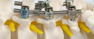
Bone exostoses of the spine and ribs
Cartilaginous and bone exostoses of the spine are a consequence of a long-term degenerative dystrophic process in the area of the intervertebral discs and endplates of the vertebral bodies. The degeneration process begins with a violation of the diffuse nutrition of cartilage tissue. It is able to receive nutrition only when the muscular frame of the back is actively working. If a person leads a sedentary lifestyle, muscles lose elasticity and performance. They cannot transport blood and nutrients dissolved in it to the cartilage tissue of the intervertebral discs.
As a result, the process of dehydration of the fibrous ring begins. It loses its shock-absorbing ability and becomes covered with a network of cracks. Calcium salts are deposited in them.
As a compensatory reaction, chondrosis and sclerosis of the endplates of the vertebral bodies begin. They increase in volume and become deformed. As a result of strengthening the blood supply point, the process of nutrition of bone tissue is enhanced. Deformation of the vertebral bodies and bases begins.
Bone exostoses of the ribs are formed as a result of traumatic exposure and curvature of the spinal column. These tight junctions fix the deformation of the chest, limit the vital capacity of the lungs and can lead to severe oxygen deficiency in the structures of the brain and vital internal organs (heart, liver, spleen).
Diagnosis is made using x-rays. For treatment, conservative methods of influence can be used, such as manual therapy, physiotherapy, shock wave therapy, etc. Surgical intervention for this localization of exostoses is used relatively rarely.
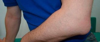
Bone exostosis of the humerus
In many cases, bone exostosis of the humerus develops with habitual dislocation, deformation of the labrum and brachial plexitis. Less common negative impact factors are injuries such as blows and bruises of soft tissues, bone fractures, etc.
Exostosis of the humerus can manifest itself with the following clinical symptoms:
- dull pain in the shoulder area when trying to move the upper limb with a wide amplitude;
- feeling of stiffness in movements in the morning;
- feeling of limited movement;
- pain that spreads along the brachial nerve;
- decreased muscle strength and decreased volume of the forearm and shoulder.
An x-ray is sufficient for diagnosis. To exclude oncological processes, a tissue biopsy can be performed followed by histological examination.
How to recognize exostosis?
The doctor can diagnose the disease during a routine appointment with the patient.
The disease is often detected during a routine examination or x-ray diagnosis, when changes are noticeable and bone growth becomes visible. Exostosis of the knee joint develops over a long period of time and is initially asymptomatic. The patient does not feel pain, there are no visible signs of inflammation, the epithelial tissue does not change color. Sometimes a person feels a slight numbness and tingling, stiffness in movements, pressure on the bone. If you have such symptoms, it is better to seek advice from a specialist.
Exostosis manifests itself not only in the joints of the lower and upper extremities. The disease affects the shoulder blades, ribs, pelvic bones, vertebrae, collarbones and even the phalanges of the fingers. The tissue under the nails can grow up to 1 cm in diameter and be accompanied by severe pain.
Treatment of osteochondral exostosis
It is necessary to begin treatment of osteochondral exostosis by establishing the causes of its occurrence. Only after all the negative impact factors have been eliminated can rehabilitation therapy begin. As long as pathogenic factors continue to have a destructive effect, there is no talk of successful treatment. Foci of deformation will arise again and again.
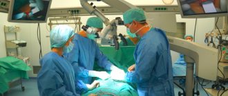
In case of a significant volume of the tumor and compression of the surrounding soft tissues, treatment of bone exostosis is provided exclusively by surgical means. During the operation, the doctor removes the pathologically formed bone tissue.
In our manual therapy clinic, treatment of osteochondral exostoses is carried out only at the initial stage. We use osteopathy, massage, kinesitherapy, reflexology, therapeutic exercises and many other methods for this. Laser treatment is effective, allowing it to actively influence the foci of exostosis formation.
If you would like personalized advice about your musculoskeletal problems, book a free orthopedic appointment at our manual therapy clinic. The doctor will review your medical documentation, conduct an examination, make a diagnosis and give all the necessary recommendations regarding the future treatment of the identified disease.
Diagnostic measures
If a person experiences knee discomfort, it is important to contact a medical facility as soon as possible. It is impossible to diagnose the disease at home. At the appointment, the doctor asks the patient how long ago the discomfort began and whether there are additional symptoms. The patient is then sent for an X-ray examination. The disadvantage of radiography is that it is impossible to accurately determine the size of the tumor. However, the results of the X-ray image show the shape of the tumor and its location. Before making a diagnosis, it is important to make sure that the patient has formed a bone growth and not a malignant neoplasm. To do this, they resort to magnetic resonance imaging.