Plantar fasciitis can be successfully treated with conservative methods in 90-95% of patients. But in some cases, the only way to get rid of a spur is through surgery.
Spur surgery is not difficult from the surgeon’s point of view, however, it is often extremely painful for patients and requires long-term rehabilitation.
If you have a heel spur, surgical treatment of which is the only way to get rid of the growth, then in this article we will talk about the types of operations and how to quickly recover and return to normal life.
When is a heel spur removed surgically?
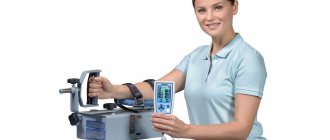
All over the world, doctors are trying to avoid heel fascia surgery by using various treatment methods. Excellent results are obtained by crushing the heel spur with a laser, shock wave therapy, and taking steroid and non-steroidal drugs to reduce inflammation.
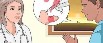
As a rule, if you follow all the doctor’s recommendations, you can overcome fasciitis in 1-6 months. The effectiveness of conservative treatment methods largely depends on how doctors treat heel spurs. If treatment with one doctor does not bring benefit, then it is advisable to contact another specialist.
Unfortunately, for some people, therapy and physical procedures do not bring a positive effect, no matter what doctor they see.
The spur growth increases in size, causing unbearable pain. The quality of life and general well-being of the patient deteriorates significantly. In this case, the only way to get rid of the spur is to surgically remove it.
Doctors consider the following indications for surgery:
- conservative treatment does not provide effect for more than 6 months;
- the person experiences excruciating pain;
- bone growth seriously limits movement;
- the patient cannot go to work, school, i.e. deprived of a normal life.
In these cases, if surgery is not performed, a heel spur, the surgical treatment of which is vital, can lead to disability, loss of social connections and professional degradation.
Heel spur: when surgery is contraindicated
Even if surgery to remove the spur is the only treatment option, it can only be performed if there are no contraindications.
The following are considered absolute contraindications:
- blood coagulation disorders;
- acute infectious diseases;
- cardiovascular diseases in the acute stage.
When operating under anesthesia, all restrictions associated with the use of anesthesia are added to the contraindications. In this case, steroid-based heel spur medication injections may provide positive results.
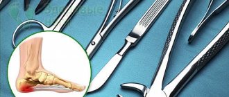
Surgical methods for removing heel spurs
Currently, orthopedic surgeons perform operations on inflamed fascia using 3 techniques:
- open surgery with dissection of the fascia;
- endoscopic surgery;
- mini-dissection of the fascia with X-ray monitoring.
All types of heel spur removal are performed using general or local anesthesia. The choice of technique and the use of one or another type of anesthesia is determined by the doctor after conducting the necessary studies.
Before the operation, blood tests (general and coagulation tests) and an X-ray of the heel spur are required. Also, if indicated, ultrasound of the extremities, studies of the cardiovascular system, etc. can be performed.
Treatment results
UVT therapy breaks down and softens calcifications, which leads to complete destruction of the bone growth.
- metabolism is normalized;
- blood circulation and lymph flow accelerate;
- heel pain disappears;
- inflammatory processes are inhibited;
- swelling decreases;
- muscle tone increases;
- tendons and ligaments are strengthened;
- regenerative ability is activated;
- legs become healthier;
- motor activity is restored.
Traditional surgical removal of heel spurs
This method is considered outdated, but it is sometimes used in poorly equipped surgical departments. The main goal of surgery is to remove the spine and relieve stress on the heel fascia caused by the pressure of the spur.
The operation is performed under general anesthesia and includes the following steps:
- Dissection of the fascia with a large longitudinal incision.
- Removal of bone-salt growth and salt deposits around it.
- Installation of drainage system.
- Suturing the incision and bandaging the foot.
Drainage is necessary to drain serous fluid; when it stops being released, the drainage is removed. The postoperative period lasts 1-3 months. After the sutures are removed, a stabilizing cast is applied to the foot to return the fascia to its original position.
For the first 3 weeks, any load on the foot is contraindicated! After this period, the patient, under the supervision of an orthopedic surgeon, begins to walk, gradually loading the heel.
Shoes begin to be worn 1 month after surgery, and the use of orthopedic insoles or heel pads is mandatory so as not to put stress on the injured heel.
Traditional open surgery has many disadvantages, the main one being the long and painful rehabilitation period. That is why this method of removing spurs is becoming a thing of the past; it is being replaced by more advanced and painless techniques.
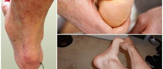
Causes
According to statistics, in 90% of cases the cause of the development of this pathology is flat feet. It is because of this disorder that the proliferation of osteophytes occurs.
In addition, other factors may influence the development of the pathological process. Let's consider the main ones:
- Heel bone injury.
- Having excess weight.
- Old age (over 50 years old).
- The presence of diseases of the musculoskeletal system.
- Disruption of metabolic processes in the body, in which salt deposition occurs.
- Poor posture, which causes gait to suffer.
- Wearing low-quality or uncomfortable shoes, perhaps the wrong size.
- Regular increased physical activity.
Hereditary predisposition should also be noted. If you have close relatives suffering from heel spurs, there is a high probability that this pathology will affect you too.
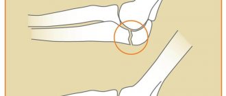
Endoscopic heel spur removal
The operation is performed after the diagnosis of the heel spur has shown its exact location and size. Removal of the growth is carried out without completely opening the calcaneal fascia.
Access to the spike is made through 2 small cuts up to 5 mm in size. Through the first incision, a microscopic trocar is inserted, which approaches the growth and wraps around it using a cannula.
A mini-camera is inserted through the second incision, the image from which is transferred to the monitor in real time. The doctor removes the spur, focusing on the screen image.
After the operation, the micro-incisions are sutured and treated with an antiseptic for 2-3 days. The patient can get up and walk on the second day after the procedure.
The load on the feet should be gradual, and when walking in shoes, arch supports or unloading heel pads for spurs must be used to protect the foot from shock loads.
Compared to traditional open surgery, the endoscopic method has the following advantages:
- painlessness and rapid relief of pain symptoms;
- scars heal within a few days;
- simpler rehabilitation period;
- return of the patient to a full life in 3-5 days.
Orthopedics and traumatology services at CELT
The administration of CELT JSC regularly updates the price list posted on the clinic’s website. However, in order to avoid possible misunderstandings, we ask you to clarify the cost of services by phone: +7
| Service name | Price in rubles |
| Appointment with a surgical doctor (primary, for complex programs) | 3 000 |
| X-ray of bones and joints of the limbs | 2 200 |
All services
Make an appointment through the application or by calling +7 +7 We work every day:
- Monday—Friday: 8.00—20.00
- Saturday: 8.00–18.00
- Sunday is a day off
The nearest metro and MCC stations to the clinic:
- Highway of Enthusiasts or Perovo
- Partisan
- Enthusiast Highway
Driving directions
Minimally invasive heel spur removal
This method of surgical removal of growth has been used relatively recently. Unlike the endoscopic method, the minimally invasive method makes only one incision no larger than 3 mm. A trocar with a cannula is inserted through this incision.
The progress of the operation is monitored using a movable X-ray tube, which accurately determines the location of the heel spike. The procedure is performed under local anesthesia.
After the spur is removed, the incision is sutured and processed. A person can step on their foot 12 hours after the intervention. This method is often used when conservative treatment of heel spurs is not effective.
The minimally invasive method is characterized by low trauma, minimal pain and quick recovery.
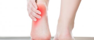
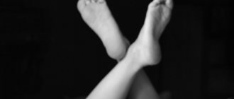
Alarming symptoms
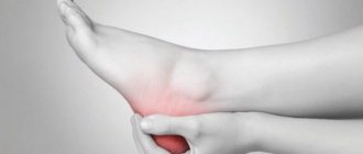
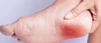
The first sign of the development of pathology is pain in the heel area. As a rule, the pain appears in the morning. At the same time, during the day, no discomfort may often be observed. Over time, the pain intensifies and can be severe. Patients describe them to the doctor as a feeling of “a nail in the heel.” The pain worsens after being at rest for a long time. After physical activity, the discomfort may subside a little, but during the evening rest the pain returns.
Additional signs include the following:
- Swelling and redness of the heel area.
- The appearance of rough calluses in the heel area.
- When palpating the affected area, sharp unpleasant sensations may be observed.
Very often, patients with heel spurs experience changes in their gait. This is due to the fact that a person experiences pain when stepping on the heel. Therefore, the patient tries not to transfer his weight to the heel. Please note that the heel spur is not visually noticeable. Therefore, you should go to the doctor as soon as you feel discomfort in the heel area.
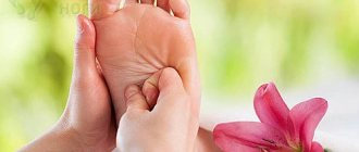
How to quickly recover after surgery to remove heel spurs?
A heel spur, surgically treated using an endoscopic or minimally invasive method, is removed immediately and does not cause pain, so you can put weight on the affected foot after 12-48 hours.
This must be done gradually so that the rehabilitation process does not drag on and complications do not arise.
Orthopedic doctors have developed a number of recommendations for post-operative recovery for people who have undergone surgery to remove a bone growth:
- You can start putting weight on your leg only after the sutures have been removed or the micro-incisions have healed.
- The load should be gradual, starting from 10-15 minutes a day.
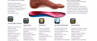
- When using shoes, incl. At home, be sure to wear orthopedic insoles for spurs or heel pads.
- Regularly examine your feet for symptoms of complications from surgery.
- Take care of your feet every day, treat the incision sites with an antiseptic, and the heels with special creams that prevent the re-formation of spur growths.
“Pyatkospor Prophylactic” can be used as preventive creams.
This cream was created specifically to prevent the appearance of thorns on the heels, contains medical bile and effectively softens keratinized areas.
Complications after surgery: how to avoid them?
Based on practical experience, doctors compiled a list of complications encountered in patients after surgery:
- foot pain;
- infection of the incision site;
- damage to nerve endings;
- drooping of the longitudinal arch of the foot;
- risk of recurrence of the growth.
To reduce the risk of complications, it is necessary to strictly follow the doctor’s instructions, properly treat surgical wounds, not overload the foot, and use good quality orthopedic devices for this.