Pregnancy and childbirth are one of the most important periods in the life of every woman. During this wonderful time, young mothers have to deal with various types of psycho-emotional and physical stress.
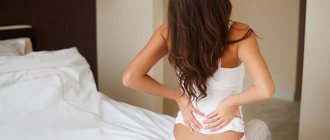
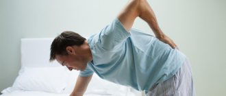
After childbirth, girls often feel a loss of strength, excessive anxiety and pain localized in different parts of the body. The rehabilitation period after childbirth lasts up to 2 months. But sometimes this time is delayed, especially if the birth was not natural, but by caesarean section.
Why does the lower back hurt after a caesarean section?
Epidural anesthesia for caesarean section can cause discomfort later.
In the first days after childbirth, a woman may be bothered by various somatic manifestations. The main rule that young mothers need to adhere to during this period is to inform the doctor about any ailment and follow his instructions.
If pain occurs later, it is important to exclude dangerous causes of its development and select therapy that will allow you to quickly and safely restore your well-being in order to enjoy a happy period of motherhood.
Post-puncture headache (PDPH)
The widespread introduction of spinal anesthesia into the practice of obstetric anesthesiology leads to a significant improvement in the outcomes of cesarean section, which is good news. But along with this, obstetric anesthesiologists are faced with PDPH. According to the available literature, PDPH appears within 1-3 days after puncture of the dura mater during spinal anesthesia in 2-3% of cases in general surgical practice and in 5-6% when performing SMA during a cesarean section and or almost always after an accidental damage to the dura mater during epidural anesthesia.
⠀
PDPH occurs when the patient moves to an upright position, often accompanied by dizziness, nausea, and nausea. In severe cases, the pain does not stop when moving to a horizontal position. According to the available literature, PDPH appears within 1-3 days after puncture of the dura mater during spinal anesthesia in 2-3% of cases in general surgical practice and in 5-6% during performing SMA during cesarean section and or almost always after accidental damage to the dura mater during epidural anesthesia.
⠀
The cause of PDPH is most often considered to be a decrease in pressure in the subarachnoid space due to the leakage of cerebrospinal fluid through a post-puncture hole in the dura mater. If the outflow of cerebrospinal fluid occurs at a rate exceeding its production, there is a possibility of displacement of intracranial structures with tension in the meninges, which is especially significant when moving to a vertical position.
⠀
The resulting pain impulses are carried along the trigeminal nerve, glossopharyngeal nerve, branches of the vagus nerve and cervical nerves. There is a high correlation between the diameter of the needle, the frequency of pain and its intensity. The location of the needle cut during puncture is of particular importance. In addition, it has been experimentally revealed that the presence of air introduced into the spinal canal is important. This significantly increases the incidence of PDPH, guaranteed to cause PDPH with just 0.2 ml of air. It is also important to remove the needle with the mandrin inserted. Its absence in a needle or attached syringe doubles the likelihood of developing PDPH.
⠀
More often, PDPH occurs in young women, more often in those with normal or reduced body weight. An increase in intra-abdominal pressure during pregnancy contributes to an increase in cerebrospinal fluid pressure and increases the rate of its flow, which leads to a more frequent development of PDPH.
⠀
There is no consensus on treatment methods for PDPH. All authors recommend strict bed rest if PDPH occurs; in 95% of cases, the effect is achieved by filling the epidural space with autologous blood; with repeated filling with autologous blood, the effect is achieved in 100%. Filling with autologous blood may be accompanied by pain during puncture of the epidural space, muscle spasm during blood injection, and the occurrence of meningeal symptoms. The described phenomena are transient and do not require additional treatment. Prescription of a 20% caffeine solution, intravenous administration of a 40% glucose solution, 25% magnesia solution, and B vitamins can be effective. IV infusion of 1200 ml of saline solutions is often used.
⠀
A relatively new direction is the use of Sphenopalantine block for the treatment of PDPH. According to published data, its effectiveness is comparable to filling the epidural space with autologous blood. According to the authors, the effect occurs within a few minutes and reaches a maximum within 1 hour after administration.
⠀
Such a large number of recommendations for the treatment of PDPH suggests that to date there is no understanding of either the mechanisms or causes of this complication.
Anesthesiologist-reanimatologist Evgeniy Anatolyevich Orlov.
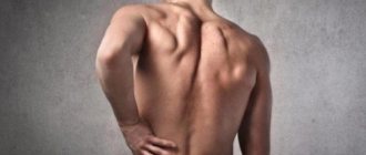
Symptoms and nature of pain
Pain is the body's way of signaling a problem. Based on its location and nature, doctors can make a presumptive diagnosis.
However, after a cesarean section, the causes of pain and its manifestations are diverse. They may depend on the specific pathology and individual sensitivity of the patient.
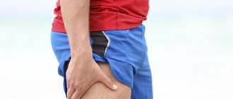
More often, women complain that the lower back begins to pull, but sometimes the pain can shoot into the shoulder blades, radiate to the limbs on the left and right, or spread to the entire back.
Diagnostics is the only way to identify the true problem and begin to treat it purposefully.
Recovery after surgery
After a caesarean section, you should not ask doctors for a speedy discharge. You need to stay in the maternity hospital as long as the doctors deem necessary. If the pain does not stop for a long time, but only intensifies, then you may have to contact a specialist. Still, what to do if the pain does not go away, but before going to see a doctor you need to relieve it?
You can try out some recommendations and understand which one suits the mother most. But even before using them, you should consult with your doctor, at least by telephone.
- Cold and hot compresses (if there are no contraindications). Both a heating pad and ice will do.
- Warm (not hot) bath with sea salt. Relieves back pain and relaxes.
- A smooth and elastic mattress, orthopedic at best.
- Yoga and Pilates help strengthen the abdominal muscles.
- Chiropractic and acupuncture can help significantly reduce pain.
- Discuss the dose of pain medication with your doctor if the new mother plans to breastfeed.
No ads 2
Causes of pain syndrome
A caesarean section is an operation to assist in childbirth and can cause complications. Most of them are diagnosed immediately or are temporary, so there is no cause for concern.
Epidural anesthesia
Back pain after epidural anesthesia occurs due to irritation of the roots.
During a CS, general anesthesia is used only if there are serious indications. Most women cope with the operation using epidural anesthesia. It has fewer contraindications and is easily tolerated.
The doctor places an injection in the back, in the space between the vertebrae and the spinal cord. After this, the patient loses sensation in the lower half of her body for a short period of time: she ceases to feel the area from the waist to the tips of her toes, which completely blocks sensations even from tactile contact for the duration of the procedure.
Statistically, complications associated with spinal anesthesia occur in no more than 0.5-1% of cases. Most often we are talking about local, point pain at the injection site. It occurs immediately after sensitivity is restored, when the woman begins to walk independently. The pain goes away on its own after a day, a maximum of several days, so treatment is usually not prescribed.
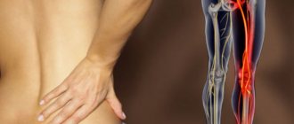
If the pain lasts longer and affects the lower back, legs, and abdomen, we may be talking about irritation of the nerve roots during the injection. This cause can be recognized by the nature of the pain: it follows the path of the nerves. In this case, the doctor usually prescribes painkillers and recommends resting the spine.
When using an epidural, psychosomatic pain often occurs. It is not dangerous, but can significantly affect the quality of life of a young mother. The symptoms can be anything, and the reason is related to the patient’s psychological discomfort due to the fact that the doctor gave an injection in the back.
Epidural anesthesia is performed under completely sterile conditions and there is little risk of infection.
Real complications from anesthesia that occur very rarely:
- injury during the procedure;
- introduced infection.
To exclude these factors, you must consult a doctor and undergo a thorough examination.
The etiology of the pain syndrome may be associated with real pathologies of the spine. Osteochondrosis, which occurs in the vast majority of people, can worsen during pregnancy.
Exacerbation of existing osteochondrosis
Increased loads on the lower back can cause exacerbation of osteochondrosis.
Pregnancy is a significant load on the musculoskeletal system. The weight increases, the fetus puts pressure on the internal organs, so the muscles that support the spinal column are stretched. The spine can become deformed, pinching the nerve roots and hurting.
The catalyst for exacerbation of osteochondrosis can be not only pregnancy, but also childbirth itself, for example, if a woman in labor takes an uncomfortable position. Due to the anesthesia, she does not feel any discomfort, but as the epidural wears off, it can become painful.
A woman's behavior after childbirth may matter. If she has not developed a muscular corset and is not used to physical activity, carrying a baby in her arms can lead to back pain. In addition, you often have to bend and unbend over the crib.
Treatment in this case is long-term, aimed at maintaining the normal position of the spine and relieving symptoms.
Hormonal changes
A woman’s body helps her survive childbirth as painlessly as possible. In order for the fetus to pass through the birth canal, the pelvic bones must move apart. To do this, the hormone relaxin is produced, which softens the joint ligaments. But it cannot act locally, affecting only the pelvic area. Therefore, discomfort in the spine is a common companion for young mothers.
Treatment of back pain
If the reason why your back hurts after a cesarean section is determined, it's time to start treatment. Today, there are a number of medications that help combat back pain. Sometimes a woman’s back pain can be so severe that the only solution to the problem is surgery.
Massage is an effective procedure that helps reduce discomfort.
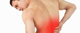
An important part of rehabilitation after a cesarean section is drug therapy, which helps maintain the patient’s condition in a normal manner. The main localization of pain after a cesarean section is in the lumbar and sacrum areas. Therefore, specialists can prescribe a course of blockades to the woman in labor.
Massage helps effectively combat pain. It can only be done after examination and permission from a doctor. Movements should be smooth and light, slowly transitioning to targeted pressure on the lumbar region. In the postpartum period, you should refrain from intense physical activity and harsh massages. Under no circumstances should you self-medicate. It is important to always seek professional help. Remember: children need healthy parents who can take care of them.
When to see a doctor
After surgery, inflammation in the uterus is possible, which causes back pain
. Whatever the cause of the pain, a woman should definitely visit a doctor. If a short period of time has passed after childbirth, you can visit the specialist who managed the pregnancy. He will prescribe studies that will eliminate the risk of inflammation of the uterus and appendages, since against the background of this process the pain can radiate to the back:
- general blood and urine analysis;
- Ultrasound of the pelvic organs.
If no abnormalities are found in the organs of the reproductive system, the doctor may recommend contacting a urologist. During pregnancy, the kidneys are under high load, so chronic pyelonephritis may worsen after childbirth.
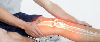
To find the cause of pain in the musculoskeletal system, you need to consult a neurologist. Magnetic resonance therapy and electroneuromyography will allow you to quickly make an accurate diagnosis.
A woman does not always have to go through a long, multi-stage diagnostic system. In the vast majority of cases, if the symptoms clearly indicate a specific pathology, it is enough to visit a therapist.
Natural childbirth
When giving birth to a child, a woman spends several hours in an obstetric chair. In this position, the tailbone is injured and the lower back arches. A woman in labor strains muscle groups that are not usually used. It is quite natural that 80% of women have back pain after childbirth.
During pregnancy, hormones gradually soften the ligaments and joints so that the baby can safely pass through the pelvic bones. Before the hormonal balance is restored, the woman experiences pain when sitting or standing for long periods of time. She may have difficulty maintaining her balance.
General recommendations after childbirth
- Young mothers are advised to lose excess weight and take care of their joints, which are still under the influence of the hormone relaxin.
- Crooked posture requires correction with exercises, massage and physiotherapeutic procedures.
- To pick up a child, it is better to sit down rather than lean towards him.
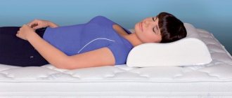
- During breastfeeding, it is recommended to constantly change sides to relieve the burden on the shoulders. Feet should rest on the floor. You can place a small pillow under your lower back.
- A woman should not lift anything larger than a baby. It is better to entrust the stroller and other heavy things to relatives.
- To carry a child, it is recommended to use slings - special devices similar to a backpack, which free up the hands and distribute the load on the back.
- During this period, you need to pay attention to all symptoms and report them to your doctor in order to notice problems in time.
Postpartum endometritis
Treatment should be comprehensive and aimed at localizing the inflammatory process, fighting infection, activating the body's defenses, detoxification and correction of homeostasis. Before starting treatment, material from the uterine cavity and vagina is taken for culture to determine the nature of the causative agents of the complication and their sensitivity to antibiotics.
Integral components of complex conservative treatment of postpartum endometritis are antibacterial, infusion and detoxification therapy, the use of uterine contractions, desensitizing and restorative therapy. To limit inflammation and activate the body's defenses, a therapeutic and protective regimen and sedative therapy are prescribed, which helps to normalize the state of the central nervous system. The patient must be protected from negative emotions and pain. A nutritious diet with a high content of proteins and vitamins is important.
Antibacterial therapy. When prescribing antibacterial therapy, it should be taken into account that infection with bacterial associations leads to the development of postpartum endometritis. It must be remembered that there are a number of strains of resistant bacteria, and in this regard, prescribe those drugs to which resistance is low. When obtaining the results of microbiological studies, it is necessary to use those antibiotics to which the detected microflora is most sensitive. At the site of infection, a concentration of the drug should be created that suppresses the growth and development of microflora.
Antibacterial therapy regimens are as follows.
The main regimen: lincomycin group (lincomycin or clindamycin) in combination with aminoglycosides (gentamicin, etc.).
Alternative modes:
- II-IV generation cephalosporins (cefuroxime, cefotaxime, ceftriaxone, cefoperazone) in combination with metronidazole or antibiotics of the lincomycin group (lincomycin or clindamycin).
- Fluoroquinolones (ciprofloxacin or ofloxacin) when combined with metronidazole or lincomycin group antibiotics (lincomycin or clindamycin).
- Carbapenems.
For late endometritis, additional oral administration of doxycycline or macrolides (azithromycin once, erythromycin, clarithromycin or spiramycin) is necessary.
Treatment can be completed 24-48 hours after clinical improvement. Further oral administration of drugs is not required, except in cases of late postpartum endometritis.
Breastfeeding during antibiotic therapy is not recommended in most cases.
- Combination of penicillins with β-lactam antibiotics: augmentin in a single dose of 1.2 g is administered intravenously 4 times a day. During hysteroscopy, 1.2 g is administered intravenously;
- unasin in a single dose of 1.5 g is administered intramuscularly 4 times a day.
- cefuroxime (zinacef, cefogen, ketocef) in a single dose of 0.75 g is administered intravenously 3 times a day;
During hysteroscopy, the following is administered intravenously: 1.5 g of cefuroxime and 0.5 g of metrogil.
- First generation cephalosporins in combination with nitroimidazoles and aminoglycosides: cefazolin in a single dose of 1 g is administered intramuscularly 3 times a day;
- metrogil in a single dose of 0.5 g 3 times a day, intravenously;
- Gentamicin in a single dose of 0.08 g is administered intramuscularly 3 times a day.
During hysteroscopy, 2 g of cefazolin and 0.5 g of metrogil are administered intravenously.
In severe cases of endometritis, thienam is prescribed intravenously at a dose of 500 mg 3-4 times a day.
To prevent candidiasis and dysbacteriosis, the treatment regimen for postpartum endometritis includes nystatin 500,000 units 4 times a day, levorin 250,000 units 4 times a day.
After the end of antibacterial therapy, it is necessary to correct the biocenosis of the vagina and intestines with therapeutic doses of probiotics (bifidumbacterin, lactobacterin, acylact, 10 doses 3 times a day for 7-10 days), growth stimulators of normal intestinal microflora (hilak forte 40-60 drops 3 times per day for a week), enzymes (festal 1-2 tablets, mezim forte 1-2 tablets with each meal).
Surgery. Surgical treatment of the uterine cavity includes hysteroscopy, vacuum aspiration of the contents of the uterus, washing its cavity with cooled solutions (8-10 ° C) of antiseptics (furacilin, 1% dioxidine, sodium hypochlorite in a volume of 1200 ml).
Rinsing the uterine cavity with antiseptic solutions is recommended to reduce the absorption of decay products and toxins in case of severe disturbances in the processes of involution, the presence of copious and purulent discharge or when the latter is delayed. The procedure is performed no earlier than 4-5 days after vaginal delivery and 5-6 days after cesarean section.
Contraindications for washing the uterine cavity are:
- postpartum endometritis after cesarean section with signs of suture failure on the uterus;
- beginning or developing peritonitis;
- the presence of purulent-inflammatory diseases in the pelvic area outside the uterus;
- extremely serious general condition of the patient, septic shock.
Before the procedure begins, the postpartum woman is placed on a gynecological chair; perform treatment of the external genitalia; the cervix is exposed using speculum and treated with Lugol's solution; the contents of the uterine cavity are sucked out using a Brown syringe for bacteriological examination; carry out careful probing to determine the length of the uterine cavity; drainage and inflow tubes connected together are inserted through the cervical canal into the uterine cavity. It is important that the inflow tube is inserted to the fundus of the uterus, which facilitates complete and uniform irrigation of the endometrial surface. In patients with postpartum endometritis after cesarean section, the tubes should be passed with extreme caution along the anterior wall of the uterus so as not to damage the sutures in the lower segment. After inserting the inflow tube to the fundus of the uterus, the outflow holes on the drainage tube should be located above the area of the internal os. A bottle with a sterile solution of furatsilin diluted 1:5000 is placed in the freezer 2-3 hours before use until the first ice crystals form in it, which indicates a decrease in temperature to +4 °C. The first portion of the cooled solution is administered in a stream over 20 minutes to quickly remove the liquid contents of the uterine cavity and achieve a hypothermic effect. After the washing liquid has cleared, the solution injection rate is set to 10 ml/min. One procedure requires 2.5-3.5 liters of solution. The total duration of lavage is 1.5-2 hours. During the procedure, the patient’s general condition and hemodynamic parameters (pulse, blood pressure) should be monitored. It is necessary to constantly monitor the free outflow of fluid from the uterine cavity. After completing the administration of furatsilin, 20-30 ml of a 1% solution of dioxidine or a single dose of the antibiotic used in this patient with novocaine (0.25% solution) or with a 0.9% solution of sodium chloride can be introduced into the uterine cavity through the inflow tube.
The general course ranges from 2-3 to 5 procedures, which can be performed daily or after the 3rd procedure - every other day. Against the background of lavage of the uterine cavity, in a number of observations, the use of only a 3-5-day course of antibacterial therapy with antibiotic-synergists is sufficient. The main criteria for deciding whether to cancel the procedure are improvement of the patient’s well-being, reduction of tachycardia, normalization of body temperature, blood parameters, cessation of pain and progressive contraction of the uterus. After the rinsing is cancelled, the postpartum mother continues to undergo restorative and nonspecific anti-inflammatory therapy for 3-5 days. The absence of relapse of the disease, progressive improvement in the patient’s condition, the disappearance of local signs of the inflammatory process against the background of normalization of laboratory parameters indicate the patient’s recovery.
When parts of the fertilized egg are retained in the uterus and become further infected, there is a danger of toxins and biologically active substances entering the patient’s body from the source of infection, which contribute to an increase in intoxication and aggravation of the course of the disease. In this case, measures should be taken to remove them by curettage or vacuum aspiration. The latter is preferred due to the lower risk of intervention. It is advisable to remove parts of the placenta in patients with a limited inflammatory process while the infection is within the uterus. If the process is more widespread and the infection is generalized, instrumental exposure is contraindicated. Removal of parts of the placenta is carried out under general anesthesia, under the control of hysteroscopy, against the background of complex use of antibiotics, infusion, detoxification and desensitizing therapy.
In the absence of a significant amount of contents in the uterine cavity, one can limit oneself only to expansion of the cervical canal under anesthesia to create a reliable outflow.
Surgical treatment of the uterine cavity for postpartum endometritis after spontaneous birth can reduce bacterial contamination of the uterine cavity. The effectiveness of surgical treatment is practically independent of the degree of initial bacterial contamination.
Infusion and detoxification therapy. Infusion therapy is designed to restore normal hemodynamics by eliminating hypovolemia, which often occurs in postpartum purulent-inflammatory diseases, and especially in postpartum women who have suffered preeclampsia, increased blood loss during childbirth or surgery.
It is advisable to compare the volume and composition of infusion therapy with colloid osmotic pressure data and osmogram indicators. On average, the volume of intravenous infusions is up to 1000-1500 ml per day for 3-5 days.
The following are used as components of infusion therapy:
- crystalloids and electrolyte metabolism correctors (5% and 10% glucose solutions, lactasol, isotonic sodium chloride solution, disol, acesol);
- plasma-substituting colloids (hemodez, rheopolyglucin, gelatinol, infucol HES 6% or 10%);
- protein preparations (FFP, 5%, 10% and 20% albumin);
- drugs that improve the rheological properties of blood (trental 10 ml, chimes 4 ml, adding to infusion media).
In a hyperoncotic state, the ratio between colloid and crystalloid solutions should be 1:2-1:3.
In normooncotic and hypooncotic conditions, this ratio should be 1:1. In the latter case, preference should be given to more concentrated solutions of albumin. The total volume of infusion therapy per day is 2.0-2.5 liters. When body temperature increases by more than 37 °C for each degree, it is recommended to increase the volume of infusion therapy by 10%.
Water and electrolyte balance should be monitored, taking into account the amount of fluid administered under the control of diuresis.
Treatment of intestinal paresis and prevention of paralytic obstruction. A special place among these therapeutic measures is occupied by the restoration of electrolyte balance. Elimination of hypokalemia, improvement of hemocirculation due to moderate hemodilution and the use of vasodilators allows one to avoid a serious outcome. Early and ongoing intervention should be nasogastric intubation. If intestinal paresis has developed, the use of hypertonic solutions in an enema is contraindicated. By replacing potassium ions, sodium aggravates hypokalemia and contributes to the progression of paresis. To restore intestinal function and empty it, the safest thing is to suction its contents through a tube, which is first inserted into the stomach and then passed into the small intestine.
Extracorporeal methods. In severe forms of postpartum endometritis, plasmapheresis may be used. The main mechanism of its therapeutic action is considered to be the removal of pathological plasma ingredients, cryoglobulins, microbes and their toxins. In addition, there is a pronounced positive effect on the hemostatic system, rheological properties of blood, and the state of the immune system, which significantly improves the course of the postpartum period in women with postpartum endometritis and accelerates reparative processes in the uterus.
Desensitizing and antihistamine therapy. In purulent-inflammatory diseases, the content of free histamine and histamine-like substances increases in the body. In addition, antibacterial therapy may also be accompanied by allergic reactions. In this regard, it is recommended to include antihistamines in the treatment of postpartum endometritis. Diphenhydramine is used 0.05 g 2 times a day orally or 1 ml of a 1% solution 1-2 times a day intramuscularly. Suprastin 0.025 g 2 times a day orally or 1 ml of a 2% solution 1-2 times a day intramuscularly.
Uterotonic drugs. Considering that in the presence of postpartum endometritis, the contractile activity of the myometrium is disrupted, it is necessary to prescribe uterine contractions. This also promotes better outflow of lochia, reduction of the wound surface, and reduces the absorption of decay products during the inflammatory process. For this purpose, it is necessary to administer 1.0 ml (5 units) of oxytocin intramuscularly 2-3 times a day or intravenously drip with 5-10% glucose solution 200.0 ml or with isotonic sodium chloride solution.
Immunocorrective drugs. Prescribe Thimalin or Taktivin 10 mcg daily for 10 days, Viferon rectal suppositories 500,000 units 2 times a day for 5 days.
Vitamin therapy. Considering that purulent-inflammatory diseases are accompanied by the development of hypovitaminosis, and also that the use of antibiotics against the background of an infectious process leads to a decrease in the content of vitamins in the body, appropriate therapy is carried out with vitamins C 250-300 mg and group B (B6 - 50 mg).
Drugs that accelerate reparative processes. Use Actovegin 5-10 ml intravenously or solcoseryl 4-6 ml intravenously drip for 5 days.
Physiotherapeutic methods of treatment. Interference current therapy according to Nemec. It is based on the use of low and medium frequency currents (about 4000 Hz) in two independent circuits using four electrodes. Low-frequency interference currents have a distinct, quickly-onset analgesic effect, improve the functional state of the neuromuscular system and peripheral circulation, promote vasodilation, accelerate and improve metabolism. In addition, rapid resorption of edema of various origins, including traumatic ones, is ensured. Carrying out physical prevention of uterine subinvolution and postpartum endometritis with interference currents according to Nemec after a cesarean section allows one to achieve the same results as when prescribing uterotonic drug therapy. However, the possibility of reducing the drug load on the body of postpartum women and reducing the overall cost of treatment makes the use of physical methods for the prevention of uterine subinvolution more justified.
Low-frequency pulse currents, galvanization of the mammary gland area, and a constant low-frequency magnetic field are recommended to be used after stopping the inflammatory reaction of the body for the purpose of early rehabilitation, eliminating the asthenic condition, and strengthening the contractility of the uterus.
Acupuncture. Recently, the method has become increasingly widespread. The beneficial effect of acupuncture on the hemostatic system in postpartum women with postpartum endometritis has been proven; a positive effect on the state of activity of factors of nonspecific resistance of the body and an immunostimulating effect have been noted.
External and intracavity irradiation using a low-intensity laser. Laser irradiation has the following beneficial properties: general stimulating, anti-inflammatory, analgesic, immunostimulating, helps normalize microcirculation, reduces intracellular and interstitial tissue edema, stimulates metabolic processes and local protective factors, reduces the pathogenicity of individual strains of microorganisms, expands the spectrum of sensitivity of microorganisms to antibiotics.
The effectiveness of complex intensive therapy for postpartum endometritis should be assessed no earlier than 7 days after the start of treatment. If the therapy is not effective, even against the background of satisfactory health of the patient, but clinical and laboratory signs of inflammation persist, it is necessary to decide on the removal of the uterus.
Epidural anesthesia
An epidural is given to numb the lower part of the body during natural childbirth or cesarean section. The medicine is injected through a catheter into a membrane of fluid that surrounds the spine. Unlike general anesthesia, the woman remains conscious.
For several days after the procedure, the woman in labor feels a stinging pain at the puncture site. But no direct relationship has been found between epidural anesthesia and chronic back pain. According to the results of clinical studies, only a third of women experienced discomfort for more than two weeks after local anesthesia.
There is a popular belief that anesthesia harms the spine and can cause chronic lower back pain. Should local anesthesia be avoided during childbirth?
Possible pain from epidural anesthesia coincides with general discomfort after childbirth. During pushing, the woman in labor has poor control over her numb body. She may suffer a tailbone injury or sprain. The effects are only felt when the anesthesia wears off.
Rehabilitation
After a natural birth using epidural anesthesia, you can follow the general recommendations for new mothers. Even if you feel weak and have back pain, it is useful to stretch and strengthen your abdominal muscles.
- It's better to start with light loads. For example, walking short distances.
- Your posture should remain straight.
- During feeding, you need to press the baby to the chest, and not lean towards him.
- When your doctor allows it, you can do exercises for your back and abs.
- Yoga and Pilates classes help improve stretching of the muscles throughout the body. You need to listen to your feelings and perform exercises that do not cause discomfort.