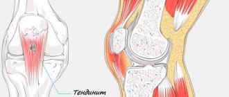
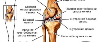
Meniscitis of the knee joint is an inflammatory disease of the menisci, which most often has nonspecific symptoms and a blurred clinical picture. During examination, doctors do not detect cystic or degenerative changes in the menisci in the patient. This is what distinguishes meniscitis from meniscopathies and cysts.
It is noteworthy that the disease most often occurs in athletes and people involved in active sports. It is detected in basketball players, football players, skiers, cyclists, athletes, figure skaters, dancers, and rugby players.
Meniscopathy develops against the background of prolonged trauma to the knee joint. Some experts call the pathology chronic traumatic meniscitis.
Development mechanism
Stability, strength, shock absorption of the knee, as well as a fairly high range of motion in it, are provided by cartilage located in the gap between the femoral condyles and the recesses of the tibia.
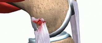
They are paired. There are two menisci – the outer (lateral) and the inner (medial). These cartilages are characterized by a crescent-shaped shape. The wide part is called the body. Narrow areas face anteriorly and posteriorly. These are called the anterior and posterior horns. The internal cartilage has a more rigid fixation by ligaments, so it is more likely to undergo changes. Violation of integrity is realized due to several basic mechanisms, which include a pronounced bruise, a fall on a bent limb (the cartilage is especially often injured when falling on a hard surface with a sharp edge - a step, a curb), as well as tucking (rotation of the lower leg with a fixed thigh or vice versa) . The damage is primarily localized in the area of the posterior horn, since it has the least strength.
Traditional methods of treatment
If you have meniscitis of the right or left knee joint and are overweight, you will need to lose weight. Due to excess body weight, additional stress is placed on the joints, both on the knees and on other parts of the body.
After consulting a doctor, you can use folk remedies:
- One of the effective methods is a tincture of elecampane roots, from which a compress is made at night. To prepare the tincture you will need 50 grams of elecampane and 125 grams of vodka. The ingredients are mixed and infused in a dark place for 14 days. In addition to compresses, the tincture can be used for rubbing.
- You can also apply a warming lotion at night. To do this, you will need 5-6 tablespoons of oatmeal, which is brewed in boiling water and cooled slightly. Then the brewed flakes are spread on a cotton cloth and applied to the sore spot. The compress can be secured on top with polyethylene.
- For compresses, you can use medical bile. It is melted and applied to the area of the affected joint for two hours. The procedure should be performed for 10 days without interruption. Then take a 5-day break and repeat everything again.
- You can make a compress from honey and alcohol. The procedure should be carried out over 2 months.
- It is recommended to take baths with medicinal herbs. A decoction of straw is suitable for this. For 6 liters of water you will need 250 grams of straw, which is boiled for 40 minutes. Afterwards the broth is filtered and added to the bath.
- Primrose herb is suitable for bathing. For 4 liters of water you will need 300 grams of the plant, which is boiled for 20 minutes. Then the broth is filtered and added to the bath.
But it should be remembered that folk remedies will only help if the knee injuries were not serious.
Causes
Changes in the structure of the internal cartilage of the knee is a polyetiological process that can develop due to the influence of several provoking factors:
- Trauma leading to the implementation of several pathogenetic mechanisms of changes developing in cartilaginous structures. It usually occurs in fairly young people, as well as in athletes - both professionals and amateurs.
- An acquired decrease in the strength of cartilage, which is observed in older people against the background of the development of degenerative changes resulting from impaired nutrition (trophism) of tissues.
- A hereditary change in the properties of cartilage, determined by a violation of the functional state of genes, while changes in integrity can occur in children against the background of low loads for their age (walking, running, squats).
- Inflammation that develops over a relatively long period of time (several years) and partially affects cartilage. It usually occurs against the background of a chronic infection or autoimmune pathology, in which antibodies are produced to body tissues.
Determining the cause of the pathological condition is necessary for adequate etiotropic therapy aimed at eliminating its effects, as well as to prevent similar injuries in the future.
Causes of meniscitis
Meniscitis of the knee joint - what is it? Let's figure it out. The cartilage tissue of the menisci is very strong. However, under excessive and prolonged loads, even it can become deformed, cracks and tears appear in it. The reason for this may be:
- Knee injuries;
- Sprains;
- Flat feet;
- Congenital pathologies of the limbs leading to their deformation;
- Injury to the menisci;
- Working on mechanisms that perform vibrating, oscillatory movements;
- Prolonged squatting.
Structure of the knee joint
Long-term exposure to the above factors contributes to the development of chronic inflammation of the cartilage plates inside the joint cavity. This is meniscitis. It can also develop after acute traumatic injury. Athletes are more often susceptible to inflammatory changes in the knee joint capsule.
If the cause of the destruction of the cartilage plates in the knee area is not treated and eliminated, cartilage dystrophy and degeneration develops.
As the process progresses, cysts appear, the elasticity of the cartilage tissue is lost, and calcification is observed.
Kinds
For the convenience of reliable detection of changes, as well as the use of the most appropriate appropriate treatment, all changes in the internal cartilage are classified according to several characteristics. According to the severity of violations, the following are distinguished:
- Incomplete (partial) rupture, characterized by the fact that the damage is usually localized (in the form of a focus), it does not affect the entire cartilage and does not lead to changes in its anatomical structure.
- A complete rupture is a severe change that affects the entire cartilage and is accompanied by the formation of a fragment.
Based on their location, the rupture of the posterior (develops most often), anterior horn and body is distinguished. Based on the main development mechanism and causative factor, the classification includes pathological processes and traumatic injuries.
Symptoms of meniscitis
Signs of inflammation of the cartilage plates are very similar to other knee pathologies. However, from some symptoms it can be understood that the inflammation is localized precisely inside the joint cavity. The main symptom of this pathology is pain.
Its features are as follows:
- The pain is periodic;
- Increases during prolonged or intense physical activity;
- Localized on the side of the knee;
- The pain can be bilateral.
The pain syndrome limits movement in the knee. There is a formation in the form of a roller, painful to the touch. An acute process can result in swelling of the knee. More often, the listed symptoms are localized on the inside of the knee due to the fact that the medial meniscus is exposed to pathological processes more often.
Clinic
Symptoms of changes that develop as a result of trauma appear acutely, their intensity is proportional to the degree of impairment. Among them:
- Pain that develops mainly on the inside of the knee and intensifies when trying to carry out active or passive movements in it.
- Restriction of mobility, which can reach a complete block when part of the cartilage is torn off and wedged. Any attempts at movement usually lead to a sharp increase in discomfort.
- Inflammatory manifestations in the form of redness, swelling of soft tissues. They lead to a visually noticeable increase in the knee. There is a local increase in temperature (the knee joint becomes hot to the touch).
In pathological disorders leading to degeneration of internal cartilage, symptoms develop gradually over a fairly long period of time. The pain may be absent at first, then it appears and intensifies with loads on the leg. Movements may be accompanied by characteristic “clicks” and “crunches,” which indicates the development of degenerative processes in cartilage tissue.
Symptoms and diagnostic methods
The disease is characterized by painful sensations that intensify with movement. Since the medial meniscus is often involved in the inflammatory process, pain usually appears on the inner surface of the knee. Note that the symptoms of this pathology are easily confused with the signs of many other diseases of the knee joint. Therefore, additional research methods are needed to make a correct diagnosis.
The diagnosis can only be confirmed using ultrasound or MRI. All other methods for this pathology have no diagnostic value.
Diagnostics
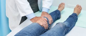
Before starting adequate treatment, the doctor must prescribe reliable instrumental diagnostics with visualization of the knee structures, including the menisci. In modern medical clinics, it is performed using X-rays, ultrasound, magnetic resonance (MRI) and computed tomography (CT) scans, as well as arthroscopy.
MRI has a high diagnostic value and is used to classify disorders according to Stoller (the classification includes four degrees of severity, determined by certain criteria).
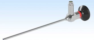
Arthroscopy is an invasive diagnostic method that involves direct examination of structures on a monitor screen by introducing an arthroscope equipped with a video camera with lighting into the joint cavity.
Causes of meniscus damage
There are 2 types of meniscal injuries:
- Traumatic - occurs after bruises, blows, sudden rotation of the lower leg (more often occurs between the ages of 20 and 40 years).
- Degenerative damage to the meniscus of the knee joint - appears when small loads are placed on the cartilage, which is weakened by constant microtraumas or diseases (they are more often diagnosed in people over 40 years of age).
| Causes of traumatic injuries | Causes of Degenerative Damage |
| External rotation of the tibia (resulting in damage to the medial meniscus) | Metabolic disorders (gout) |
| Internal rotation of the lower leg (resulting in injury to the lateral meniscus) | Rheumatism (damage to the connective tissue of the body involving the joints) |
| Kick to the knee | Chronic intoxication (long-term use of drugs) |
| Sharp flexion-extension | Regular loads |
| Sharp extension of the joint from a bent position (for example, cartilage can be damaged by squatting with a barbell) | Microtraumas |
| Other types of injuries | Repeated injuries |
Principles of treatment
The main goal of therapeutic measures is to correct disorders of the cartilage of the knee, as well as its functional state. They are complex and aimed at all parts of the pathogenetic mechanism, and may include several areas:
- Conservative (non-surgical) measures.
- Operation.
- Recovery.
The scope of treatment is determined by the treating specialist based on the results of a clinical subjective examination and additional instrumental diagnostics.
Treatment without surgery
If, based on a comprehensive diagnosis, a mild injury to the internal meniscus of the knee joint has been identified, treatment may only include conservative measures without surgery. These include:
- Providing rest, which is achieved through the use of tight bandages with an elastic bandage or the application of a plaster splint.
- Prescription of medications of various groups - anti-inflammatory (glucocorticosteroids or non-steroidal drugs) medications to reduce the intensity of inflammation, chondroprotectors that promote the restoration of cartilage tissue, as well as preventing its further destruction, vitamins to improve the course of metabolic processes.
- Physiotherapeutic procedures, which are of particular importance in the gradual chronic development of disorders. They include magnetic therapy, electrophoresis with medications, ozokerite.
A set of conservative treatment measures allows one to achieve a good therapeutic effect and avoid surgery, but only in the case of incomplete rupture of the internal cartilage.
Operation
If there is a complete tear that involves the entire medial meniscus of the knee, treatment usually involves surgery. Its main purpose is plastic surgery of altered structures or removal of part of the cartilage if it cannot be restored. Modern surgery is performed using 2 main methods:
- Open access intervention with wide tissue incisions is rarely used today, as it leads to significant trauma. It is usually prescribed for significant combined changes that require the removal of a large volume of tissue, as well as in connection with the need for implantation.
- Arthroscopic technique is a minimally invasive operation. Today it is increasingly used in well-equipped medical clinics. During its implementation, an arthroscope, special small-sized instruments and manipulators are inserted into the joint cavity, which requires small incisions. Under visual control on the monitor, the doctor performs the necessary plastic manipulations.
Thanks to the introduction of arthroscopic surgery into clinical practice, it was possible to achieve a significant reduction in the morbidity of surgical treatment and reduce the length of time required for:
- tissue regeneration and restoration of mobility;
- the entire course of treatment and a person’s stay in a medical institution.
Postoperative recovery
After surgical procedures, regardless of the method of their implementation, the patient remains in the hospital of a medical institution for a certain period of time. A set of measures is prescribed aimed at speedy tissue regeneration of the postoperative wound area and prevention of complications:
- Limitation of leg mobility.
- Periodic treatment of surgical suture materials with special antiseptics, which are usually used in the form of a solution.
- Prescribing antibacterial agents to prevent infection.
- The use of hemostatic medications to prevent bleeding, especially after open surgery.
The average postoperative period is determined by the method of performing the operation. It varies from 3-5 to 10 days.
Possible complications
If the injury is not treated, then there is a huge risk, for example, against the background of meniscitis of the left knee joint, the appearance of a cyst. Most often, such a growth appears on the back wall of the knee; fluid collects inside the cyst itself. Although the growth may appear on the edge or inside the meniscus. Under any circumstances, a cyst increases the likelihood of rupture several times. People with rheumatism and osteoporosis, as well as those who have a physiological predisposition to this pathology, are at risk for the appearance of cysts. Subsequently, after the development of the cyst, arthrosis appears, which inevitably leads to deformation of the joints as a whole.
Rehabilitation
Carrying out rehabilitation measures is no less important than the main course of treatment. It involves resting the knee immediately after surgery or conservative therapy, followed by special activities that involve certain movements.
As the tissues recover, the range of motion and load gradually increase. This makes it possible for the cartilage tissue to adapt to loads and subsequently restore the functional state of the knee.
Knee meniscitis: causes, symptoms, treatment
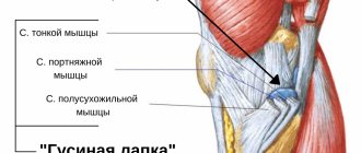
The knee joint is anatomically complex due to the static and dynamic loads placed on it. The joint, thanks to its features, provides the functions of support and walking. This is possible, among other things, thanks to the menisci. Menisci look like plates, which include cartilage and connective tissue. They are located between the tibia and femur. Frequent injuries lead to inflammatory changes in the cartilage, which causes the development of meniscitis of the knee joint.
The menisci absorb movement of the lower extremities and prevent friction of the articular surfaces. The plates help reduce the impact of static and dynamic loads on cartilage tissue during movements.
Treatment at home
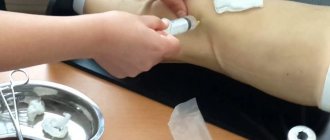
Carrying out therapeutic measures at home is only possible if non-surgical treatment is prescribed. If there is a need for parenteral (intramuscular, subcutaneous or intravenous) administration of drugs, the patient receives treatment on an outpatient basis. To do this, he visits the manipulation room of a medical institution, where he undergoes the necessary manipulations.
An attempt to independently treat damage to the internal cartilage without undergoing the necessary set of diagnostic tests can cause the development of various complications, including pinching of part of the damaged meniscus with its wedging into the joint space. Such complications lead to the need for subsequent surgery.
Symptoms of a kneecap meniscus injury
You can understand that a meniscus tear has occurred by your own feelings. A sign of rupture is a characteristic cracking sound. In the first minutes, pain is not felt; the person may not even understand that he has been injured. There is no pain when moving, even with full support on the injured leg. And if the load does not weaken, for example, the athlete continues training, then the danger that a fragment will fall into the joint space increases.
Symptoms increase gradually and appear approximately 2 days after injury. In the initial phase, the patient has a significant limitation in the extension of the diseased limb. A characteristic sign of injury is swelling, which is then accompanied by other signs of damage:
- restriction of freedom of movement of the joint, stiffness, inability to straighten the leg -
- sudden complete blockade of knee mobility -
- instability of the knee joint -
- increased swelling -
- increased sensitivity in the area of injury -
- pain.
The location of the injury can be determined by the nature of the pain. When the inner meniscus ruptures, shooting pain occurs on the inside of the knee. In addition, point discomfort occurs above the place of its attachment. Pain also occurs when turning a bent knee, or with excessive force when bending the leg. There is a weakening of the thigh muscles on the front surface. With partial or complete destruction of the outer meniscus, severe pain is felt in the outer part of the cup, when the knee turns inward. In addition, there are special symptoms specific to such injuries.
- When lifting the leg straight at the knee, atrophy of the quadriceps femoris muscle on the inside and strong tension of the sartorius muscle (otherwise known as the sartorius symptom) become clearly visible.
- Pressing on a leg bent at a right angle while passively extending it causes increased pain - this is how Baikov’s symptom manifests itself.
- Discomfort and increased pain are observed during the usual leisurely descent from the stairs. This phenomenon is called the “staircase symptom” (or otherwise, Pelman’s symptom).
- Even with normal walking at a calm pace, a “clicking” symptom can be observed, and an attempt to sit with crossed legs causes discomfort and increased pain.
- Rauber's - detected on x-rays 2-3 months after injury and consists of the growth of awl-like formations on the condyles of the knee joint.
Changes also occur inside the joint. Synovial fluid accumulates in the joint cavity, and the articular cartilage gradually breaks down, exposing the surface of the bones in the joint.
Forecast
The prognosis with correct and timely treatment of changes in the medial meniscus is generally favorable. After non-surgical treatment or surgery with mandatory subsequent implementation of restorative and general recommendations, the functions of the knee are usually completely restored. An exception is the need to remove part or all of the cartilage, followed by implantation.
The use of modern therapy methods in specialized medical clinics, including arthroscopic surgery, has made it possible to significantly shorten the postoperative and rehabilitation period, and make the therapy itself as comfortable as possible for the patient.
Chronic form of pathology
If for some reason there is no treatment, then meniscitis can take a chronic form. Most often, in such cases, cartilage tissue is deposited in the knee. The chronic form may occur due to repeated injuries, constant awkward positioning, working on certain machines, or sprains. Women may develop what is called valgus knee position. In such cases, the pain occurs periodically and can worsen after a long walk or when the knee bends strongly. Some patients experience a ridge or thickening in the joint space area.
