Connections of the skull bones
The bones of the skull are connected to each other mainly through continuous connections : syndesmoses (ligaments, fontanelles, sutures, impactions), and to a lesser extent - synchondroses. Cartilaginous joints – synchondroses – are characteristic of the bones of the base of the skull. They are represented by fibrous cartilage. In children, temporary synchondrosis is clearly expressed - sphenoid-occipital, synchondrosis sphenooccipitalis. There are also permanent synchondrosis: stony-occipital synchondrosis, synchondrosis petrooccipitalis, sphenoid-petrosal synchondrosis, synchondrosis sphenopetrosa.
Only the temporomandibular joint
In newborns, syndesmoses are represented by connective tissue membranes called fontanelles; fonticuli are non-ossified areas of the membranous skull (desmocranium), which are located in places where future sutures are formed (Fig. 4.19 A, B).
In areas where several bones meet, there are 6 fontanelles. The largest of them is the anterior (frontal) fontanel, fonticulus anterior (frontalis), located between the two parts of the frontal bone and the parietal bones. It has a diamond shape and becomes overgrown in the 2nd year of life. The posterior (occipital) fontanel, fonticulus posterior (occipitalis), is located between the two parietal bones and the occipital bone, has a triangular shape, and closes in the 2nd month of life. The anterior and posterior fontanelles are unpaired. In addition to them, there are paired fontanelles.
Wedge-shaped, fonticulus sphenoidalis, paired, located in the anterior section of the lateral surfaces of the skull, between the frontal, parietal, sphenoid and temporal bones. They ossify almost immediately after birth. The mastoid fontanel, fonticulus mastoideus, paired, is located posterior to the sphenoid, at the junction of the occipital, parietal and temporal bones. Overgrows at 2-3 months of life. Due to the presence of fontanelles, the newborn's skull is very elastic; its shape can change as the fetal head passes through the birth canal during childbirth.
In an adult, the syndesmoses of the skull are represented by sutures, suturae, - connections of the edges of the bones of the brain and facial skull with layers of connective tissue. There are jagged and scaly sutures between the roof bones. A serrated suture, sutura serrata, is present between the parietal bones ( sagittal suture, sutura sagittalis); between the parietal and frontal ( coronal suture, sutura coronalis); between the parietal and occipital ( lambdoid suture, sutura lambdoidea). With the help of a scaly suture, sutura squamosa, the scales of the temporal bone are connected to the parietal bone and the greater wing of the sphenoid bone. The bones of the facial skull are connected through flat (harmonic) sutures, sutura plana. The specific names of the sutures are made up of the names of the connecting bones, for example, sutura frontozygomatica, sutura frontoethmoidalis, etc.
Synchondroses are cartilaginous joints characteristic of the bones of the base of the skull. They are represented by fibrous cartilage.
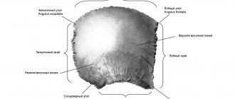
Rice. 4.19. Newborn skull.
A – side view: 1 – fonticulus anterior; 3 – fonticulus sphenoidalis; 2 – fonticulus posterior; 4 – fonticulus masoideus; B – top view: 1 – sutura frontalis; 2 – tuber frontalis; 3 – fonticulus posterior; 4 – os occipitale; 5 – sutura lambdoidea; 6 – tuber temporale; 7 – sutura sagittalis; 8 – os temporale; 9 – sutura coronalis; 10 – os frontale; 11 – fonticulus anterior.
1. Temporary synchondrosis between the body of the sphenoid bone and the main part of the occipital bone - sphenoid-occipital synchondrosis, synchondrosis sphenooccipitalis (clearly expressed in children).
2. Permanent synchondrosis:
– between the pyramid of the temporal bone and the main part of the occipital bone – stony- occipital synchondrosis, synchondrosis petrooccipitalis;
– between the greater wing of the sphenoid bone and the pyramid of the temporal bone, sphenoid-petrosal synchondrosis, synchondrosis sphenopetrosa;
– cartilage covering the torn hole, foramen lacerum.
As a person ages, cartilage is replaced by bone. An example of synostosis in an adult is the connection between the bodies of the occipital and sphenoid bones, between the sacral vertebrae, and the halves of the lower jaw.
Synovial joints of the skull, artt. synoviales cranii – temporomandibular joint, right and left, artt. temporomandibulares dexter et sinister (Fig. 4.20, 4.21).
1. Temporomandibular joint, right and left, artt. temporomandibulares dexter et sinister
2. Bones forming the joint: temporal bone, os temporale; lower jaw, mandibula; articular surfaces: mandibular fossa, fossa mandibularis and articular tubercle of the temporal bone tuberculum artuculare ossis temporalis; head of the lower jaw caput mandibulae.
3. The joint capsule is attached:
– on the temporal bone: in front – along the tuberculum articulare; behind – along the fissura petrotympanica;
– on the lower jaw: along the collum mandibulae.
It is thin in front and dense in the back, so jaw dislocations are possible only anteriorly.
4. In appearance – simple, art. simplex, complex, art. composita, combined, art. combinatoria, with the joint of the same name on the opposite side.
5. According to the shape of the articular surfaces – condylar, art. bicondylaris.
6. By the number of axes of rotation - biaxial.
7. Movements:
– around the frontal axis: raising and lowering the lower jaw;
– movement of the frontal axis: movement of the jaw back and forth;
– around the vertical axis: rotation, rotatio (movement of the jaw to the right and left); when rotating, the jaw moves to the side: on its side, rotation occurs in the fossa; on the opposite side there is a displacement of the jaw onto the tubercle.
8. Fixing apparatus: lateral ligament, lig. laterale; awl-mandibular ligament, lig. stylomandibulare; sphenomandibular ligament, lig. sphenomandibulare.
9. An auxiliary element of the joint is the articular disc, discus articularis. The suspensory apparatus of the lower jaw - the wedge- and stylomandibular ligaments (ligg. sphenomandibulare et stylomandibulare), are extracapsular ligaments.
10. Blood supply, venous and lymphatic drainage, innervation:
| Blood supply | Venous and lymphatic drainage | Innervation |
| – a. auricularis profunda from a. maxillaris. | 1. Rete articulare mandibulare plexus venosus pterygoideus ' v. jugularis interna. 2. The outflow of lymph is carried out in the nodi lymphatici parotidei et cervicales profundi. | – n. auriculotemporalis (branch of n. mandibularis from n. trigeminus) |
Morpho-functional features of the joint:
– the presence of an articular disc, discus articularis, – an auxiliary element of the joint;
– the joint is incongruent (articular surfaces do not correspond to each other);
– the joint cavity is divided into upper and lower isolated floors;
– discrepancy between the shape of the joint (condylar) and the range of movements (three axes of movement: around the frontal axis - lowering and raising the lower jaw; moving the frontal axis forward - moving the lower jaw forward and returning back; around the vertical axis - rotation);
– presence of extra-articular ligaments (spheno- and stylomandibular)
| Rice. 4.20. Temporomandibular joint, art. temporomandibularis (sagittal section). 1 – processus articularis (condylaris) mandibulae; 2 – caput mandibulae; 3 – capsula articularis; 4 – porus acusticus externus; 5 – discus articularis; 6 – fossa mandibularis; 7 – tuberculum articulare; 8 – m. pterygoideus lateralis; 9 – processus temporalis ossis zygomatici; 10 – processus coronoideus. | Rice. 4.21 Ligaments of the temporomandibular joint (view from the medial side). 1 – ligamentum laterale (articulatio temporomandibularis); 2 – capsula articulationis temporomandibularis; 3 – ligamentum sphenomandibulare; 4 – ligamentum stylomandibulare; 5 – foramen mandibulae; 6 – arcus zygomaticus; 7 – sinus sphenoidalis; 8 – fossa hypophysialis. |
Connection system
CONNECTIONS OF THE SKULL BONES
The bones of the skull are connected to each other mainly by continuous connections - sutures and synchondroses.
Only the lower jaw forms a paired temporomandibular joint with the temporal bone. The temporomandibular joint (articulatio temporomandibularis) is formed by the mandibular fossa of the temporal bone and the head of the condylar process of the mandible (Fig. 122, 123, Table 23). In front of the fossa is the articular tubercle. Between the articular surfaces there is a biconcave articular disc (discus articularis) of oval shape, formed by fibrous cartilage, which divides the joint cavity into two sections: upper and lower. In the upper floor, the articular surface of the temporal bone articulates with the upper surface of the articular disc. The synovial membrane of this floor (membrana synovialis superior) covers the inner surface of the capsule and is attached to the edges of the articular cartilage. In the lower floor the head of the mandible and the lower surface of the articular disc articulate. The synovial membrane of the lower floor (membrana synovialis inferior) covers not only the capsule, but also the posterior surface of the neck of the condylar process, located inside the capsule.
The free articular capsule on the temporal bone is attached anteriorly to the articular tubercle, and posteriorly at the level of the petrotympanic fissure. On the condylar process, the articular capsule is attached anteriorly along the edge of the head, and posteriorly 0.5 cm below the head of the mandible. The articular capsule is fused along the entire circumference with the articular disc. The capsule is thin at the front, but at the back it thickens and is strengthened by several ligaments (Fig. 124). The fan-shaped lateral ligament (ligamentum laterale), which strengthens the joint capsule on the lateral side, starts from the base of the zygomatic process of the temporal bone. The fibers of this ligament run posteriorly and inferiorly and are attached to the posterolateral surface of the neck of the condylar process. The sphenomandibular ligament (ligamentum sphenomandibulare), located on the medial side of the joint, begins on the spine of the sphenoid bone and is attached to the lingula of the lower jaw. The stylomandibular ligament (ligamentum stylomandibulare) starts from the styloid process of the temporal bone and is attached to the inner surface of the posterior edge of the ramus of the mandible near its angle. The ligament is located medial and posterior to the temporomandibular joint. Both of these extra-articular ligaments are separated from the joint capsule by fatty tissue.
Rice. 122. Temporomandibular joint, right. View from the lateral side, the zygomatic bulb has been removed, the articular capsule is opened:
1 - Stylomandibular ligament; 2 — Hcad of mandible; 3 - Aiticular tuberc; 4 — Joint capsule; Articular cap- sule; 5 — Articular disc; 6 - Temporal bone, squamous part; 7 - Lateral pterygoid, upper hcad; superior liead; 8 - Lateral pterygoid, lowerhead; inferior head; 9 - Medial pterygoid; 10—Buccinator
Rice. 123. Temporomandibular joint, left, capsule is opened, all surrounding tissues are removed, side view:
1 - Articular tubercle; 2 — Joint capsule; Articular capsule; 3 — Articular disc; 4—Head of mandible; 5—Stylomandibular ligament
Temporomandibular joint elliptical complex biaxial combined. The right and left joints function together, making movements around the vertical and frontal axes. Around the frontal axis, the lower jaw rises and falls; around the vertical axis, the lower jaw makes lateral movements to the right and left. Due to the extensive articular surface on the temporal bone, the articular processes, along with the entire lower jaw, move forward and backward. The articular processes of the lower jaw, when the lower jaw moves forward, are displaced onto the articular tubercles, and when the jaw moves backward, they return to their original position - into the articular fossae.
When lowering the lower jaw, the chin protuberance moves downward and somewhat posteriorly, describing an arc with its concavity facing posteriorly and upward. This movement can be divided into three phases. In the first (slight lowering of the lower jaw), movement around the frontal axis occurs in the lower floor of the joint, the articular disc remains in the articular fossa. In the second (significant lowering of the lower jaw), against the background of the ongoing hinge movement of the articular heads in the lower floor of the joint, the cartilaginous disc, together with the head of the articular process, slides forward and emerges onto the articular tubercle. The condylar processes of the mandible move forward approximately 12 mm. In the third (maximum lowering of the jaw), movement occurs only in the lower floor of the joint around the frontal axis, the articular disc at this time is on the articular tubercle.
With further strong opening of the mouth, the head of the lower jaw may slip from the articular tubercle anteriorly, into the infratemporal fossa, and dislocation in the temporomandibular joint. The mechanism for raising the lower jaw repeats the stages of its lowering in reverse order. If the lower jaw moves forward, the movement occurs only in the upper floor of the joint. The articular processes, together with the articular discs, slide forward and extend onto the tubercles in both the right and left temporomandibular joint.
With a lateral displacement of the lower jaw, the movements in the right and left temporomandibular joints are not the same. So, when the lower jaw moves to the right in the left temporomandibular joint, the articular head together with the disc slides forward and comes out onto the articular tubercle, i.e. sliding occurs in the upper floor of the joint. At this time, in the right joint, the articular head rotates around a vertical axis passing through the neck of the condylar process. When the lower jaw moves to the left, the head, together with the articular disc, slides forward in the right joint, and rotation around the vertical axis occurs in the left.
Rice. 124. Extra-articular ligaments of the temporomandibular joint, right, internal view:
1 - Pterygoid process, laleral piate; 2 —- Pterygospinotis ligament; 3 - Mandibular notch; 4 - Stylomandibular ligament; 5 - Sphenomandibular ligament; 6 - Pterygoid process, medial piate
Table 23. Temporomandibular joint
| Articular surfaces | Joint capsule | Articular ligaments | Type of joint, axis of motion | Function and muscles acting on the joint |
| Mandibular fossa of the temporal bone, head of the condylar process of the mandible. There is an articular disc between the articular surfaces | Thin, fused along the entire circumference with the articular disc | The lateral ligament connects the base of the zygomatic process of the temporal bone with the posterolateral surface of the neck of the condylar process. The sphenomandibular ligament connects the spine of the sphenoid bone to the lingula of the mandible. The stylomandibular ligament connects the styloid process of the temporal bone with the inner surface of the posterior edge of the ramus of the mandible near its angle | Ellipsoidal complex biaxial combined. Around the frontal axis - raising and lowering the lower jaw, around the vertical axis - lateral movements of the jaw to the right and left | Raising the lower jaw, muscles: temporal; raising the angle of the lower jaw: chewing, medial pterygoid. Promotion of the lower jaw forward - both lateral pterygoid muscles, with unilateral contraction - displacement of the lower jaw to the opposite side |
The lowering of the lower jaw is carried out by contraction of the paired digastric, mylohyoid and sublingual muscles. Lifting the jaw is also performed by the paired temporalis, masseter and medial pterygoid muscles. The lower jaw is pushed forward by the lateral pterygoid muscles and the anterior bundles of the masticatory muscles, and the lower (posterior) bundles of the temporal muscles return to their original position. Lateral movements of the lower jaw to the right and left are performed by the lateral pterygoid muscle of the opposite side with unilateral contraction.
The capsule of the temporomandibular joint is supplied with blood by the branches of the maxillary artery, venous blood flows into the venous network surrounding the joint, and then into the mandibular vein. Lymph flows into the deep parotid and then into the deep cervical lymph nodes. Innervation is provided by the auriculotemporal nerve (a branch of the mandibular nerve).
Continuous connections of the skull bones are represented by fibrous connections - sutures in an adult and interosseous membranes (syndesmoses) in newborns. There are synchondroses in the area of the base of the skull.
The bones of the roof of the skull are connected to each other through serrated and scaly sutures. The medial edges of the parietal bones are connected to each other by a serrated sagittal suture (sutura sagittalis). The anterior edges of the parietal bones are connected to the posterior edge of the frontal bone by a serrated coronal suture (sutura coronalis). The posterior edges of the parietal bones form a serrated lambdoid suture (sutura lambdoidea) with the anterior edge of the occipital bone. The scales of the temporal bone connect with the parietal bone and the greater wing of the sphenoid bone, forming a scaly suture (sutura squamosa). In the area of the facial part of the skull, the sutures are even, flat, or harmonious (Table 24).
The skull also has non-permanent (temporary) sutures formed as a result of late fusion or non-fusion of individual ossification points. For example, sometimes the upper part of the occipital squama is separated by a transverse suture entirely or partially from the rest of the occipital bone. Sometimes both halves of the frontal bone do not fuse. In these cases, the sagittal suture (the so-called metopic) starts from the glabella or slightly higher. In the presence of the intermaxillary, or incisive, bone, an incisive suture is formed. When the parietal bone is doubled, there is an interparietal suture. In addition, there are non-permanent sutures: squamomastoid, sphenoid-maxillary (between the pterygoid process of the sphenoid bone and the body of the upper jaw). In the second half of a person’s life, most sutures completely or partially heal. In this case, the connective tissue plate between the bones of the skull is replaced by bone tissue.
Cartilaginous joints, or synchondroses, of the skull (Table 25) are located in the area of its base; they are formed by fibrous cartilage. Typically, as a person ages, cartilage tissue is replaced by bone tissue. At the site of sphenoid-occipital synchondrosis, synostosis forms by the age of 20.
Table 24. Flat seams of the skull
| Name of seams | Bone connections (structures) |
| Occipitomastoid (sut. occipitomastoidea) | The occipital edge of the mastoid process of the temporal bone - with the mastoid edge of the occipital squama of the occipital bone |
| Parietomastoid (sut. parietomastoidea) | Mastoid angle of the parietal bone - with the parietal notch of the squamosal part and the adjacent portion of the mastoid process of the temporal bone |
| Sphenoparietal (sut. sphenoparietalis) | Wedge-shaped angle of the parietal bone - with the parietal edge of the sphenoid bone |
| Sphenoid-frontal (sut. sphenofrontalis) | Frontal edge of the greater and lesser wings of the sphenoid bone - with the orbital part of the frontal bone |
| Sphenoethmoidalis (sut. sphenoethmoidalis) | Sphenoid crest of the sphenoid bone - with the posterior edge of the perpendicular plate of the ethmoid bone |
| Wedge-squamous (sut. sphenosquamosa) | Squamous edge of the greater wing of the sphenoid bone - with the wedge-shaped edge of the scaly part of the temporal bone |
| Wedge-vomeral (sut. sphenovomeralis) | The lower surface of the body and the beak of the sphenoid bone - with the upper surface of the wings of the vomer |
| Sphenozygomatic (sut. sphenozygomatica) | Zygomatic edge of the greater wing of the sphenoid bone - with the frontal process of the zygomatic bone |
| Frontonasal (sut. frontonasalis) | Nasal edge of the frontal bone - with the upper edge of the nasal bones |
| Frontoethmoidalis (sut. frontoethmoidalis) | Orbital and nasal parts of the frontal bone - with corresponding edges of the ethmoid bone |
| Frontomaxillary (sut. frontomaxillaris) | Nasal part of the frontal bone - with the frontal process of the maxilla |
| Frontolacrimalis (sut. frontolacrimalis) | Orbital part of the frontal bone - with the upper edge of the lacrimal bone |
| Frontozygomatic (sut. frontozygomatica) | Zygomatic process of the frontal bone - with the frontal process of the zygomatic bone |
| Zygomaticomaxillaris (sut. zygomaticomaxillaris) | Zygomatic bone - with the zygomatic process of the maxilla |
| Ethmoidomaxillary (sut. ethmoidomaxillaris) | The lower edge of the orbital plate of the ethmoid bone - with the orbital surface of the body of the upper jaw |
| Ethmoidolacrimal (sut. ethmoidolacrimalis) | Orbital plate of the ethmoid bone - with lacrimal bone |
| Temporozygomatic (sut. temporozygomatica) | Zygomatic process of the temporal bone - with the temporal process of the zygomatic bone |
| Internasal (sut. internasalis) | The medial edges of the nasal bones facing each other |
| Nasomaxillary (sut. nasomaxillaris) | Lateral edge of the nasal bone - with the frontal process of the maxilla |
| Lacrimal-maxillary (sut. lacrimomaxillaris) | The lower edge of the lacrimal bone (posteriorly) - with the orbital surface of the upper jaw |
| Lacrimal-conchalis (sut. lacrimoconchalis) | The lower edge of the lacrimal bone (in front) - with the lacrimal process of the inferior turbinate |
| Intermaxillary (sut. intermaxillaris) | Alveolar processes of both upper jaws |
Table 25. Synchondose poses of the cheppe
| Name of synchondrosis | Connectable bones |
| Sphenooccipital (synchondrosis sphenooccipitalis) | Posterior surface of the body of the sphenoid bone - with the basilar part of the occipital bone |
| Wedge-shaped stony (synchondrosis sphenopetrosa) | Body of the main bone - with the angular spine of the petrous part of the temporal bone |
| Stony-occipital (synchondrosis petrooccipitalis) |