10.10.2014
Maltseva Marina Arnoldovna
Head of the consultation department - neurologist, specialist in the field of extrapyramidal pathologies, doctor of the highest category
A fracture of the base of the skull is one of the most severe traumatic brain injuries. This condition is accompanied by damage to one or even several bones that form the base of the skull. Damaged in this case may be:
- occipital bone;
- temporal bone;
- ethmoid bone;
- sphenoid bone.
A fracture of the base of the skull can occur from a variety of serious physical injuries: car accidents;
- when a person falls from a great height;
- when hitting the face in the area of the lower jaw or base of the nose.
In most patients, the base fracture occurs from the arch. Statistics say that the number of such patients reaches 59%.
What is a basal skull fracture?
A basal skull fracture is a very severe traumatic brain injury (TBI) in which one or more of the bones that make up the base of the skull: the occipital, sphenoid, temporal or ethmoid, breaks. The cause of such damage is usually a significant impact: it could be an accident, a fall backward from a height, a direct blow from something heavy to the head or face in the lower jaw area.
Fracture of the bones of the base of the skull accounts for 4% of the number of diagnosed traumatic brain injuries. A combination of fractures of the base and vault of the skull is also possible, which occurs in 50-60% of patients with this injury.
Survival rate for skull fracture
Of decisive importance is how quickly and competently first aid is provided for a fracture and hospitalization with subsequent medical measures is carried out. TBI is often accompanied by heavy bleeding, which can lead to death in the first hours after injury or cause a prolonged coma, the prognosis of which is extremely unfavorable. In this case, there is a high probability of lifelong disability, when basic life functions are impaired and intelligence is seriously affected.
For fractures without displacement, single cracks that do not require surgical intervention, the prognosis is relatively favorable.
The mortality rate for fractures of the bones of the base of the skull ranges from 24 to 52%, depending on the severity and complexity of the injury and its subsequent complications.
Complications
Fractures of the base of the skull are accompanied by brain contusion of varying severity; they can cause massive subarachnoid hemorrhage, the formation of a brain hematoma, damage or compression of the brain stem with respiratory and cardiac disorders. When an infection occurs, meningitis develops. In the long-term period, the formation of a liquor fistula is possible - a fistula that is the cause of permanent rhinoliquorhea or otoliquorrhea. Dangerous late complications are brain abscess and meningitis, which can occur several months or years after injury with damage to the air sinus. In case of severe displaced fractures, cosmetic deformations are observed as a result; in case of damage to the nerve trunks, paresis of the corresponding nerves (usually the facial one) is observed.
Signs and symptoms of a skull fracture
Symptoms depend on the severity, location of the fracture, and the extent of damage to brain structures. Loss of consciousness can take any form, from short-term fainting at the time of injury to long-term coma. The more severe the damage, the more severe the damage, but with intracranial hematoma there may be a period of lucidity preceding loss of consciousness, which should not be mistaken for the absence or mildness of injury.
Common signs of a fracture include:
- Expanding headaches due to developing cerebral edema;
- Vomiting, aspiration of vomit, or spontaneous leakage of stomach contents into the lungs;
- Symmetrical bilateral hemorrhages around the eyes in the form of “spectacles”;
- Different diameters and lack of pupillary response;
- Respiratory and circulatory disorders in case of compression of the brain stem;
- Leakage of cerebrospinal fluid (cerebrospinal fluid), mixed with blood, from the nose and (or) ears;
- Cardiac disorders: arrhythmia, tachycardia, bradycardia, high or low blood pressure;
- Excitement or immobility;
- Confusion;
- Involuntary urination.
Fractures of the temporal bone pyramid can be longitudinal, transverse, or diagonal. With longitudinal fractures, the middle and inner ear and the facial nerve canal are affected. Symptoms: bleeding from the ear and leakage of cerebrospinal fluid due to a ruptured eardrum, hemorrhage in the area of the temporal muscle and behind the ear, partial hearing loss. Bleeding increases when you turn your head, so this is strictly prohibited.
A transverse fracture of the temporal bone is characterized by complete loss of hearing, disturbances in the functioning of the vestibular apparatus, paralysis of the facial nerve, and loss of taste.
- Clinical signs of anterior fossa fracture: nosebleeds, nasal liquorrhea (cerebrospinal fluid discharge through the nose), hemorrhages around the orbits and under the conjunctiva. Bruises appear 2-3 days after the injury, which fundamentally distinguishes them from ordinary bruises that appear as a result of direct blows to the face. Sometimes so-called subcutaneous emphysema occurs: when the cells of the ethmoid bone are damaged, air penetrates into the subcutaneous tissue, causing blisters to form on the skin.
- Clinical signs of a middle cranial fossa fracture: unilateral ear bleeding; a sharp decrease in hearing or complete deafness; release of cerebrospinal fluid due to a ruptured eardrum, dysfunction of the facial nerve, bruising in the area of the temporal muscle and behind the ear; partial loss of taste. Middle fossa fractures account for 70% of skull base injuries.
- Clinical signs of a fracture of the posterior cranial fossa: simultaneous damage to the auditory, facial and abducens nerves; bruising behind the ears on one or both sides; when the caudal nerves are pinched or ruptured, the tongue, palate, and larynx are paralyzed and the functioning of vital organs is disrupted.
- Fractures of the base of the skull are also characterized by damage to the olfactory or optic nerve. With such fractures, the meninges are ruptured, as a result of which a communication channel is formed between the brain matter and the external environment through the nasal and oral cavities, middle ear or orbit. Splinter fractures are especially dangerous in this regard: bone fragments can injure arteries and veins. Communication with the external environment leaves the brain open to infections and microbes and can lead to the development of encephalitis, meningitis or brain abscess.
Diagnostics
The diagnosis of a fracture of the base of the skull is made taking into account the results of an examination by a neurosurgeon and data from hardware studies. The examination program is determined by the severity of the victim’s condition, the need for emergency resuscitation measures and the technical equipment of the hospital. Differential diagnosis is carried out with other TBIs; sometimes (when injured in a state of severe intoxication), it is additionally required to distinguish between symptoms caused by traumatic injury and the toxic effects of drugs or alcohol on the brain. The following techniques are used:
- Questioning, general examination. The doctor finds out from the patient or accompanying persons the circumstances of the injury and the characteristics of the condition (in particular, the presence of a “light gap” followed by a sharp deterioration, indicating the formation of a hematoma) from the time of injury to the moment of admission to the hospital. The neurosurgeon assesses the degree of impairment of consciousness and general clinical indicators (pulse, respiration, blood pressure), detects symptoms indicating the presence and localization of a head injury (bleeding and liquorrhea, typical bruises), and identifies associated injuries that require advisory and therapeutic assistance from other specialists.
- Neurological examination. Due to disturbances of consciousness, the development of life-threatening conditions is often carried out using a simplified method, which involves studying all indicators taking into account topographic reference (face, arms, legs, torso). The doctor determines the severity of TBI using the Glasgow scale and pays attention to objective signs indicating the location of the damage, including brainstem disorders and disorders of the cranial nerves.
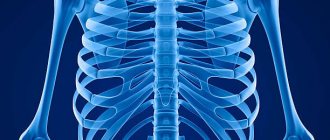
- Radiography. All patients undergo survey photographs of the skull in two projections; in case of damage to the occipital bone, a posterior semi-axial projection is additionally used; in case of possible damage to the temporal bone, Schuller radiographs are prescribed. When interpreting images, it is taken into account that small cracks may not be visualized; an indirect sign of injury in such cases is darkening of the pterygoparietal sinus or mastoid process.
- Echoencephalography. Along with radiography, it is included in the list of mandatory diagnostic procedures. Typically, ECHO-EG is performed by a neurosurgeon in the emergency room; later it can be performed dynamically in the ward or in the operating room. It is an accessible non-invasive study that allows you to detect dislocation symptoms, M-echo displacement and other signs that indicate the presence of structural changes in the brain.
- CT scan. It makes it possible to detail the data obtained during echoencephalography and is included in the recommended modern programs for creating a “step-by-step neuroimage”. Cerebral CT is used to clarify the nature and location of the fracture, more accurately assess the severity of damage to intracerebral structures, and identify cerebral edema. Sometimes a cerebral MRI is prescribed as an alternative.
- Spinal puncture. Due to the risk of brainstem herniation, lumbar puncture is used only in the absence of signs of dislocation and displacement of the M-echo on ECHO-EG. Performed to confirm traumatic subarachnoid hemorrhage and determine its severity. Supplemented by a study of cerebrospinal fluid. At an early stage, an increase in the number of red blood cells is detected in the cerebrospinal fluid, usually correlating with the severity of TBI. Subsequently, xanthochromia is detected due to hemolysis of red blood cells.
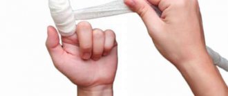
First aid for a skull fracture
If you suspect a fracture, you should immediately call an ambulance. If the victim’s condition is satisfactory and he is conscious, then he should be placed on his back (without a pillow), his head and upper body immobilized and secured, and an antiseptic bandage applied to the wound. If hospitalization is delayed, dry ice can be applied to the head. If there are no breathing problems, you can give the victim diphenhydramine or analgin.
In an unconscious state, the victim should be laid on his back in a half-turn position and his head should be turned slightly to the side to avoid aspiration in case of vomiting, loosen tight clothes, remove existing glasses, dentures, and jewelry. To secure the body, place a cushion of clothing or a blanket under one side of the body.
In case of acute respiratory distress, artificial respiration is performed through a mask. Cardiovascular drugs (sulfocamphocaine, cordiamine), glucose solution, Lasix are administered. In case of heavy bleeding and a sharp drop in pressure, Lasix is replaced with intravenous administration of polyglucin or gelatinol. During motor excitation, a solution of suprastin is injected intramuscularly.
Painkillers should be used with caution as they may complicate bleeding. The use of narcotic painkillers is contraindicated; they aggravate respiratory disorders.
Classification of skull base fractures
Fractures vary:
- According to the damaged bones of the same name;
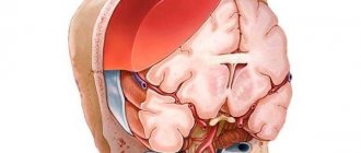
- Along the cranial fossae of the inner surface of the skull: anterior, middle and posterior;
- In relation to the external environment;
- By the presence or absence of bone displacement.
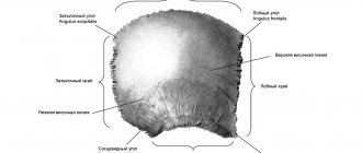
The occipital and sphenoid bones are part of the brain section of the skull. The temporal bones form the cranial vault and house the hearing organs: the pyramid of the temporal bone contains the tympanic cavity and the inner ear. The anterior fossa is formed by the frontal bone, the plate of the ethmoid bone, and is separated from the middle fossa by the edges of the sphenoid bone. The middle fossa is formed by the sphenoid and temporal bones. The posterior fossa is formed by the occipital bone, the posterior part of the sphenoid bone.
Non-displaced fractures are classified as open TBIs and have a favorable prognosis. If the fracture is accompanied by blood loss or leakage of cerebrospinal fluid, it is considered an open penetrating type TBI.
Treatment of a skull fracture
For an accurate and detailed diagnosis of injury, magnetic resonance imaging (MRI) or computed tomography (CT) is used. Depending on the severity and complexity of the damage, treatment can be conservative or surgical.
Conservative treatment
Conservative methods are indicated for mild to moderate injuries, when liquorrhea can be eliminated non-surgically.
It is necessary to observe strict bed rest, the head should be in an elevated position - this helps to reduce the secretion of cerebrospinal fluid. Treatment includes dehydration therapy (aimed at reducing fluid content in the organs), for this purpose, lumbar punctures (taking cerebrospinal fluid from the spinal cord at the lumbar level) are performed every 2-3 days, and subarachnoid insufflations (introduction into the subarachnoid space of the spinal cord) are carried out in parallel. the same amount of oxygen. Drugs that reduce the production of cerebrospinal fluid are also used - diuretics Diacarb, Lasix.
Physical activity is limited for six months. The victim must be registered with a traumatologist and neurologist, and be observed by an otolaryngologist and an ophthalmologist.
Particular attention should be paid to the prevention of intracranial complications of a purulent nature. For this purpose, sanitation of the nasopharynx, oral cavity and middle ear is carried out using antibiotics. In the presence of purulent complications, intramuscular or intravenous injections are supplemented by the introduction of antibiotics into the epidural space (endolumbar). For this, kanamycin, chloramphenicol, monomycin, and polymyxin are used. Also, endolumbar administration of kanamycin is carried out 2 days after the cessation of liquorrhea. The best way to select a drug is by culture of the cerebrospinal fluid flora or a smear taken from the nasal mucosa.
Surgery
Surgery is necessary in the following cases:
- Detection of a comminuted fracture;
- Damage or compression of brain structures;
- Leakage of cerebrospinal fluid through the nose, which cannot be stopped by conservative methods;
- Recurrences of purulent complications.
Surgical treatment is used in the presence of bleeding, hematoma or bone fragments that may pose a direct threat to life. In this case, trephination (opening) of the skull is performed, and after the operation, the bone tissue defect is closed with the removed bone or a special plate (in most cases). This is followed by long-term rehabilitation.
On topic: 12 folk methods for home treatment
General symptoms
Main complaints: severe focal pain in the forehead area; nausea; vomiting that does not bring relief; dizzy; short or prolonged loss of consciousness; with a depressed fracture, distortion of the frontal bone is noticeable; visual impairment, the consequence of which is double vision; excitability or passivity to what is happening; incoherence of consciousness; involuntary urination; secretion of cerebrospinal fluid through the nasal passage.
Associated symptoms of these fractures are concussion, severe bruising, and compression. After experiencing trauma, a person does not always realize what happened to him. And the injury itself, as a rule, occurs with accompanying damage to the body. A slight loss of consciousness is typical. The patient may not remember all events. Based on the duration of unconsciousness, a conclusion is made about the severity and complexity of the victim’s condition.
httpv://www.youtube.com/watch?v=embed/a8b5vhXG-hY
Clinical symptoms of a fracture of the frontal bone: nausea and vomiting, jumps in blood pressure and pulse, possible tinnitus, as well as disorientation. When providing emergency medical care, it is not advisable to give a person plenty of fluids, as there is a risk of brain swelling and fluid accumulation in the head area. Alcohol is strictly contraindicated; it is necessary to give the patient peace and quiet. He can be very sensitive to irritating external factors: bright lighting, sharp sounds. This dangerous diagnosis must be confirmed by a traumatologist and neurologist after examining the x-ray image. Such a thorough examination is associated with the further development of post-traumatic neurosis, up to attacks of epilepsy.
Brain contusion is the result of hemorrhage (bleeding) and destruction of the brain matter. There are three degrees of injury severity:
- mild – short-term loss of consciousness, lasting up to 15-25 minutes. The patient complains of weakness, drowsiness, malaise, and sometimes there is short-term amnesia.
- medium – loss of consciousness ranges from a couple of minutes to several hours, pronounced amnesia, cardiac dysfunction, meningeal symptoms are observed, the patient has a sensitivity disorder, paresis and paralysis develop.
- severe – prolonged stay in a comatose state, a sharp increase in body temperature, various neurological disorders, changes in muscle tone. Treatment must be carried out in a hospital, under the supervision of specialists. Further recovery depends on its effectiveness.
Compression progresses against the background of compression of cerebral tissues and increased intracranial pressure. This entails the death of brain cells. About 50% of cases end in death of the organism. Symptoms: sleep disturbance, acute pain in the head area, the appearance of hallucinations, which are replaced by apathy. Diagnosed based on clinical presentation and tomography results. The prognosis is not always comforting; disorders of motor, mental, and speech functions are observed.
httpv://www.youtube.com/watch?v=embed/a2mOnvQjVUY