Aseptic necrosis of the femoral head (AFH) is a pathological condition characterized by necrosis of the bone marrow of the femoral head. The chronic process eventually leads to extensive osteoporosis and osteonecrosis due to impaired local circulation.
The disease was described almost a hundred years ago, but its detailed study continues today. Medical statistics show that men get sick almost 8 times more often than women. The disease is detected between the ages of 20 and 40 years.
In half of the patients, the pathological process is detected on both sides. In 15% of cases, ANFH is combined with aseptic necrosis of the femoral condyles and the head of the humerus.
Causes of bone infarction
- Symmetrical lesions are typical.
- Bone infarction or ischemic bone necrosis
- Typically diagnosed after age 40 as an incidental finding.
- Bone infarction most often affects the metaphyses of long bones (usually the femur, tibia, or humerus)
- May extend far into the diaphysis (up to 20 cm in length)
- Infarction of the pituitary bone alone is rare
- Impaired blood supply to the bone (obstruction, compression, or rupture of blood vessels)
- Vascular occlusion
- Polycythemia vera (increased blood viscosity)
- Hemoglobinopathies
- Hypocorticism
- Pancreatitis
- Frostbite
- Severe burns
- Radiation therapy
- Decompression sickness (nitrogen emboli).
Stages of necrosis of the femoral head
Aseptic necrosis of the femoral head develops in stages. Experts distinguish the stages of the pathological process:
- Stage I – the onset of the disease – lasts about six months. At this stage, the spongy core of the head slowly breaks down. Patients complain of pain, which intensifies after exercise. The condition improves in the lying position. Periods of remission and exacerbation alternately occur.
- Stage II – impression fracture – lasts up to six months. The images reveal deformation and compression of the altered bone beams. Patients note that the pain becomes constant and does not subside when lying down. The volume of thigh muscle tissue is reduced by up to 20%.
- Stage III - sequestration - lasts about two years. The femoral head flattens and the joint space increases. The process of resorption of necrotic bone particles begins. In their place, connective and cartilaginous tissue is formed. Patients report severe pain and difficulty moving. Almost all patients use a cane, as it is impossible to walk otherwise. The shortening of the injured limb is visually determined.
- Stage IV - reparation - is characterized by the disappearance of fragmentation of the bone structure. The images show the normal outlines of the head of the femur, cyst-like areas of clearing due to incomplete restoration of the structure.
- Stage V – deforming arthrosis – the final stage, characterized by the appearance of osteophytes and the formation of cystic cavities. A flat head of the femur is visually determined, which does not correspond to the articular capsule. Patients cannot move independently and complain of acute pain running from the buttocks to the lower back.
Which method of diagnosing bone infarction to choose: MRI, CT, X-ray, ultrasound
Selection Methods
- X-ray examination, MRI.
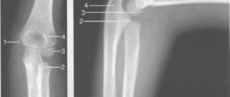
Radiological signs of bone infarction
- In the early stage - osteopenia and loss of trabecularity of bone tissue with a surrounding sclerotic ring
- Areas of sclerosis that form in the area of bone necrosis and the formation of relatively new bone tissue
- In the late stage, irregular areas of shading or calcification (sea pebble, grape, target, or ring pattern), usually located at the periphery
- The bone infarct area can be up to 20 cm long
- With an infarction in the area of the epiphysis, a tongue-shaped or wedge-shaped darkening spreads from the base of the epiphysis to the articular surface.
What will CT scans of bone show during a heart attack?
- Destruction of trabeculae
- Usually an accidental find.
Why is scintigraphy performed if a bone infarction is suspected?
- In the early stage of the disease - a decrease in the accumulation of radionuclide at the site of necrosis (“cold spot”)
- Later, an increase in radionuclide accumulation also appears (a “cold in a hot” spot).
What MRI images of bone will show during a heart attack
- In the early stage of the disease - edema (hypointense on T1-weighted image and hyperintense on T2-weighted image)
- Late demarcation along the periphery of the affected bone (hypointense on T1-weighted image; on T2-weighted image - hyperintense line towards the necrosis zone, consistent with granulation tissue)
- Hypointense line towards healthy bone (sclerosis, fibrosis): double line sign
- Accumulation of contrast agent in the peripheral zone
- In old bone infarcts, the signal intensity of the necrotic zone is equivalent to adipose tissue
- The peripheral zone is typically sinuous, resembling a garland.
a, b Mature bone marrow infarction.
(a) Sagittal proton density-weighted MRI with fat suppression. The image shows a garland-shaped sclerotic margin and a central area with adipose marrow signal. Multiple necrotic zones are located mainly in the metadiaphyseal region, but several are located directly next to the joint, and therefore there is a risk of flattening of the articular surfaces; b ) X-ray examination demonstrates a pronounced sclerotic margin and a central area of decreased radiolucency in the distal femur and tibia. The proximal femur shows an area of partial gross sclerosis that is sometimes difficult to distinguish from an enchondroma.
Treatment of ANFH
Avascular necrosis occurs due to hemodynamic disturbances. The disease is terrible, its consequences can be severe. It is very important to make a diagnosis in time, then there is a chance to save the leg and avoid surgical intervention. Comprehensive treatment of osteonecrosis depends on the stage and severity of the disease. At the onset of the disease, conservative therapy is used, later on they resort to surgery and endoprosthetics.
Goal of treatment:
- resumption of hemodynamics in the affected area;
- bone cell regeneration;
- restoration of limb functionality;
- relief from pain.
Treatment tactics by periods of illness
The approximate treatment regimen for osteonecrosis depends on the severity of the process. The patient must know its basic principles; each point of treatment is agreed upon with the patient, because only through the joint and concerted efforts of the doctor and the patient can the desired effect be achieved.
Initial stage of ANGBC
The initial stage of the disease lasts from several days to six months. This is the stage when blood vessels are injured. During this period, it is still possible to reverse the process by improving local trophism and stimulating the growth of bone tissue. Great emphasis in this period is placed on a gentle protective regime, thanks to which the head of the joint is unloaded.
Recommendations:
- The orthopedic regimen consists of following measures aimed at reducing the load on the affected joint. How to achieve this?
- it is necessary to reduce the time spent on your feet;
- use a stick or crutches while moving;
- standing for long periods of time should be avoided;
- whenever possible you need to rest, preferably in a lying position;
- eliminate the transfer of cargo;
- Exercise therapy:
- be careful when choosing physical exercises, avoid jumping, running, twisting in the hip area;
- be sure to do strength exercises to avoid congestion in the vessels and to activate blood flow (up to 30-40 minutes a day);
- All exercises must be coordinated with your doctor or exercise therapy instructor.
- Medicines (only as prescribed by a doctor):
- NSAIDs;
- vasodilators;
- if necessary, blockade with anesthetic,
- vitamin and mineral supplements.
- Massage – both general and local (the qualifications and experience of a specialist are extremely important);
- MBST
Second stage of ANGBC
Six months after the onset of the disease, intense bone destruction begins, and the femoral head becomes deformed.
Recommendations:
- Exercise stress:
- daily measured walking for half an hour to an hour (preferably Scandinavian) without getting tired;
- going up and down stairs;
- forward and reverse riding on an exercise bike (at a calm pace) or a calm bike ride (without going uphill);
- visiting the pool (water aerobics classes without sudden movements, swimming).
- Strengthening therapeutic exercises.
Therapeutic exercises are necessary in any period of illness. It is important that the set of exercises is compiled by an instructor and that at first he observes their correct implementation.
- Drug therapy:
- dilating vessels;
- promoting bone cell regeneration.
- Massage
Massage can also be beneficial in the second stage. But hand movements should be light, after the massage the patient should feel pleasant warmth
- Femoral head decompression - this intervention can help the patient and slow down the disintegration of the femoral head.
3rd stage
The disease enters the third stage usually after 8-9 months. The head is almost destroyed and the question of surgical intervention arises.
Recommendations:
- Exercise therapy.
- Massage.
- Drug therapy.
- Surgical intervention.
Physical therapy, drug therapy, massage and physiotherapy at this stage are auxiliary measures. The emphasis in this period is already on operations.
What diseases have symptoms similar to bone infarction?
Enchondroma
— Lobed image in the form of bunches of grapes
— Small, dotted or comma-shaped areas (calcifications) with a decrease in signal intensity, scattered in the central part of the lesion
Chondrosarcoma
— Highly differentiated forms are similar to enchondroma; if the diagnosis is in doubt, a biopsy is necessary
Infectious process (early stage)
— Lack of periosteal reaction
- Signal intensity on MRI is similar to edema
— Even a fresh infarction is often clearly limited and does not have a reaction from surrounding tissues
Material and methods
The authors used open Internet resources: electronic scientific library (elibrary), SciVerse (Science Direct), Scopus, PubMed and Discover. Keywords for searching sources of information: bone marrow edema, bone marrow, computed tomography, magnetic resonance imaging, bone fractures, joint injuries, trabecular edema, bone marrow edema, BME, MR images, MRI.
The article presents the main results of MRI studies of injuries and pathologies of bones (joints) in publications of foreign and domestic specialists, with an emphasis on diagnosing patients with RCM at different times of imaging. A number of examples present the comments of the authors of this publication on the forensic medical significance of the identified changes in the bone marrow.
Diagnostics
Diagnosing bone marrow cancer requires a comprehensive approach. The first step is to collect an anamnesis, assess existing complaints and conduct a physical examination. Next, the patient is sent for examination. Myeloma can be suspected based on a bone x-ray; leukemia can be suspected based on a general blood test.
To confirm the diagnosis, a morphological study of the tumor substrate is required. For this purpose, a biopsy or bone marrow puncture is performed. In addition, additional tests are carried out aimed at clarifying the molecular genetic profile of cancer. This is necessary to determine treatment tactics and select the optimal chemotherapy method.
1.Bone marrow
Bone marrow
has no direct relation to the human nervous system.
However, its importance is so great that it is reflected in the name. The internal contents of the bones are a vital organ called the “brain,” which emphasizes the high functional significance of the bone substance. As you know, the work of the entire body is organized in such a way that a constant exchange of substances is necessary between all parts of the body, organs and tissues. This function is performed by blood. It is in the bone marrow that the constant renewal of blood components occurs - the process of formation of new blood cells of three types: erythrocytes, platelets and leukocytes.
The second unique characteristic of bone marrow is the presence in its composition of stem cells that can transform into cells of any organ or any tissue inherent in a given organism. This feature is currently being actively studied and used in the most innovative methods for treating diseases that until recently were considered incurable (primarily oncology).
The weight of bone marrow tissue in the entire body is about three kilograms. Most of them are in the bones of the pelvis, sternum, ribs, skull and spine. The tissue is a soft, spongy substance. The contents of bones are two types of bone marrow: red and yellow. Red bone marrow
carries all the functional load for blood synthesis. Yellow is an excipient. Bone marrow tissue is functionally very labile and responds by increasing the production of certain blood components in response to processes occurring in the body (infections, bleeding, somatic disorders). The liver, spleen and lymph nodes are auxiliary organs of hematopoiesis, also involved in maintaining optimal blood composition.
A must read! Help with treatment and hospitalization!
Relapse
The likelihood of recurrence depends on the type of cancer. For example, most forms of acute leukemia respond well to therapy and have a high probability of complete cure. Chronic leukemia tends to develop slowly, but is very difficult to cure. Myeloma is also an incurable form of bone marrow cancer. The time to relapse with standard therapy is about 29 months, and with tandem transplantation it is about 42 months.
If a relapse develops, treatment tactics are determined depending on the period of relapse-free survival. Typically, if more than 6-12 months have passed, first-line chemotherapy regimens can be used. For shorter periods, the tactics are changed and drugs of the second and subsequent lines of therapy are prescribed, to which the cancer has not yet developed resistance.