Joints of the upper limb
Sternoclavicular joint. Formed by the sternal end of the clavicle and the sternal notch. A simple saddle joint with an articular disc, thanks to the disc, moves like a ball-and-socket joint. The volume of movement is limited. Strengthened by ligaments: interclavicular, costoclavicular, anterior and posterior sternoclavicular.
Acromial - clavicular. A simple joint of a flat shape, which can turn into synchondrosis, is strengthened by ligaments: coracoclavicular, acromioclavicular. Movements are very limited, mainly performed around the sagittal axis - sliding.
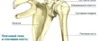
Shoulder joint. Formed by the head of the humerus and the glenoid cavity of the scapula. The joint is spherical, poorly congruent, with a wide articular lip along the edge of the cavity. Strengthened by the coracobrachial ligament. The movements of the joint are usually combined with the movement of the scapula, which together allows the limb to describe a hemisphere. The tendon of the long head of the biceps muscle passes through the joint cavity, which strengthens the joint. At the joint, the shoulder moves around three axes (flexion, extension, adduction and abduction, pronation and supination).
Elbow joint. Formed by the humerus, ulna and radius bones. It includes three joints: the humeroulnar
(trochlear, connects the shoulder block and the trochlear notch of the ulna),
brachioradial
(spherical, between the head of the humeral condyle and the articular cavity of the radius),
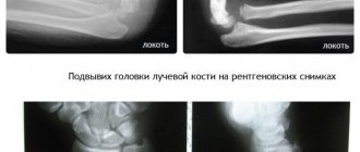
proximal radioulnar joint
(cylindrical, head of the radius and radial notch of the ulna). Since there is a membrane stretched between the radius and ulna bones, the sagittal axis in the humeral-radial joint is not used. As a result, flexion - extension of the order of 1400 is possible - humeroulnar + humeroradial, pronation - supination of the order of 1400 (can be up to 1800) - brachioradialis and radioulnar. Ligaments of the elbow joint: ulnar and radial - collateral, annular ligament of the radius.
Distal radioulnar joint. Cylindrical. It also participates in pronation - supination of the forearm, and forms a combined cylindrical joint with the proximal one.
Wrist joint. Articulation of the radius and 3 carpal bones: scaphoid, lunate, triquetrum. The disc under the ulna fuses with the articular surface of the ulna. This is a complex elliptical biaxial joint: flexion and extension, adduction and abduction.
Midcarpal joint. Between the two rows of carpal bones there is a complex, irregularly shaped surface (flat).
The ligamentous apparatus of the hand is very complex, the main ligaments are the collateral ligaments of the wrist - the radial and ulnar. The transverse ligament - holds the wrist - forms the carpal tunnel on the palmar side, through which nerves and blood vessels pass.
Carpometacarpal joints. Between the bones of the second row of the wrist and the bases of the metacarpal bones. All are flat, inactive 5-100, with the exception of the thumb. The saddle joint has a separate articular cavity and 2 axes of rotation, 40-600. can withstand a load of 60-100 kg, but dislocations are possible, which often become habitual.
Metacarpophalangeal joints. Spherical, 3 axes of rotation, but pronation and supination are carried out only passively. Strengthened by collateral and palmar ligaments.
Interphalangeal joints. Block-shaped, 1 axis of rotation. Flexion - extension - 110-1200 in the proximal, 80-900 in the distal. 3 ligaments: medial, lateral, palmar.
Radiation anatomy of the elbow joint
Radiation anatomy of joints
- X-ray anatomy of the elbow joint
- Normal elbow anatomy
- MRI anatomy of the elbow joint
- Ultrasound anatomy of the elbow joint
- Radiation criteria for normal structures of the elbow joint
If you are interested in ultrasound scanners, we can recommend scanners from Sonoscape. The website sonopro.ru has a large selection of ultrasound scanners at affordable prices. The company's managers will help with the choice and provide full information and technical support.
X-RAY ANATOMY OF THE ELBOW JOINT
The elbow joint is one of the most complex joints in its anatomical structure. It is formed by the distal epiphysis of the humerus and the proximal articular ends of both bones of the forearm. The distal metaphysis of the humerus has two epicondyles - a relatively large and steep medial one and a flatter lateral one. In the middle part of the dorsal and palmar surface of the metaphysis of the humerus there are two fossae - the coronoid and the olecranon fossa, separated by a thin bony septum that forms the bottom of these fossae.
The distal epiphysis of the humerus has a complex shape. Its lateral part is the head of the condyle of the humerus (or the so-called lateral condyle of the humerus), and the medial part has the shape of a block. The head of the radius entering the joint is flat and round and articulates with the head of the condyle of the humerus and with the radial notch of the proximal end of the ulna. The head of the radius passes into the neck, which has a well-defined tuberosity with a convex outer surface. The proximal end of the ulna has a trochlear notch and two processes. The coronoid process is characterized by its small size. It is located at the palmar surface of the trochlear notch. The massive olecranon process forms the superodorsal portion of the proximal end of the ulna. The triceps tendon is attached to its dorsal surface, which plays the role of an apophysis. On the radial side of the proximal end of the ulna, immediately below the trochlear notch, is a semicylindrical radial notch that articulates with the lateral surface of the head of the radius.
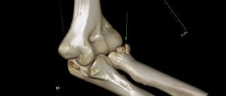
Thus, the elbow joint includes three joints that have a common joint cavity: the humeroradial, humeroulnar and proximal radioulnar joints (Fig. 19.26).
Rice. 19.26. X-ray of the elbow joint in an adult.
1 - humerus; 2 - head of the humerus (lateral condyle of the humerus); 3 - olecranon process of the ulna; 4 - medial epicondyle of the humerus; 5 - head of the radius; 6 - posterior “fat body”; 7 - anterior “fat body”.
After the formation of the elbow joint is completed, it is possible to evaluate all radiological indicators of its anatomical structure. On an anteroposterior radiograph, such indicators include, first of all, the ratio of the spatial positions of the shoulder and forearm, which is characterized by the magnitude of the angle formed by the intersection of the longitudinal axes of the named segments of the upper limb. Normally, the angle is open to the radial side when its value is 175-162°. The contours and structure of the metaepiphyses of the bones forming the elbow joint are smooth, smoothly rounded, with a greater or lesser degree of convexity. The structure of the distal metaphysis of the humerus is characterized by the presence of so-called arcades - a system of very powerful arcuate lines of force, convexity facing upward, as well as the display of the bottom of the coronoid fossa and the olecranon fossa in the form of an oval area of low optical density and the upper edge of the second fossa - in the form of an arcuate strip. The architectonics of the structure of the distal epiphysis of the humerus and the proximal metaphyses of the bones of the forearm is formed by systems of vertically oriented lines of force.
On radiographs in the lateral projection, the spatial position of the distal meta-epiphysis of the humerus is characterized by the magnitude of the angle formed at the intersection of the longitudinal axis of the diaphysis and the line connecting the bottom of the fossae of the humerus with the center of the articular surface of the lateral condyle of the humerus. The standard values for this angle (open to the ventral side) are 35-45°. The distal epiphysis of the humerus appears as four circles on a lateral radiograph. The largest and most ventrally located corresponds to the lateral condyle of the humerus, the smallest and most clearly defined - the notch between the shafts of the trochlea.
For the shoulder-ulnar joint, an indicator of the norm of anatomical relationships is the uniformity of the width of the x-ray joint space projected between the contour of the trochlear notch of the ulna and the lower part of the contour of the circle corresponding to the lateral shaft of the trochlea of the distal epiphysis of the humerus. The criterion for the norm of anatomical relationships in the brachioradial joint is the location of the center of the articular fossa of the head of the radial bone at the level of the border between the first and second quadrants of the head of the condyle of the humerus (counting from the ventral edge of the articular surface of the head). The given indicator is valid only if an x-ray is taken with the forearm positioned to the humerus at an angle close to 90°.
Stages of ossification of the bones of the elbow joint Age from 3 months to 1 year is characterized by the following changes.
During the first 9-12 months after birth, the metaepiphyses of the bones forming the elbow joint generally retain the degree of ossification achieved by the end of intrauterine development. Apart from the increase in the size of the diaphyses and the cartilaginous patterns of the epiphyses and apophyses, only minor ossification of the radial neck occurs. During this period, both epicondyles of the humerus, its distal epiphysis, the head and part of the neck of the radius, the entire coronoid process, as well as the dorsal and partially upper part of the olecranon process of the ulna have a cartilaginous structure.
Rice. 19.27. Radiographs of the elbow joint.
a — 1 year: 1 — humerus; 2 - metaphysis of the humerus; 3 - ulna; 4 - radius; 5 - ossification nucleus of the lateral condyle of the humerus.
b - 1 year. The ossification nucleus of the lateral condyle of the humerus appears: 1 - nucleus of the lateral condyle of the humerus.
c, d — 3 years: 1 — nucleus of the head of the lateral condyle of the humerus; 2 - area of the medial epicondyle; 3 - nucleus of the radial head.
From 1 to 4 years of age, the main manifestation of enchondral bone formation is the onset of ossification of the distal epiphysis of the humerus and the head of the radius (Fig. 19.27). In the distal epiphysis of the humerus during these periods, only the head of the condyle and partially the lateral shaft of the trochlea become ossified. The center of ossification of the head of the radial bone appears at the age of 3 years and is localized in its central section; at the age of 4 years, ossification of the medial epicondyle of the humerus may begin, although its average duration is 6-7 years. The cartilaginous structure of both epicondyles of the humerus is retained for up to 4 years; the entire medial shaft of the trochlea of the distal epiphysis of the humerus and about half the volume of cartilaginous models of the lateral shaft and head of the condyle; the predominant part of the head and about 1/3 of the length of the neck of the radius; the coronoid process and the superodorsal part of the olecranon process of the ulna.
An indicator of the correspondence of the local bone age to the passport age in children aged 1 year is the presence of centers of ossification of the head of the condyle and the lateral shaft of the trochlea of the distal epiphysis of the humerus, in children 3 years old - the presence of an ossification nucleus of the head of the radius.
7-11 years old. The age of 6 years is the period when ossification of the medial epicondyle of the humerus begins (Fig. 19.28). At 7 years of age, multiple centers of ossification of the medial shaft of the trochlea of the distal epiphysis of the humerus appear, localized mainly in the lateral two-thirds of its cartilaginous model. At about 8 years of age they fuse with each other, and at the same time the first ossification nucleus of the apophysis of the olecranon process of the ulna appears (Fig. 19.29). By the age of 10, almost the entire medial shaft of the trochlea has ossified and ossification of the upper part of the olecranon process of the ulna begins due to the appearance of one, sometimes two separate centers of ossification. During this age period, the ossification of the head of the radius also ends and the degree of ossification of the head of the condyle and the lateral shaft of the trochlea of the distal epiphysis of the humerus increases significantly, the architectonics of the bone structure of the metaphyses and partially the epiphyses of the bones forming the elbow joint is finally formed.
The cartilaginous structure by 11-12 years is preserved by: the lateral epicondyle of the humerus; marginal sections of the medial shaft of the block of the distal epiphysis of the humerus; a small area of the distal epiphysis of the humerus between the ossified parts of the lateral and medial shafts of the trochlea; about 1/2 the volume of the olecranon process of the ulna and most of the coronoid process, metaepiphyseal and apophyseal growth zones (Fig. 19.30).
Rice. 19.28. X-rays of the elbow joint (6 years). The nucleus of the medial epicondyle appears .
a: 1 - nucleus (apophysis) of the medial epicondyle; 2 - core (epiphysis) of the head of the radial bone; 3 - nucleus (epiphysis) of the lateral condyle of the humerus; 4 - distal metaphysis of the humerus, b: 1 - nucleus (apophysis) of the medial epicondyle; 2 - core (epiphysis) of the head of the radial bone; 3 - nucleus (epiphysis) of the lateral condyle of the humerus; 4 - ulna.
Rice. 19.29. X-rays of the elbow joint (6 years). The nucleus of the medial epicondyle appears.
a - 7 years: 1 - nucleus of the lateral condyle of the humerus; 2 - nucleus of the medial epicondyle; 3 - nucleus of the radial head; 4 - anterior “fat body”; 5 - coronoid process of the ulna; 6 - olecranon process of the ulna.
b — 8 years: 1 — small multiple nuclei of the epiphysis of the humerus; 2 - the ossification nucleus of the apophysis of the olecranon process of the ulna appears.
Rice. 19.30. X-ray of the elbow joint (11 years).
1 — multiple nuclei of the apophysis of the olecranon process of the ulna; 2 - growth zone of the condyles of the humerus.
When assessing the relationship between the spatial positions of the shoulder and forearm in this age group, it should be taken into account that the standard indicators of the angle between the longitudinal axes of these segments are 175°. An indicator of the correspondence of the local bone age to the passport age in children 7 years old is the presence of ossification nuclei of the medial shaft of the block of the distal epiphysis of the humerus and the medial epicondyle; in children 8-9 years old - complete ossification of the head of the radial bone and the presence of an ossification nucleus of the apophysis of the olecranon process of the ulna; in children 9 - Schlet - the presence of two (three) ossification nuclei of the apophysis of the olecranon process (see Fig. 19.30).
Rice. 19.31. X-rays of the elbow joint (14 years).
1 - fusion of the ossification nuclei of the olecranon process of the ulna; 2 - coronoid process of the ulna; 3 - medial epicondyle; 4 - lateral epicondyle.
12-14 years old. During this age period, ossification of the metaepiphyseal bones forming the elbow joint is completed (except for synostosis of the metaepiphyseal and apophyseal growth zones). All centers of ossification of the medial and middle sections of the medial shaft of the block of the epiphysis of the humerus merge and centers of ossification of its marginal sections appear, merging with the main part of the shaft by 14, less often by 15 years. The dorsal and proximal ossification nuclei of the apophysis of the olecranon process of the ulna reach the size of its cartilaginous model. Ossification of the lateral epicondyle of the humerus and the coronoid process of the ulna occurs.
By the age of 14, the cartilaginous structure is preserved by: a small strip of cartilaginous tissue between the medial and lateral shafts of the epiphysis of the humerus, a similar cartilaginous layer between the ossified dorsal and upper parts of the apophysis of the ulnar process of the ulna, and metaepiphyseal growth zones (Fig. 19.31).
At 15-17 years of age, synostosis of the metaepiphyseal and apophyseal growth zones begins and generally ends.
When assessing the relationship between the spatial positions of the shoulder and forearm, the same standard indicators are used as in adults.
Normal elbow anatomy
The elbow joint is represented by three joints: humeroulnar, humeroradial and radioulnar. All three joints communicate with each other and are surrounded by a common capsule. In addition, the head of the radius is surrounded by an annular ligament, which holds it to the ulna. The coronoid process and ulnar collateral ligament play an important role in stabilizing the elbow joint. The biceps brachii and brachioradialis muscles contribute to flexion, the triceps and ulnaris muscles contribute to extension. Pronation is carried out by the pronator teres and quadratus, while supination is carried out by the supinator and biceps muscle.
The muscles that act on the elbow joint can be divided into 4 groups:
- - anterior group - biceps and brachialis muscles;
- - lateral group - supinator, brachioradialis muscle and wrist extensors;
- - medial group - pronator teres, wrist flexors and palmaris longus;
- - posterior group - triceps and ulnar muscles.
The main large artery is the brachial artery. It lies anterior to the brachialis muscle and medial to the medial muscle and divides into the radial and ulnar arteries just inferior to the elbow joint.
The major nerves that cross the ulnar region are:
- - median nerve (n. medianus), running anterior to the brachial muscle;
- - radial nerve (n. radialis), located in the area of the elbow joint between the brachial and brachioradialis muscles;
- - ulnar nerve (n. ulnaris), which passes behind the medial epicondyle. The ulnar nerve groove is located along the posteromedial surface of the humerus.
The extensor muscles and their tendons begin in the region of the lateral epicondyle of the humerus, the flexor muscles - at the medial epicondyle. This is of particular importance in the development of muscle insertion tendinopathies, such as in tennis and golf athletes.
The aponeurosis of the biceps muscle plays an important role. It begins medially and somewhat distally from the biceps brachii tendon and crosses the brachial artery and median nerve (passing obliquely over the brachial artery and median nerve). In the region of the cubital fossa, which is bounded laterally by the brachioradialis muscle and medially by the pronator teres muscle, the biceps tendon lies laterally, the brachial artery lies adjacent to the tendon, and the median nerve lies medially.
The radial artery in most cases is a continuation of the brachial artery, and the ulnar artery departs from the brachial artery at a right angle. The lateral saphenous vein and the medially located main vein are the saphenous veins of the ulnar region. The median nerve passes between the head of the pronator teres and the ulnar artery, just beneath the ulnar head of the pronator teres. In an extended position, the internal, external supracondyles and olecranon are on the same horizontal line; in a bent position, they are located in such a way that they are the vertices of an isosceles triangle.
Rice. 19.32. CT scan of the elbow joint in the axial plane.
a: 1 - head of the radius; 2 - ulna; 3 - m. brachioradialis; 4 - t. pronator teres.
b: 1 - lateral epicondyle of the humerus; 2 - medial epicondyle of the humerus; 3 - m. brachialis; 4 - tendon of the triceps; 5 - fatty tissue (anterior “fat body”).
The ulnohumeral joint is a trochlear (helical) joint that has a trochlear notch with a smooth ridge into which the block of the humerus slides. In an extended position, the elbow joint forms the cubitus valgus. The articular surface of the head of the radius and the capitate eminence are partially congruent. The annular ligament covers the articular circumference of the head of the radius and is attached to the anterior and posterior edges of the radial notch of the ulna. Its width is about 10 mm.
The articular surfaces are covered with hyaline cartilage. The articular capsule in the anterior and posterior sections is thin. It is reinforced in front by fibers from the brachialis muscle and in the back by fibers from the olecranon muscle. Laterally, the capsule is strengthened by the collateral ligament, which holds the joint in place.
The internal joint capsule forms synovial folds over the extrasynovial fat in the ulnar, radial, and coronoid fossae (Fig. 19.32). A meniscus-like dense fold is constantly projected onto the humeroradial joint. The bursa is found in the olecranon region of both epicondyles of the humerus and the head of the radius. An accessory bursa may be observed under the extensor carpi radialis brevis muscle, as well as under the anconeus muscle.
MRI anatomy of the elbow joint
The elbow joint is examined in the coronal, sagittal and axial planes. Since the elbow joint is trochlear, the optimal position for examining the axial and coronal (Figs. 19.33, 19.34) planes is extension. In the sagittal plane (Fig. 19.35), the anatomical structures are also well identified with the elbow joint flexed. Recommended planes are shown in table. 19.5.
Rice. 19.33. MRI of the elbow joint. Axial plane.
a: 4 - m. brachialis; 8 - medial epicondyle; 10 - olecranon process of the ulna; 17 - m. brachioradialis; 19 - t. anconeus; 20 - nervusulnaris; 21 - tendon of the biceps brachii; 22 - t. pronator teres.
b: 1 - head of the radius; 2 - ulna; 3 - n. medianus; 4 - arteria, vena, n. radialis; 17 - m. brachioradialis; 21 - tendon m. biceps brachii; 22 - m. pronator teres.
Rice. 19.34. MPT of the elbow joint. Coronal plane.
1 - head of the radius; 2 - lateral condyle of the humerus; 3 - trochlea (medial condyle) of the humerus; 4 - m. brachialis; 5 - tendon of the t. extensorisdigitorum; 6 - coronoid process of the ulna; 7 - ligamentum collateral ulnare; 8 - medial epicondyle of the humerus.
Joint capsule. Usually not visible unless there is effusion or thickening. Normally, it is difficult to separate the capsule from the brachialis muscle anteriorly and from the triceps tendon posteriorly. Fatty layers between the synovial lines and fibrous layers of the capsule are visible posteriorly in the antecubital fossa and anteriorly in the coronoid fossa of the humerus. On sagittal sections, the fossae form an image resembling a “waist” figure.
Bursae of the elbow joint. The bursae of the elbow joint are divided into superficial and deep. Knowing their location is very important, since it is necessary to differentiate them from cysts and other pathological conditions. The superficial bursae are: medial epicondylar, lateral epicondylar, olecranon bursa (Fig. 19.36) The olecranon bursa potentially has three typical locations: subcutaneous, intratendinous and subtendinous. The subtendinous bursa is better visible in transverse and sagittal sections and can be mistaken for fluid in joint effusion, but if the fluid is not visible anterior to the joint, then it is more likely to be bursitis. Damage to the subcutaneous bursa in the area of the internal and external epicondyle must be differentiated from changes in the ligamentous apparatus. Normally, these bags are not visible, they can be seen in the presence of an inflammatory process, and they are clearly visible on T2-weighted images.
Arteries are difficult to differentiate from veins located nearby.
Nerves. Visualization of the nerve depends on the amount of periarticular fat. The median and radial nerves are better visualized in proximal, transverse sections. The ulnar nerve is better visible in transverse sections just dorsal to the internal epicondyle.
Rice. 19.35. MRI of the elbow joint. Sagittal plane.
a: 4 - m. brachialis; 9 — block of the humerus; 10 - olecranon process of the ulna; 11 - tendon m. brachialis; 12 - t. biceps brachii; 13 - t. triceps brachii; 14 — diaphysis of the humerus; 15 - diaphysis of the ulna; 16 - posterior “fat body”; 17 - anterior “fat body”,
b — elbow joint, sagittal plane (with signal suppression from fat): 11 — tendon m. brachialis; 16 - posterior “fat body” (the signal from fat is suppressed); 18 - anterior “fat body”; 19 - coronoid process of the ulna; 20 - olecranon process of the ulna,
c — elbow joint, sagittal plane (through the lateral condyle): 1 — head of the radius; 2 - lateral condyle of the humerus; 4 - m. brachialis; 12 - t. biceps brachii; 13 - t. triceps brachii; 14 - diaphysis of the humerus; 16 - m. extensor digitorum; 17 - t. brachio-radialis; 18 - t. extensor carpi ulnaris; 19 - t. anconeus.
d — MPT of the elbow joint, sagittal plane: 1 — head of the radius; 2 - lateral condyle of the humerus; 16 - m. extensor digitorum; 17 - t. brachioradialis; 18 - t. extensor carpi ulnaris; 19 - t. anconeus.
Table 19.5. Recommended planes for MRI examination of the anatomical structures of the elbow joint
| Structure type | Anatomical structures | Recommended cuts |
| Bone structures | Humerus, radius and ulna | Sagittal/coronal |
| Joints | Humeral-ulnar joint Radioulnar joint Internal articular structures and articular surfaces Hyaline cartilage Articular capsule | Sagittal/coronal Axial (oblique)/coronal Sagittal/coronal Sagittal/coronal Sagittal/coronal |
| Bone | Humeral trochlea Head of the radius Ulnar groove of the humeral trochlea Ulnar groove of the radius Coronoid process of the ulna Ulnar process and cubital fossa with fat layer | Sagittal/coronal Coronal/axial Sagittal Axial Sagittal Sagittal |
| Ligaments | Ulnar collateral ligament Radial collateral ligament Annular ligament of the radius | Coronal/axial Coronal/axial Axial |
| Bags | Subtendinous bursa of the olecranon process Supracondylar bursa | Sagittal/axial Axial/sagittal |
| Muscles and tendons | Attachment of the biceps and triceps muscles Attachment of the ulnar muscle All four muscle groups of the elbow region | Sagittal/Axial Sagittal Axial |
| Vessels and nerves | Arteries/veins Median nerve Radial nerve Ulnar nerve | Axial Axial Axial Axial |
Ultrasound anatomy of the elbow joint
Structures subject to ultrasound evaluation in the elbow joint include: the joint cavity, joint capsule, articular cartilage, muscle tendons, medial and lateral supracondyles, and ulnar nerve. Ultrasound of the elbow joint is performed from four standard approaches: anterior, medial, lateral and posterior.
The study is carried out by longitudinal and, less commonly, transverse scanning along the bone landmarks of the joint: the medial and lateral epicondyles of the humerus. Along the anteromedial surface, the bony landmarks are the tuberosity of the radius and the coronoid process of the ulna. When scanning with an anteromedial approach, the distal part of the biceps brachii tendon, the brachialis tendon, as well as the vessels of the coronoid fossa and the joint capsule are assessed. The anterolateral approach allows one to assess the condition of the lateral humeral condyle and the head of the radius. The annular ligament cannot be reliably visualized due to the oblique direction of its fibers.
Rice. 19.36. Synovial bursae of the elbow joint.
1 - olecranon bursa; 2 - intratendinous bursa; 3 - supratendinous bursa; 4 - tendon m. triceps.
When scanning from the posterior approach, the olecranon process serves as a bony landmark. The olecranon process, triceps tendon, olecranon bursa, olecranon fossa, and ulnar nerve are assessed, which can be identified by transverse scanning in the recess between the medial epicondyle along the posterior surface and the olecranon process. The triceps tendon is attached to the proximal part of the olecranon process, forming a bursa (bursa olecrani) at the site of attachment (Fig. 19.36).
The technique for performing ultrasound in children and adolescents does not differ from that in adults, however, one should remember the multiple centers of ossification of the epiphyses and apophyses of the bones that form the elbow joint (Fig. 19.37-19.41). On ultrasound, the thickness of hyaline cartilage and epiphyseal cartilage sums to form a thicker hypoechoic layer than in adults, which is characteristic of all incompletely ossified epiphyses in all joints.
Rice. 19.37. Ultrasound of the elbow joint (7 years).
a - coronally through the lateral condyle of the humerus:
I - nucleus of the head of the lateral condyle of the humerus; 2 - metadiaphysis of the humerus; 3 - nucleus of the radial head; 4 - proximal metaphysis of the radius; 5 - tendon m. extensor digitorum and radial collateral ligament; 6 - m. extensor carpi radialis. b — anterolateral approach through the lateral condyle:
1 - nucleus of the head of the lateral condyle of the humerus; 2 - region of the growth zone of the humerus; 3 - nucleus of the radial head; 4 - proximal metaphysis of the radius; 5 - metaphysis of the humerus; 6 - joint capsule; 7 - radial collateral ligament and tendon fibers m. extensor digitorum.
Rice. 19.38. Ultrasound of the elbow joint (11-12 years), medial coronal approach.
1 - nucleus of the medial epicondyle; 2 - metaphysis of the humerus; 3 - small nuclei of ossification of the block; 4 - ulna; 5 - tendon.
The olecranon bursa is located at the insertion of the triceps tendon.
muscles and consists of three sections: subcutaneous, intertendinous and subtendinous. The biceps tendon bursa is located behind the tendon at its insertion into the radial tuberosity. The supracondylar medial and lateral bursae lie under the tendons above the corresponding epicondyles.
Radiation criteria for normal structures of the elbow joint
- — the angle of the elbow joint during extension is about 162° (open to the radial side) (in children 175°);
- - the total thickness of the cortical layer of the humerus (in the middle third) is 5-10 mm;
- — the width of the joint space of the elbow joint in the posterior and lateral projection is 3 mm;
- – the presence at the level of the distal metaepiphysis of the humerus of a section of fatty tissue located on the anterior surface indicates the absence of effusion in the cavity of the elbow joint.
Rice. 19.39. Ultrasound of the elbow joint (11-12 years), lateral coronal approach.
1 - nucleus of the apophysis of the lateral epicondyle; 2 - lateral condyle of the humerus; 3 - head of the radius; 4 - radial collateral ligament; 5 - growth zone between the apophysis and lateral epicondyle.
Rice. 19.40. Ultrasound of the elbow joint (7 years), posterior scanning approach.
1 - humerus, posterior surface; 2 - nucleus of the condyle of the humerus; 3 - joint capsule and ulnar fossa; 4 - epiphysis (head) of the radius; 5 - metadiaphysis of the radius (posterior surface).
Rice. 19.41. Ultrasound of the elbow joint (5 years), medial approach.
1 - area of the epiphyseal cartilage (non-ossified trochlea nuclei); 2 - ulna; 3 - growth zone of the condyle of the humerus; 4 - metadiaphysis of the humerus.
Will physical therapy help?
Physiotherapeutic methods are good in the first and second stages of arthrosis. They reduce pain, strengthen periarticular muscles and increase mobility. Depending on the clinical picture and location of osteoarthritis, the patient may be prescribed a course of the following procedures:
- electrophoresis with individually selected medications;
- paraffin therapy (stimulates blood circulation in the joint);
- laser therapy (destroys osteophytes and prevents the formation of new ones);
- mud compresses (eliminate atrophic changes in tissues);
- acupuncture (reduces pain and restores muscle tone);
- massage and therapeutic exercises.
Most physiotherapeutic methods are allowed only during remission
For shoulder pain, this simple set of exercises will help improve joint mobility: