Osteochondropathy of the calcaneus is a disease characteristic of the female half of humanity. In particular, it affects teenage girls. Affects one or both legs.
Haglund's disease, or Haglund's deformity, is also called in the medical world, since the first description of the disease was given in 1907 by the Swedish orthopedic surgeon P. Haglund. It is believed that osteochondropathy occurs infrequently in young children, but can manifest itself at the age of 9 years. The disease can be cured completely without resorting to surgical methods . With only one caveat, that getting rid of the disease will begin in the early stages.
Causes of the disease
Schintz disease most often affects children: boys aged 9–11 years, girls aged 7–8 years.
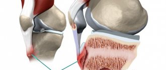
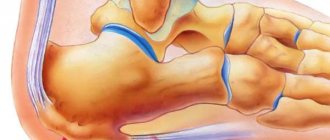
Adults who are actively involved in sports and dancing are also susceptible to pathology. Refers to diseases of adolescence and childhood; it is very rare in adults. The pathological process can involve either both or one limb. Unilateral lesions are diagnosed much more often than bilateral ones. The calcaneus is the largest bone of the foot; in its structure it belongs to the spongy bones. It bears a significant part of the load on the foot when running, walking and jumping, participates in the formation of several joints, and is the site of attachment of ligaments and tendons. On the back surface of the bone there is a protruding area - the calcaneal tubercle, which is affected in Schinz's disease. In the middle part, the Achilles tendon is attached to this tubercle, and in the lower part - the long plantar ligament.
Doctors have not yet identified the final cause that leads to the appearance of this pathology. But there are several factors that can contribute to the development of the disease:
- repeated heel injuries, sometimes minor;
- constant stress on muscles;
- diseases of the endocrine system associated with hormonal imbalance;
- diseases of the cardiovascular system;
- practicing certain sports, during which overstrain of the tendons of the muscles of the sole of the foot occurs;
- disruption of the body's absorption of calcium;
- vascular diseases;
- neurotrophic disorders;
- genetic predisposition;
- microtraumas in the area of the heel tubercle due to insufficient blood supply.
Due to excessive loads, vascular tone is disrupted, the bone area ceases to receive sufficient nutrients, and aseptic necrosis develops (bone destruction without inflammation and the participation of infectious agents).
In children and adults, the development of this pathology is provoked by the same factors.
There are five stages of Schinz's disease:
- Aseptic necrosis. The nutrition of the bone area is disrupted, and an area of necrosis occurs.
- Impression (depressed) fracture. The dead area cannot withstand normal loads and is “squeezed through.” Some areas of the bone are wedged into others.
- Fragmentation. The affected part of the bone is divided into separate fragments.
- Resorption of necrotic tissue.
- Repair. At the site of necrosis, connective tissue is formed, which is subsequently replaced by new bone.
Osteochondropathies
Ruben Garnikovich Minasyan
Osteochondropathies are a group of cyclical, long-term diseases, which are based on malnutrition of bone tissue with its subsequent aseptic necrosis.
Clinical manifestations of osteochondropathy are associated with resorption and replacement of damaged areas of bone. Diagnosis of osteochondropathy is based on ultrasound, x-ray and tomographic data. Treatment includes immobilization, physiotherapy, vitamin therapy, exercise therapy. According to indications, surgical treatment can also be performed. Osteochondropathies develop in patients of childhood and adolescence, more often affecting the bones of the lower extremities, and are characterized by a benign chronic course and a relatively favorable outcome.
COURSE OF OSTEOCHONDROPATHY.
First stage. Necrosis of bone tissue. Lasts up to several months. The patient experiences mild or moderate pain in the affected area, accompanied by impaired limb function. Palpation is painful. Regional lymph nodes are usually not enlarged. X-ray changes during this period may be absent.
Second stage . "Compression fracture." Lasts from 2-3 to 6 or more months. The bone “sags”, damaged bone beams wedge into each other. Radiographs reveal homogeneous darkening of the affected parts of the bone and the disappearance of its structural pattern. When the epiphysis is damaged, its height decreases and an expansion of the joint space is detected.
Third stage . Fragmentation. Lasts from 6 months to 2-3 years. At this stage, the dead areas of bone are reabsorbed and replaced by granulation tissue and osteoclasts. Accompanied by a decrease in bone height. Radiographs reveal a decrease in bone height, fragmentation of the affected parts of the bone with a chaotic alternation of dark and light areas.
The fourth stage of osteochondropathy . Recovery. Lasts from several months to 2 years. The shape and, somewhat later, the bone structure are restored.
The full cycle of osteochondropathy takes 2-4 years. Without treatment, the bone is restored with more or less pronounced residual deformation, which subsequently leads to the development of deforming arthrosis.
Osteochondropathy of the hip joint. (Legg-Calvé-Perthes disease). Affects the head of the hip bone. It most often develops in boys aged 4-9 years. The occurrence of osteochondropathy may be preceded by injury to the hip joint. It begins with a slight lameness, which is later joined by pain in the area of the injury, often radiating to the knee joint. Gradually, movements in the joint become limited. Upon examination, mild atrophy of the muscles of the thigh and lower leg, limitation of internal rotation and abduction of the hip are revealed. Painful reaction when loading the greater trochanter. Often the affected limb is shortened by 1-2 cm, caused by upward subluxation of the hip. Osteochondropathy lasts 4-4.5 years and ends with restoration of the structure of the femoral head. But without treatment, the head takes on a mushroom shape. The shape of the head does not correspond to the shape of the acetabulum, and deforming arthrosis develops over time. For diagnostic purposes, ultrasound and MRI of the hip joint are performed. To ensure restoration of the shape of the head, it is necessary to completely unload the affected joint. Treatment of osteochondropathy is carried out in a hospital with bed rest for 2-3 years. Skeletal traction may be applied. The patient is prescribed physiotherapy, vitamin therapy and climate therapy. Constant exercise therapy is of great importance to maintain the range of motion in the joint. If the shape of the femoral head is abnormal, osteoplastic operations are performed.
Osteochondropathy of the heads of the II and III metatarsal bones . (Keller's disease). It most often affects girls and develops at the age of 10-15 years. Keller's disease begins gradually. Periodic pain occurs in the affected area, lameness develops, which goes away when the pain disappears. Upon examination, slight swelling is revealed, sometimes - hyperemia of the skin on the dorsum of the foot. Subsequently, shortening of the second and third fingers develops, accompanied by a sharp limitation of movements. Palpation and axial load are sharply painful. This osteochondropathy does not pose a significant threat to subsequent dysfunction of the limb and the development of disability. Outpatient treatment with maximum load on the affected part of the foot is indicated. Patients are given a special plaster boot, vitamins and physical therapy are prescribed.
Osteochondropathy of the navicular bone of the foot. (Keller's disease). Rarely develops. It most often affects boys aged 3-7 years. Initially, pain in the foot appears for no apparent reason, and lameness develops. Then the skin on the back of the foot turns red and swells. Treatment is outpatient. The patient is limited in the load on the limb, in case of severe pain, a special plaster boot is applied, and physical therapy is prescribed. After recovery, it is recommended to wear shoes with arch supports.
Osteochondropathy of the tibial tuberosity . (Osgood-Schlatter disease). The disease develops at the age of 12-15 years, boys are more often affected. Swelling gradually appears in the affected area. Patients complain of pain that worsens when kneeling and walking up stairs. The function of the joint is not impaired or only slightly impaired. Treatment is conservative and carried out on an outpatient basis. Limitation of the load on the limb is prescribed; in case of severe pain, a plaster splint is applied for 6-8 weeks, physiotherapy (electrophoresis with phosphorus and calcium, paraffin baths), and vitamin therapy. The disease progresses favorably and ends with recovery within 1-1.5 years.
Osteochondropathy of the calcaneal tuberosity. Schintz disease develops very rarely, usually affecting children aged 7-14 years. Accompanied by the appearance of pain and swelling. Treatment is outpatient, load limitation, calcium electrophoresis and thermal procedures.
Partial osteochondropathy of articular surfaces. Typically develop between the ages of 10 and 25 in men. Usually in the knee joint area. An area of necrosis appears on the convex articular surface. Subsequently, the damaged area can separate from the articular surface and turn into an “articular mouse” (a loose intra-articular body). Diagnosis is carried out by ultrasound or MRI of the knee joint. In the first stages, conservative treatment is carried out: rest, physiotherapy.
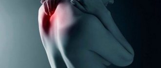
Osteochondropathy of the vertebral apophyses. Common pathology. Scheuermann-Mau disease occurs in adolescence, most often in boys. Accompanied by kyphosis of the middle and lower thoracic spine (round back). The pain may be mild or completely absent. Sometimes the only reason to visit an orthopedist is a cosmetic defect. Diagnosis of this type of osteochondropathy is carried out using radiography and CT scan of the spine. Additionally, to examine the condition of the spinal cord and ligamentous apparatus of the spinal column, an MRI of the spine is performed. Osteochondropathy affects several vertebrae and is accompanied by severe deformation that remains for life. To maintain the normal shape of the vertebrae, the patient must be provided with rest. For most of the day, the patient should remain in bed in a supine position (in case of severe pain, immobilization is performed using a posterior plaster bed). Patients are prescribed back and abdominal muscle massage and therapeutic exercises. With timely and correct treatment, the prognosis is favorable.
Osteochondropathy of the vertebral body. Calve's disease develops at the age of 4-7 years. The child, for no apparent reason, begins to complain of pain and a feeling of fatigue in the back. Upon examination, local pain and protrusion of the spinous process of the affected vertebra are revealed. Radiographs reveal a significant (up to 1/4 of normal) decrease in vertebral height. Usually one vertebra in the thoracic region is affected. Treatment is only inpatient. Rest, therapeutic exercises, and physiotherapy are indicated. The structure and shape of the vertebra is restored within 2-3 years.
With wishes of health,
Yours Ruben Minasyan
Symptoms and course of the disease
Osteochondropathy of the calcaneus, or Haglund-Schinz disease, is associated with excessive stress on the leg muscles and tendon injuries. Typically, the disease develops at puberty, although an earlier onset is possible - cases of Schinz's disease have been described in patients 7-8 years old. It starts gradually.
Among the main symptoms of Schinz's disease are:
- pain in a vertical position when resting on the heel. Pain syndrome occurs mainly after exercise (running, long walking, jumping);
- "bouncing gait";
- absence of pain at night and at rest;
- swelling, swelling of soft tissues, skin atrophy, but there are no signs of inflammation (hyperemia, characteristic pressure, burning or distension);
- atrophy of the lower leg muscles;
- skin hyperesthesia and increased tactile sensitivity of the affected area;
- pain in the foot when flexing and extending;
- lameness of the affected leg while walking;
- Palpation of the heel tubercle area is painful.
The severity of the disease may vary. In some patients, the pain remains moderate, and support on the leg is slightly impaired. In the other part, the pain progresses and becomes so unbearable that support on the heel is completely excluded. Patients are forced to walk, relying only on the middle and forefoot, and they need to use a cane or crutches.
In children, sometimes symptoms may disappear after the foot stops growing.
There is no need to be afraid of the disease, since its correct treatment most often leads to a positive result without complications.
Getting rid of osteochondropathy using traditional methods and prevention
At home, the patient applies warm compresses to the foot and heel. Use Dimexide (purchased at the pharmacy without a prescription): dilute in a 1:1 ratio with water and apply lotions to the sore spots using gauze or a cloth. To create a compress, place a bag on top, wrap it with a bandage and put on a wool sock. Keep the product for 50 minutes - 1 hour.
Warm salt baths have an analgesic effect: 100 g of salt, preferably sea salt, is dissolved in hot water poured into a basin and the heel is immersed there for half an hour. Afterwards, the foot is doused with warm running water, dried and a sock is put on.
Therapeutic physiotherapeutic procedures with paraffin and ozokerite can also be carried out at home. Dissolve both products in equal parts in a water bath, pour the mixture into a mold and let cool slightly. Next, the base for the application is made: you need to put a blanket on the floor, on top - oilcloth and polyethylene, on which to lay out the product. Place the heel on the prepared paraffin and wrap it in stages in polyethylene, oilcloth and a blanket. Keep your leg like this for up to half an hour.
Physical therapy is also indicated. Exercises for the feet that will strengthen the muscles, promote blood flow and relieve pain.
Exercise #1:
- lie on the floor on your side so that your sore leg is below;
- place your arm bent at the elbow under your head;
- move the leg that is at the top back;
- lift the sore foot and rotate it in different directions;
- Duration of the exercise: 1-1.5 minutes.
Exercise No. 2. Standing on the floor, lift your toes and try to spread them as far as possible, like a fan. In this case, the foot and heel should remain motionless. Stay like this for 15 seconds. Rest a little and repeat the exercise 3 more times.
Exercise No. 3. Sitting on a chair, place your heels on the floor and place your knees straight. Raise your toes alternately, bringing them to their starting position. Of course, the heel should not move during this. Let your feet rest for a few seconds and continue training your toes for another 5 minutes.
Exercise No. 4. Sit on a chair and close your soles for a while, relax. Repeat 15 times with breaks.
Exercise No. 5. Sit on the floor, keeping your legs extended. Place the sore foot on the knee of the healthy leg and, straining, make rotational movements.
After the symptoms of the disease have ceased to bother you, it is necessary to constantly take preventive measures so that osteochondropathy does not begin to develop again.
Wear loose, flat shoes.
Continue with physical therapy. Don't overcool your feet or put too much strain on them.
Share link:
Treatment of the disease
The choice of treatment method for the disease largely depends on the patient’s condition and the degree of complexity of the pathology.
Conservative treatment
Conservative treatment is carried out in an emergency room or outpatient orthopedic appointment. During the period of exacerbation, rest (ensuring complete immobility with the help of a plaster splint), physiotherapy aimed at improving local blood circulation, and wearing orthopedic shoes with an extended heel are necessary. In case of severe pain, short-term fixation with a plaster splint is possible. The patient is referred for ozokerite, electrophoresis of novocaine with analgin, ultrasound and microwave therapy. To reduce pain, use ice and prescribe drugs from the NSAID group. Taking vasodilators and vitamins B6 and B12 is also indicated.
Surgery
In some cases, with unbearable pain and lack of effect from conservative therapy, surgical intervention is performed. In case of severe deformation of the posterior part of the calcaneus, surgical intervention is performed.
Surgical intervention, depending on the degree of deformation, consists of:
- in the resection of osteochondral growths of the calcaneal tuber;
- shortening the calcaneus using osteotomy;
- neurotomy of the saphenous and tibial nerves and their branches. This operation consists of complete transverse cutting of the nerve trunk. It should be borne in mind that this operation not only completely relieves the patient of pain, but also leads to loss of skin sensitivity in the heel area.
Calcaneal epophysitis: symptoms and treatment
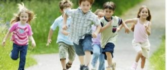
Children tend to run, jump, and kick the ball around the football field. Unfortunately, this is what causes some of them to develop heel pain. Pain in the foot occurs for various reasons.
The child could get bruised or get a callus. Or it may be caused by a disease of the calcaneus - epophysitis. This is pathology. It is accompanied by an inflammatory process in the distal heels. In this case, the chondral layer may be damaged, which will lead to further ruptures of the fibrous parts of the connective tissue. According to statistics, the majority of patients are boys under fourteen years of age.
Description and causes of the disease
The heel has two centers of ossification. But since bones are just being formed in childhood, they don’t exist as such yet. There are only cartilages. They harden over time and become bones.
One of the ossification centers develops immediately after the baby is born. The other undergoes changes during the first seven years of a child's life. At this time, only cartilage tissue separates them.
Children tend to move a lot. However, due to increased loads, there is a danger of micro-fracture of the connecting fibers. If this happens, inflammation begins. And if the loads do not stop, this process intensifies. Over time, the Achilles tendon becomes inflamed. And if the foot bends incorrectly, an injury results. It is this that opens the way to pathology—epophysitis.
Why does everything happen this way? There is no clear answer. Perhaps these are the consequences of repeated minor injuries received in physical education classes or during various sports games.
The tendency of a certain number of children to develop calcaneal epophysitis for unknown reasons also plays a role. This location, together with stress caused by jumping or running, increases the risk of disease.
The way you move is also important. It happens that children roll their feet excessively on the inside while walking and running. This can lead to weakened and pulled muscles. The result is that the depreciation does not work well, which means microtrauma.
Symptoms
Recognizing the disease is not always easy, because its course is not much different from other inflammations inherent in the heel. You need to start from the fact that the pain is felt precisely on the sides and back of the heel. The pain intensifies when the child runs, jumps and simply walks. Sometimes it hurts even when you touch the inflamed area.
Another symptom is that the movements of the foot become limited. Redness of the skin in the heel area may indicate advanced disease. There is one more detail. The possibility of micro-fractures was discussed above. So, if they exist, then swelling appears and the temperature in the affected node rises.
Because of all this, the patient needs to constantly keep the foot straightened. And this causes a lot of inconvenience, since it greatly limits mobility. But the worst thing is that if for some reason epophysitis is not treated, it can end in lameness. In view of all that has been said, it is obvious that if pain occurs in the heel, you should consult a doctor.
Treatment
Heels are one of the most vulnerable places in a person. They bear the heaviest load while walking or running.
A huge number of capillaries and nerve endings come out here, so self-medication of the pain that arises here is categorically unacceptable. Otherwise, you can get the opposite effect - increased pain.
There are still reasons why it is better to consult a doctor. We saw that it would not be possible to give the correct diagnosis on our own. There are several other rare problems that can also cause heel pain. So, let's go to a pediatric orthopedist.
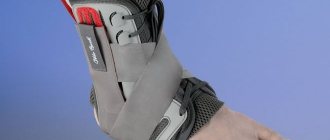
If the diagnosis of epophysitis is confirmed, you will be advised to ease the load on the epophysis of the child’s heel bone and reduce the tension of the tendons and ligaments attached to it. In other words, you need to provide support for the foot and stabilize the back of it during movement.
There are several means:
- Heel pads;
- Standard insoles;
- Custom insoles.
The effect of individual insoles is determined by the fact that they are made from a cast made according to the shape of the patient’s feet, which means they fully correspond to the shape of the foot of a particular child. Heel pads and standard insoles are not so effective, since they are made according to a standard design and simply cannot fully meet the needs of your child.
After unloading the painful area, so-called conservative treatment is applied. Its task: to reduce swelling and pain. Most often this is:
- Ice cooling;
- Exercises aimed at stretching the Achilles tendon;
- Eliminating inflammation ointments;
- Exemption from physical education.
It is important not only to solve current problems, but also to try to avoid them in the future. To do this, in particular, consult with a podiatrist about the type and quality of shoes the patient wears. It needs to be of high quality and not irritate the foot.
Conclusion
Epophysitis is a disease that can bring a lot of trouble and complicate life already in childhood.
Because of it, children are deprived of the opportunity to actively develop by doing physical education or simply playing with their peers. Do not hesitate to get a medical examination if your child complains of heel pain. Do not waste time and money on treatment. To date, it is believed that there are no long-term complications associated with childhood epophysitis. There will be difficulties if the disease turns out to be advanced. Author: K.M.N., Academician of the Russian Academy of Medical Sciences M.A. Bobyr
Diagnosis of the disease
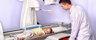
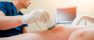
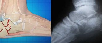
Diagnosis of Schinz's disease is based on radiological results (fragmentation and hardening of the apophysis, roughness of the tubercle of the calcaneus) and clinical data: the child's complaints and medical history (when did the symptoms appear, were there any foot injuries, etc.). In severe forms of the disease, radiographic signs such as separation of fragments of the marginal bone and an increase in the distance between the calcaneus and the apophysis are clearly visible. In doubtful cases, comparative radiography of both calcaneal bones is performed or patients are referred to a CT scan of the calcaneus or MRI of the calcaneus.
It is imperative to carry out a differentiated diagnosis with diseases such as osteomyelitis, periostitis, bursitis, tuberculosis of the calcaneus. If the disease is observed in older people, it is necessary to exclude the so-called “heel spur”. X-ray, MRI and CT help to definitively differentiate Schinz's disease from other diseases. In doubtful cases, consultation with an oncologist or TB specialist may be required.
Prices
| Disease | Approximate price, $ |
| Prices for diagnosing childhood arthritis | 2 000 — 3 000 |
| Prices for diagnosing childhood epilepsy | 3 100 — 4 900 |
| Prices for pediatric neurosurgery | 30 000 |
| Prices for treatment of childhood epilepsy | 3 750 — 5 450 |
| Prices for treatment of umbilical hernia in children | 9 710 |
| Disease | Approximate price, $ |
| Prices for hip replacement | 23 100 |
| Prices for clubfoot treatment | 25 300 |
| Prices for Hallux Valgus treatment | 7 980 |
| Prices for knee joint restoration | 13 580 — 27 710 |
| Prices for scoliosis treatment | 9 190 — 66 910 |
| Prices for knee replacement | 28 200 |
| Prices for treatment of intervertebral hernia | 35 320 — 47 370 |